

1. Introduction
Ichthyosaurs are Mesozoic marine reptiles that evolved a diversified array of pelagic forms, although only a handful of them have features considered as permitting deep-diving habits. One of these taxa, the Callovian–?Berriasian Ophthalmosaurus, is often regarded as a hyperspecialized exception among ichthyosaurs (Bakker, Reference Bakker, Caldwell and Kauffman1993), having enormous eyes (Motani, Rothschild & Wahl, Reference Motani1999; Humphries & Ruxton, Reference Humphries and Ruxton2002; Fernández et al. Reference Fernández, Archuby, Talevi and Ebner2005), ontogenetically reduced dentition (McGowan, Reference McGowan1976), vertebral regionalization (Buchholtz, Reference Buchholtz2001; Massare et al. Reference Massare, Buchholtz, Kenney and Chomat2006) and atrophied hind fins (Andrews, Reference Andrews1910).
The recent recognition of a clade of derived ichthyosaurs grouping ‘Ophthalmosaurus-like’ forms, ophthalmosaurine ophthalmosaurids, called this into question by suggesting that ichthyosaurs closely related to Ophthalmosaurus survived the Jurassic–Cretaceous boundary extinction and extended up to the late Albian (Fischer et al. Reference Fischer, Maisch, Naish, Liston, Kosma, Joger, Krüger, Pardo-Pérez, Tainsh and Appleby2012). The similarity between the Callovian–?Berriasian Ophthalmosaurus and the Hauterivian Acamptonectes densus in basicranium and shoulder girdle anatomy indicated a remarkable conservatism of morphology within ophthalmosaurines (Fischer et al. Reference Fischer, Maisch, Naish, Liston, Kosma, Joger, Krüger, Pardo-Pérez, Tainsh and Appleby2012), although significant differences in vertebral regionalization exist between these forms (Massare et al. Reference Massare, Buchholtz, Kenney and Chomat2006; Fischer et al. Reference Fischer, Maisch, Naish, Liston, Kosma, Joger, Krüger, Pardo-Pérez, Tainsh and Appleby2012). Accordingly, it is currently unclear whether ophthalmosaurines occupied several ecological niches or not during their long history (Bajocian to latest Albian period; up to 72 Ma), and how widespread the peculiarities of Ophthalmosaurus are among ophthalmosaurines.
In order to better characterize the evolutionary history of ophthalmosaurine ophthalmosaurids, we (1) describe a new taxon, Leninia stellans gen. et sp. nov., from a poorly sampled interval and location: the lower Aptian of western Russia, (2) determine its phylogenetic position, and (3) review ophthalmosaurine ecology across time and compare ocular characteristics among ophthalmosaurids.
2. Materials and methods
2.a. Institutional abbreviations
CM – Carnegie Museum of Natural History, Pittsburgh, PA, USA; GLAHM – The Hunterian Museum, University of Glasgow, Glasgow, UK; IRSNB – Royal Belgian Institute of Natural Sciences, Brussels, Belgium; MJML – Museum of Jurassic Marine Life, Ashfield, Kimmeridge, Dorset, UK; SSU – Geological Museum of Saratov State University, Saratov, Saratov Region, Russian Federation; UISU – Ulyanovsk State University Museum, Ulyanovsk Region, Russian Federation; UW – University of Wyoming, Laramie, Wyoming, USA; YKM – Ульяновский областной краеведческий мУзей им И.А. Гончарова [Ulyanovsk Regional Museum of Local Lore named after I. A. Goncharov], Ulyanovsk, Ulyanovsk Region, Russian Federation.
2.b. Geography and stratigraphy
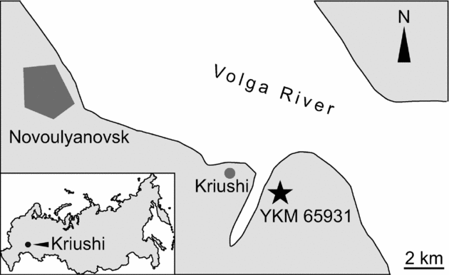
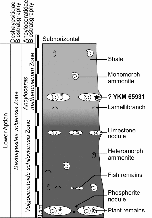
The described material was found on the right bank of the Volga River, 2 km southeast from Kriushi village, Sengiley district of Ulyanovsk Region, Russian Federation (Fig. 1). Only a small part of the lower Aptian is exposed in this locality and is contained within the Deshayesites volgensis Zone (Baraboshkin & Mikhailova, Reference Baraboshkin and Mikhailova2002) (Fig. 2). The section is described in the online Supplementary Material at http://journals.cambridge.org/geo.
Location of YKM 65931's discovery site, Kriushi area, Ulyanovsk Region, Russia. Geographic coordinates of the site: 54°5′53.38″N; 48°33′25.91″E.

Stratigraphic log at YKM 65931's discovery site, Kriushi locality, Russia. As the specimen was found in a limestone nodule on the riverbank, its position within the stratigraphic column is tentative.

The specimen YKM 65931 was not found in situ but embedded in a large limestone nodule lying on the shoreline. Field observations by I. M. Stenshin & G. N. Uspensky suggest this nodule is similar to those of the ‘upper nodule bed’ and would thus belong to the lower A. matheronianum Zone. This age, based on the biostratigraphy of heteromorph ammonites, is, however, tentative; therefore, we date this specimen with no further precision than Deshayesites volgensis Zone, because the whole section is contained within this zone in the Kriushi area.
2.c. Phylogeny
We coded Leninia in the phylogenetic matrix of Fischer et al. (Reference Fischer, Maisch, Naish, Liston, Kosma, Joger, Krüger, Pardo-Pérez, Tainsh and Appleby2012) with one additional character (see online Supplementary Material at http://journals.cambridge.org/geo). However, only 23.07% of the characters (12/52) could be coded owing to the absence of teeth and postcranial skeleton in the holotype. We used exact parsimony searches of TNT v1.1 (Goloboff, Farris & Nixon, Reference Goloboff, Farris and Nixon2010) to analyse the character matrix (exact algorithm, 10000 trees in memory) and calculate the Bremer support, Jacknife (removal probability of 36, 1000 replications) and bootstrap (standard, 1000 replications) values. We generated the phylogenetic tree using Winclada v.0.9 (Nixon, Reference Nixon1999) (detailed character state distribution in all optimizations, Bremer support, bootstrap and Jacknife values are available in Figs S1–S2 in the online Supplementary Material at http://journals.cambridge.org/geo). Characters were not weighted and, except for characters 17, 39 and 45, were not ordered (see Fischer et al. Reference Fischer, Maisch, Naish, Liston, Kosma, Joger, Krüger, Pardo-Pérez, Tainsh and Appleby2012 for an explanation of these characters).
2.d. Optics
We attempted to apply the calculations and methods of Motani, Rothschild & Wahl (Reference Motani, Rothschild and Wahl1999) and Fernández et al. (Reference Fernández, Archuby, Talevi and Ebner2005) to assess the visual acuity and ecology of Leninia. We calculated precise orbital, sclerotic and sclerotic aperture areas using a plugin for the freeware program Inkscape v0.48.2 (Albert et al. Reference Albert, Andler, Bah and Barbry-Blot2012) on high-quality photographs. Then, rather than simply making an average of the dorsoventral and anteroposterior lengths, we recalculated precise diameters from the measured areas, assuming that the measured areas were those of circles (d = 2*√[area/π]), in order to cope with diagenetic deformations. We used a similar technique to calculate the eye parameters of Sveltonectes insolitus (IRSNB R269). Because the skull of Leninia is crushed transversely, the axial length of the eye (a parameter in the estimation of the minimum f-number; Motani, Rothschild & Wahl, Reference Motani, Rothschild and Wahl1999) could not be assessed. We refrained from estimating this variable, as we feel the method used by Motani, Rothschild & Wahl (Reference Motani1999) is too subjective and hardly replicable since there is no constraint on the placement of the eyeball in the coronal plane. The suggestion of Fernández et al. (Reference Fernández, Archuby, Talevi and Ebner2005) to use the bending of the sclerotic plates as a proxy for eye size is also problematic because it involves a spherical eye, which is probably not the case in ichthyosaurs (Motani, Rothschild & Wahl, Reference Motani1999; Maisch & Matzke, Reference Maisch and Matzke2003; Fernández et al. Reference Fernández, Archuby, Talevi and Ebner2005). Therefore, we feel that the minimum f-number cannot be satisfactorily calculated in ichthyosaurs yet. Moreover, it should be noted that Motani, Rothschild & Wahl (Reference Motani, Rothschild and Wahl1999) examined a single specimen of O. natans (CM 878) to calculate the eye parameters but incorporated several specimens of O. icenicus (from the NHMUK) to analyse avascular necrosis. This is questionable, given the fact that the most recent phylogenies of ophthalmosaurids (including this one) do not recover Ophthalmosaurus as monophyletic (Druckenmiller & Maxwell, Reference Druckenmiller and Maxwell2010; Fischer et al. Reference Fischer, Maisch, Naish, Liston, Kosma, Joger, Krüger, Pardo-Pérez, Tainsh and Appleby2012). Accordingly, we simply compare the absolute sclerotic aperture of ophthalmosaurids as a proxy for dim light vision but we do not assess their deep-diving habits.
3. Systematic palaeontology
Order ICHTHYOSAURIA Blainville, Reference Blainville1835
Family Ophthalmosauridae Baur, Reference Baur1887
Subfamily Ophthalmosaurinae Baur, Reference Baur1887 sensu Fischer et al. Reference Fischer, Maisch, Naish, Liston, Kosma, Joger, Krüger, Pardo-Pérez, Tainsh and Appleby2012
Leninia stellans gen. et sp. nov.
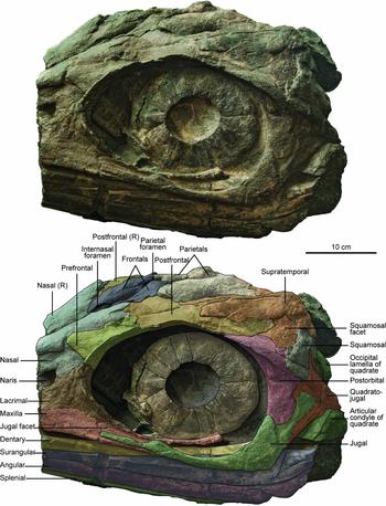
Photograph and interpretation of the holotype specimen of Leninia (YKM 65931) in lateral view.

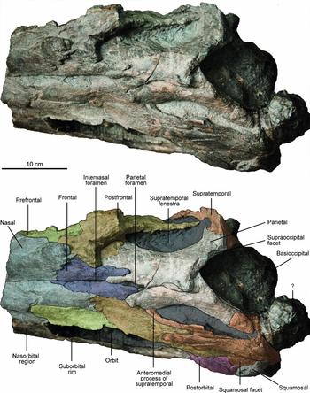
Photograph and interpretation of the holotype specimen of Leninia (YKM 65931) in dorsal view.

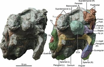
Photograph and interpretation of the holotype specimen of Leninia (YKM 65931) in posterior view. Abbreviations: ECA – concave extracondylar area; Foram. – foramina; Hy. pr. – hyoid process of the stapes; Postorb. – postorbital; Prefr. – prefrontal; QJ – quadratojugal; Sq. – squamosal; St. pr. – stapedial process of the supratemporal.

Holotype. YKM 65931, an articulated but incomplete skull preserved in three-dimensions.
Type horizon and locality. Deshayesites volgensis Zone, lower Aptian, Lower Cretaceous of the Kriushi locality, Sengiley district, Ulyanovsk Region, Russia.
Etymology. The museum where YKM 65931 is housed is located within the Lenin Memorial and Lenin school complex in Ulyanovsk; accordingly, the generic name reflects the geohistorical location of the find. The specific name refers to the peculiar frontal–parietal suture, which is star-shaped in the holotype.
Diagnosis. Leninia stellans is characterized by the following potential autapomorphies within Ophthalmosauridae: posterior process of the maxilla extending as far as the middle orbit (reminiscent of Ichthyosaurus intermedius; Maisch, Reference Maisch1997); presence of an anterior process on the prefrontal covering the nasal; frontal–parietal suture formed by the means of elongate medial forked processes of the frontal and the parietal; presence of an anterolateral process of the supratemporal contacting the parietal. Leninia stellans is also characterized by a combination of features shared by some other ophthalmosaurids (see Description, Section 4).
Note. Additional remains resembling the holotype of Leninia have been found in other Aptian localities near Ulyanovsk and Saratov; these fossils (UISI 115, a partial braincase; SSU 104a/28, a partial skull; SSU 14/53, a partial skull) are, however, fragmentary and cannot be determined unambiguously at the present time.
4. Description
4.a. Skull roof
4.a.1. Maxilla
The posterior end of the maxilla is preserved, but the narial lamella and its possible contribution to the narial aperture cannot be described because of the poor preservation of that region. The anterior cross-section suggests that the maxillary dental groove is extremely shallow relative to the size of the skull (height: 16 mm). Posteriorly, the maxilla forms a long palatal plate ventral and medial to the jugal. Unusually, this plate extends at least up to the middle-orbit level, whereas it ends more anteriorly in other ophthalmosaurids (e.g. Ophthalmosaurus icenicus, Platypterygius australis; Kear, Reference Kear2005; A. Kirton, unpub. Ph.D. thesis, Univ. Newcastle upon Tyne, 1983). The medial part of that plate is broken and pushed dorsally into the orbit by the dorsal surface of the mandible (Fig. 3).
4.a.2. Lacrimal
The posteroventral process of the lacrimal lies on the dorsal surface of the palatal plate of the maxilla. The lacrimal extends dorsally above the level of the narial aperture and contacts both the prefrontal and the nasal. The nasal–lacrimal contact is elongated and the lacrimal forms the posterior edge of the naris; therefore, it excludes the prefrontal from the narial aperture, unlike in Aegirosaurus and Sveltonectes (Bardet & Fernández, Reference Bardet and Fernández2000; Fischer et al. Reference Fischer, Clément, Guiomar and Godefroit2011b , respectively).
4.a.3. Jugal
The anterior process and the shaft of the jugal are extremely reduced in dorsoventral height (shaft is 13 mm high at middle orbit). Anteriorly, it forms a transversely compressed ramus covering the lateral surface of the maxilla. The shaft is rounded in cross-section. The posterior plate of the jugal is well developed and expends dorsally up to the middle-orbit level, even more than in O. icenicus, and as in Mollesaurus (Fernández, Reference Fernández1999).
4.a.4. Postorbital
The postorbital is of usual shape, with a prominent dorsal orbital rim and a posteroventral plate articulating with the quadratojugal and the jugal. As in Ophthalmosaurus (e.g. Andrews, Reference Andrews1910), the postorbital is reduced in anteroventral length (44 mm) with respect to the size of the orbit (anteroposterior length ≈ 265 mm).
4.a.5. Quadratojugal
The quadratojugal is covered by all elements of the postorbital region. Nevertheless, it appears more robust and longer anteroposteriorly than in Ophthalmosaurus icenicus (Andrews, Reference Andrews1910; A. Kirton, unpub. Ph.D. thesis, Univ. Newcastle upon Tyne, 1983). It articulates dorsally and anterodorsally with the squamosal. The anterior surface of the quadratojugal is thin, forming a concave lateral articular surface beneath the postorbital, the squamosal and the jugal. An angle is present between the squamosal and the postorbital/jugal facet, as in P. hercynicus (Fischer, Reference Fischer2012), but it is more conspicuous in Leninia. Posterodorsally to these large facets, the quadratojugal thickens and becomes pillar-shaped. Posteroventrally, this pillar slightly extends posteriorly to form the processus quadratus. As described by Kirton in O. icenicus (A. Kirton, unpub. Ph.D. thesis, Univ. Newcastle upon Tyne, 1983), there is a concave area for ligamentous attachment dorsomedially to the processus quadratus.
4.a.6. Squamosal
The squamosal is present, unlike in P. australis (Kear, Reference Kear2005) and P. americanus (Romer, Reference Romer1968). It is, however, incomplete; only its posteroventral part is preserved, but its general shape can be assessed because it covers numerous bones of the postorbital region (postfrontal, postorbital, quadratojugal), leaving a shallow facet textured by a series of subtle dorsoventral ridges. However, we refrained from coding character 14 (squamosal shape) for YKM 65931, but instead put the polymorphism 0/1 (as state ‘2’ stands for the absence of a squamosal). The squamosal covers the postfrontal extensively and the posterodorsal lamella of the postorbital. The squamosal–quadratojugal contact is not conspicuous.
4.a.7. Nasal
The nasal participates in the dorsal edge of the naris and presumably contacts the lacrimal over a long distance since the prefrontal is excluded from the narial aperture (see below). Dorsally to the naris, the nasal forms a subtle lateral wing as in Acamptonectes (Fischer et al. Reference Fischer, Maisch, Naish, Liston, Kosma, Joger, Krüger, Pardo-Pérez, Tainsh and Appleby2012) and some derived platypterygiines (P. australis, P. bannovkensis; Kear, Reference Kear2005 and V. Fischer, pers. obs., respectively). As in Acamptonectes (Fischer et al. Reference Fischer, Maisch, Naish, Liston, Kosma, Joger, Krüger, Pardo-Pérez, Tainsh and Appleby2012), the prefrontal facet is set directly posteriorly to the lateral wing, but there is no clear evidence for a foramen in that region. The posterior part of the nasal is medially incomplete, but the nasal seems to be excluded from the internasal foramen by the frontal.
4.a.8. Prefrontal
The prefrontal forms a thick anterior process constricting the nasal, unlike in other ophthalmosaurids (e.g. Andrews, Reference Andrews1910; Kear, Reference Kear2005) where the prefrontal is restricted to the anterodorsal orbital rim. The lacrimal–prefrontal suture is straight, unlike in Simbirskiasaurus (Ochev & Efimov, Reference Ochev and Efimov1985; V. Fischer, pers. obs.), O. icenicus (A. Kirton, unpub. Ph.D. thesis, Univ. Newcastle upon Tyne, 1983) and possibly Sveltonectes (Fischer et al. Reference Fischer, Clément, Guiomar and Godefroit2011b ); this suture is placed lower than in Athabascasaurus (Druckenmiller & Maxwell, Reference Druckenmiller and Maxwell2010) and higher than in Aegirosaurus (Bardet & Fernández, Reference Bardet and Fernández2000).
4.a.9. Frontal
The frontal is roughly triangular. It encloses the so-called internasal foramen. A long posterior process interdigitating with the forked process of the parietal forms the anterior margin of the parietal foramen (Fig. 4). The frontal is strongly reduced laterally and does not participate in the anterior margin of the supratemporal fenestra unlike in many platypterygiine ophthalmosaurids (Fischer et al. Reference Fischer, Clément, Guiomar and Godefroit2011b ; Fischer, Reference Fischer2012), but as in Athabascasaurus (Druckenmiller & Maxwell, Reference Druckenmiller and Maxwell2010) and Ophthalmosaurus (Gilmore, Reference Gilmore1906; A. Kirton, unpub. Ph.D. thesis, Univ. Newcastle upon Tyne, 1983).
4.a.10. Postfrontal
The anterior part of the postfrontal contacts the nasal over a short distance whereas its anteromedial part contacts the frontal over a long distance. As in Ophthalmosaurus (Gilmore, Reference Gilmore1906; A. Kirton, unpub. Ph.D. thesis, Univ. Newcastle upon Tyne, 1983) and unlike in Sveltonectes (Fischer et al. Reference Fischer, Clément, Guiomar and Godefroit2011b ), P. australis (Kear, Reference Kear2005), P. hercynicus (Fischer, Reference Fischer2012) and Athabascasaurus (Druckenmiller & Maxwell, Reference Druckenmiller and Maxwell2010), the postfrontal does not form a Y-shaped anterior process. The postfrontal is excluded from the lateral margin of the supratemporal fenestra by an elongated anteromedial process of the supratemporal, which constitutes an autapomorphy. However, the postfrontal seems to contact the parietal internally, as suggested by the broken supratemporal on the right side. Medially, the postfrontal forms a long process interdigitating with the supratemporal. The posterolateral part of the postfrontal is sheet-like and contacts the postorbital laterally and the squamosal posteriorly.
4.a.11. Parietal
The anterior margin of the parietal forms a forked process receiving the posterior process of the frontal (Fig. 4). The parietal contacts the anteromedial process of the supratemporal, a unique condition among ichthyosaurs. Both parietals are crushed against each other medially; there is, however, no evidence of a parietal crest. The posteromedial notch, receiving the dorsal surface of the supraoccipital, is markedly concave, even more than in Athabascasaurus (Druckenmiller & Maxwell, Reference Druckenmiller and Maxwell2010); however, the moderate lateral crushing of the skull does not permit satisfactory assessment of the shape of this notch in YKM 65931. The parietal is covered posterolaterally by the supratemporal.
4.a.12. Supratemporal
The supratemporal is extensive and forms the entire lateral and posterior edges of the supratemporal fenestra. Anteromedially, the supratemporal forms a long finger-like process contacting the anterolateral part of the parietal. A similar process is present in O. icenicus (A. Kirton, unpub. Ph.D. thesis, Univ. Newcastle upon Tyne, 1983), but it does not contact the parietal anteriorly in this taxon. Laterally to that process, the supratemporal receives the posteromedial finger-like process of the postfrontal. Ventrolaterally to that zone, the supratemporal is sheet-like and covers the postfrontal. It is, however, separated from the postorbital by the postfrontal and the squamosal, unlike in Caypullisaurus, where the supratemporal contacts the postorbital (Fernández, Reference Fernández2007). A squamosal facet is present posterolaterally. Posteriorly, the supratemporal is expanded, closing the dorsolateral part of the basicranium. A few foramina are present. Posteroventrally, the supratemporal forms a process contacting the stapes (Fig. 5), a feature previously regarded as unique to O. icenicus (A. Kirton, unpub. Ph.D. thesis, Univ. Newcastle upon Tyne, 1983) and O. natans (Gilmore, Reference Gilmore1906).
4.a.13. Pterygoid
Both pterygoids are present but can only be seen in cross-section slightly posterior to the dorsal lamella and are distorted by the lateral compression of the skull. The lateral lamella is thin and extensively covers the quadrate.
4.b. Basicranium
4.b.1. Basioccipital
The extracondylar area of the basioccipital is partly reduced, and forms a wide concave area external to the articular condyle (Fig. 5), which is an ophthalmosaurine synapomorphy (Fischer et al. Reference Fischer, Maisch, Naish, Liston, Kosma, Joger, Krüger, Pardo-Pérez, Tainsh and Appleby2012). As in adult Acamptonectes but unlike in Mollesaurus (Fernández, Reference Fernández1999) and Ophthalmosaurus (Fischer et al. Reference Fischer, Maisch, Naish, Liston, Kosma, Joger, Krüger, Pardo-Pérez, Tainsh and Appleby2012), this groove is continuous ventrally, although a ventral notch is present. There is a depression anterior to the peripheral edge of the concave area, as in Acamptonectes (Fischer et al. Reference Fischer, Maisch, Naish, Liston, Kosma, Joger, Krüger, Pardo-Pérez, Tainsh and Appleby2012) and Ophthalmosaurus (A. Kirton, unpub. Ph.D. thesis, Univ. Newcastle upon Tyne, 1983). The occipital condyle is bulbous, slightly deflected peripherally, and the notochordal pit is set in the dorsal half of the condyle. Unfortunately, the dorsal surface of the basioccipital is not available for study, so the shape of the floor of the foramen magnum, which may be diagnostic in ophthalmosaurids (Fischer et al. Reference Fischer, Maisch, Naish, Liston, Kosma, Joger, Krüger, Pardo-Pérez, Tainsh and Appleby2012), cannot be assessed on this specimen.
4.b.2. Stapes
As in Acamptonectes, the occipital head of the stapes is markedly expanded dorsoventrally (≈53 mm high) and the shaft is a slender rod (≈ 25 mm high) (Fig. 5). A large hyoid process is present, but this feature appears variable in ophthalmosaurids (Fischer et al. Reference Fischer, Maisch, Naish, Liston, Kosma, Joger, Krüger, Pardo-Pérez, Tainsh and Appleby2012). The quadrate head of the stapes is not preserved.
4.b.3. Quadrate
The quadrate is ear-shaped as in most ophthalmosaurids. An occipital lamella is present, unlike in P. hercynicus (Kolb & Sander, Reference Kolb and Sander2009; Kuhn, Reference Kuhn1946) and some specimens referable to Platypterygius sp. (V. Fischer, pers. obs.).
4.c. Lower jaw
The posterior part of the lower jaw is preserved but lacks distinctive or diagnostic features. A coronoid process is present, although the presence of this process varies intraspecifically in some ophthalmosaurids (Fischer et al. Reference Fischer, Maisch, Naish, Liston, Kosma, Joger, Krüger, Pardo-Pérez, Tainsh and Appleby2012). The fossa surangularis is present but extremely reduced. As in all ophthalmosaurids (Motani, Reference Motani1999), the angular is markedly exposed laterally.
4.d. Sclerotic ring
The left sclerotic ring is complete and almost undistorted. It is composed of 14 intermeshing trapezoidal plates. Each individual plate bears subtle striations on its lateral surface, and the internal edge is crenulated. The external edge is bent medially. The sclerotic aperture is moderately small compared to the orbital area (11.03%, see Section 5), suggesting that the specimen was mature (Fernández et al. Reference Fernández, Archuby, Talevi and Ebner2005).
5. Results and discussion
5.a. Phylogenetic position
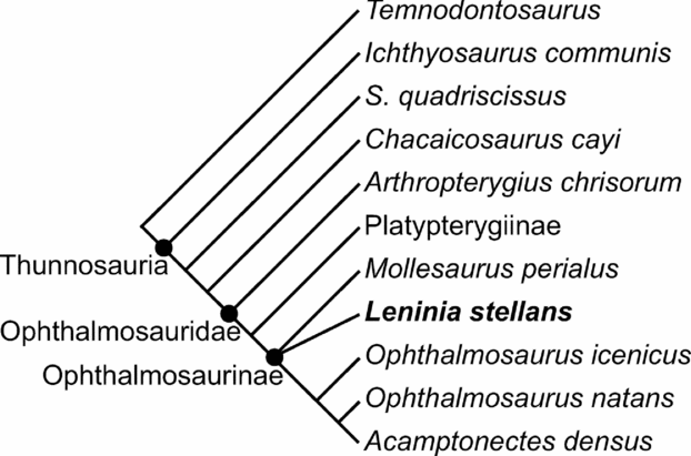
The cladistic analysis (a single most parsimonious tree, 103 steps long, consistency index: 0.55, retention index: 0.67) recovers Leninia as a basal ophthalmosaurine ophthalmosaurid, forming a polytomy at the base of the subfamily with the Bajocian taxon Mollesaurus perialus (Fig. 6). This polytomy is actually an ‘irresolvable’ node, because there is no character state in the dataset that could decipher which taxon is more derived than the other. In other words, the branch separating Leninia from Mollesaurus is of length = 0. This uncertainty and the surprisingly basal position of Leninia among ophthalmosaurines are possibly due to the large amount of missing data for both Mollesaurus and Leninia. Moreover, the skull of the only other Cretaceous ophthalmosaurine, Acamptonectes, is poorly known, preventing detailed comparison with that of Leninia. The large amount of missing data of the basal ophthalmosaurine Mollesaurus perialus is also the main reason why the clade Ophthalmosaurinae is supported by a very small number of unambiguous synapomorphies in the analysis undertaken by Fischer et al. (Reference Fischer, Maisch, Naish, Liston, Kosma, Joger, Krüger, Pardo-Pérez, Tainsh and Appleby2012). The inclusion of Leninia in the phylogenetic analysis does not modify this scheme as it adds a large number of uncertainties on postcranial anatomy as well. Additionally, while the exclusion of Mollesaurus from the data matrix provides additional unambiguous synapomorphies to diagnose the clade Ophthalmosaurinae, it does not improve the support of the tree, quite the contrary. Nevertheless, our analysis adds an unambiguous and non-homoplastic synapomorphy uniting ophthalmosaurines: the presence of a supratemporal–stapes contact (character state 52.1; see online Supplementary Material at http://journals.cambridge.org/geo), whereas they were only united by a single homoplastic unambiguous synapomorphy in the analysis of Fischer et al. (Reference Fischer, Maisch, Naish, Liston, Kosma, Joger, Krüger, Pardo-Pérez, Tainsh and Appleby2012). Despite the morphological similarities between Ophthalmosaurus and Cretaceous ophthalmosaurines, more data are still needed to clarify the tempo of character acquisition in basal ophthalmosaurines.
Simplified version of the single most parsimonious tree (length = 102 steps, consistency index = 0.55, retention index = 0.67) resulting from the cladistic analysis. All nodes received a Bremer support of 2, except the clade Ophthalmosaurus natans + Acamptonectes densus (1). The bootstrap and Jacknife values are low; the clade Ophthalmosauridae receives values > 50%: bootstrap = 54%, Jacknife = 53%. See online Supplementary Material at http://journals.cambridge.org/geo for optimizations.

5.b. Evolutionary history of ophthalmosaurines
Only two groups of marine animals evolved a very large eye size: the modern giant and colossal squids and ichthyosaurs (Nilsson et al. Reference Nilsson, Warrant, Johnson, Hanlon and Shashar2012). The size of the eye is related to ecology, where large eyes usually indicate adaptations for feeding and/or predator detection in low light setting, such as the deep marine realm (Motani, Rothschild & Wahl, Reference Motani, Rothschild and Wahl1999; Humphries & Ruxton, Reference Humphries and Ruxton2002; Fernández et al. Reference Fernández, Archuby, Talevi and Ebner2005; Nilsson et al. Reference Nilsson, Warrant, Johnson, Hanlon and Shashar2012). Whereas relative eye size is linked to ontogeny and/or possible evolutionary mechanisms such as paedomorphosis (Fernández et al. Reference Fernández, Archuby, Talevi and Ebner2005), absolute eye size determines visual acuity (Motani, Rothschild & Wahl, Reference Motani, Rothschild and Wahl1999; Humphries & Ruxton, Reference Humphries and Ruxton2002; Fernández et al. Reference Fernández, Archuby, Talevi and Ebner2005; Nilsson et al. Reference Nilsson, Warrant, Johnson, Hanlon and Shashar2012). The absolute size of the sclerotic aperture is a good estimation of the size of the cornea (and thus the dilated pupil) and is likely to remain rather constant during ontogeny (Fernández et al. Reference Fernández, Archuby, Talevi and Ebner2005). Because it approaches the size of the cornea, the sclerotic aperture can give clues on the ability to see in low light conditions. Using this variable, among others, Motani, Rothschild & Wahl (Reference Motani1999) postulated that Ophthalmosaurus could dive to depths in excess of 500 m and still rely on vision to hunt for prey or detect predators.
These extreme adaptations of Ophthalmosaurus are considered as exceptional among ichthyosaurs (Bakker, Reference Bakker, Caldwell and Kauffman1993; Fernández et al. Reference Fernández, Archuby, Talevi and Ebner2005). However, numerous ichthyosaurs closely related to Ophthalmosaurus, ophthalmosaurine ophthalmosaurids, are now known in strata ranging from the Bajocian to the Albian (Fernández, Reference Fernández1999; Fischer et al. Reference Fischer, Maisch, Naish, Liston, Kosma, Joger, Krüger, Pardo-Pérez, Tainsh and Appleby2012), and it is possible that a large sclerotic aperture is characteristic for the entire ophthalmosaurine clade; the peculiar anatomy of Ophthalmosaurus would therefore not be an exception but characteristic of a long-lived clade of ichthyosaurs. The skull of Leninia appears similar to that of Ophthalmosaurus or Mollesaurus in overall architecture as well as in morphological details, suggesting that ophthalmosaurines kept a similar skull shape and visual acuity throughout their evolution. To test this, we looked at the eyes and dental groove of Leninia, and compared them to those of other ophthalmosaurids.
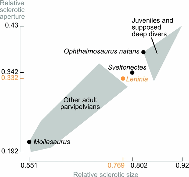
When the sclerotic aperture diameter is plotted against the sclerotic area diameter (both relative to orbital area as in Fernández et al. Reference Fernández, Archuby, Talevi and Ebner2005; see Fig. 7), Leninia and Sveltonectes fall outside the convex polygon encompassing adult and supposedly non-deep-diving parvipelvian ichthyosaurs and is located in between this group and the convex polygon grouping juveniles and ‘deep-diving’ forms (Ophthalmosaurus and Eurhinosaurus, according to Fernández et al. Reference Fernández, Archuby, Talevi and Ebner2005). The interpretation of this result is hazardous because two factors, ontogeny and ecomorphology, affect the position on the graph. Because the specimens of Sveltonectes and Leninia used here appear osteologically mature, this graph suggests that the relative eye size of these taxa was in between ‘deep-diving’ and ‘non-conclusively deep-diving’ forms. However, as mentioned above, the absolute size of the sclerotic aperture is far more important to assess dim light vision than is the relative size. This remark is also important for Mollesaurus, which is set on the bottom-left corner of the ‘adult parvipelvian’ group, yet possesses a sclerotic aperture similar to other ophthalmosaurines (see below).
Sclerotic ring and sclerotic aperture diameters of various parvipelvian ichthyosaurs relative to the orbit diameter, modified from Fernández et al. (Reference Fernández, Archuby, Talevi and Ebner2005). Grey areas represent the convex polygon encompassing the two groups in the Fernández et al. paper: juveniles + supposed deep divers (Ophthalmosaurus, Eurhinosaurus) versus other adult parvipelvians. Both Sveltonectes and Leninia fall outside these groups, despite great differences in terms of absolute sclerotic apertures. This suggests parvipelvians actually display a continuum of relative sclerotic dimensions that may evolve with age, as suggested by Fernández et al. (Reference Fernández, Archuby, Talevi and Ebner2005).

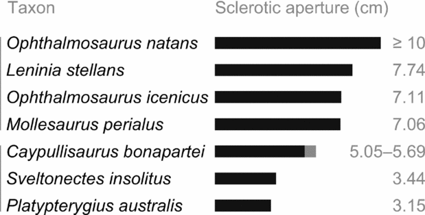
The absolute size of the sclerotic ring of Leninia (area of 252.5 cm2, diameter of 17.93 cm) is among the largest ever measured in ichthyosaurs, although much smaller than those of Temnodontosaurus (23.5 cm) and O. natans (22 cm) (Motani, Rothschild & Wahl, Reference Motani1999). The absolute size of the sclerotic aperture of Leninia is 7.74 cm long in diameter with a total area of 47.07 cm2; this falls close to Mollesaurus (diameter: 7.06 cm; Fernández et al. Reference Fernández, Archuby, Talevi and Ebner2005). Fernández et al.'s dataset of absolute sclerotic aperture diameters does not contain adult specimens of Ophthalmosaurus; similarly, Motani, Rothschild & Wahl (Reference Motani, Rothschild and Wahl1999) do not provide the sclerotic aperture of the specimens they measured. Nevertheless, we recorded a sclerotic aperture diameter of slightly below 10 cm in photographs of O. natans (CM 878, from Fernández et al. Reference Fernández, Archuby, Talevi and Ebner2005; UW 24816, from Wahl, Reference Wahl2009) and of 7.11 cm in Kirton's (A. Kirton, unpub. Ph.D. thesis, Univ. Newcastle upon Tyne, 1983) detailed reconstruction of Ophthalmosaurus icenicus. Whereas these calculations are rough, they suggest that the sclerotic aperture of both Mollesaurus and Leninia fall within the range of those of the largest-eyed animals ever, Ophthalmosaurus and the modern giant squid Architeuthis (Nilsson et al. Reference Nilsson, Warrant, Johnson, Hanlon and Shashar2012), and appears significantly higher than in most platypterygiine ophthalmosaurids (Fig. 8): Sveltonectes: 3.44 cm; Platypterygius australis: 3.15 cm (calculated on a photograph of AM F116939 from Kear, Reference Kear2005); Caypullisaurus bonapartei: 5.05 to 5.69 cm (adults; Fernández et al. Reference Fernández, Archuby, Talevi and Ebner2005). Moreover, it should be noted that the theoretical modelling undertaken by Nilsson et al. (Reference Nilsson, Warrant, Johnson, Hanlon and Shashar2012) indicated that the range of vision is roughly similar with a 7 or 10 cm wide pupil, but that such a pupil size provides a vision range increased by 10–20 m (depending on depth and the object to detect) when compared to the already large pupils of platypterygiine ichthyosaurs.
Sclerotic aperture diameter of ophthalmosaurids. Vertical bars on the left denote two groups: ophthalmosaurine (top) and platypterygiine (bottom) ophthalmosaurids. The aperture of ophthalmosaurines is usually markedly higher than that of platypterygiines, regardless of total size, and is within the range of that of the modern giant squid (Nilsson et al. Reference Nilsson, Warrant, Johnson, Hanlon and Shashar2012).

No teeth are preserved within the holotype of Leninia. However, the minute depth of the maxillary dental groove (16 mm high) at the level of the naris suggests a markedly reduced dentition (in apicobasal size) for this moderately large taxon, at least in this part of the snout. A similarly reduced dentition is present in other ophthalmosaurines such as in Ophthalmosaurus natans (Gilmore, Reference Gilmore1902, Reference Gilmore1905, Reference Gilmore1906; V. Fischer., pers. obs. on CM material), Acamptonectes densus (Fischer et al. Reference Fischer, Maisch, Naish, Liston, Kosma, Joger, Krüger, Pardo-Pérez, Tainsh and Appleby2012) and Mollesaurus (Fernández, Reference Fernández1999). The dentition of Ophthalmosaurus icenicus is subject to debate. This taxon has widely been considered as edentulous in the past (e.g. McGowan, Reference McGowan1976), but this was due to the weak attachment of teeth within the groove, as numerous specimens of this taxon are preserved with teeth, even adults (A. Kirton, unpub. Ph.D. thesis, Univ. Newcastle upon Tyne, 1983; Zammit, Reference Zammit2012; V. Fischer, pers. obs. on GLAHM and MJML material). Interestingly, teeth of O. icenicus do not appear as small and slender as in other ophthalmosaurines, which may indicate a slightly different diet for this taxon.
Ophthalmosaurines appear conservative in cranial anatomy; large eyes and reduced dentition probably appeared early in the evolution of the group and were apparently conserved throughout their long history (Middle Jurassic – Early Cretaceous). Similar sclerotic apertures and absence of profound modifications in tooth size and skull shape suggest that ophthalmosaurines conserved similar ecological niche(s), as colonization of a particular ecological niche is usually linked to numerous craniodental modifications, as recently demonstrated for marine crocodyliforms (e.g. Pierce, Angielczyk & Rayfield, Reference Pierce, Angielczyk and Rayfield2009; Young et al. Reference Young, Brusatte, de Andrade, Desojo, Beatty, Steel, Fernández, Sakamoto, Ruiz-Omeñaca and Schoch2012). Moreover, the metabolic cost of growing and maintaining eyes of such size is so high that it is extremely unlikely to be a plesiomorphic feature devoid of eco/ethological significance (Nilsson et al. Reference Nilsson, Warrant, Johnson, Hanlon and Shashar2012). On the contrary, the other ophthalmosaurid subfamily, Platypterygiinae, evolved a much more disparate assemblage of skull shapes and feeding guilds within the same time interval, with small piscivorous/teuthophageous forms (Sveltonectes: Fischer et al. Reference Fischer, Clément, Guiomar and Godefroit2011b ), large opportunistic predators (Platypterygius: Kear, Boles & Smith, Reference Kear, Boles and Smith2003; Brachypterygius: A. Kirton, unpub. Ph.D. thesis, Univ. Newcastle upon Tyne, 1983; McGowan, Reference McGowan1976) as well as forms in between these extremes (Aegirosaurus: Fischer et al. Reference Fischer, Clément, Guiomar and Godefroit2011a ), but seemingly lacked the deep-diving/low light vision adaption of the eye that is found in ophthalmosaurines. Additional and independent methods to evaluate deep-diving behaviour such as analyses of bone histology and avascular necrosis (Motani, Rothschild & Wahl, Reference Motani1999; Rothschild, Xiaoting & Martin, Reference Rothschild, Xiaoting and Martin2012) could be useful to further investigate the evolutionary trends outlined here.
6. Conclusion
A new ichthyosaur, Leninia stellans, from lower Aptian deposits of Russia highlights a remarkable conservatism of skull shape and eye size among ophthalmosaurine ophthalmosaurids throughout their long history (Middle Jurassic – Early Cretaceous), suggesting these ichthyosaurs kept similar ecological niche(s) during this time interval. These similarities are not biased by phylogeny because the features like eye architecture and dentition are not linked to the characters used in the cladistic analysis. However, the fragmentary nature of the basal ophthalmosaurines Mollesaurus and Leninia does not permit clarification of the tempo of character acquisition at the base of this subfamily. Nevertheless, this contribution highlights distinct evolutionary histories for the ophthalmosaurid subfamilies, by indicating that the peculiar morphology of Ophthalmosaurus – often considered as a hyperspecialized exception – is actually found within an increasing number of closely related Jurassic and Cretaceous ichthyosaurs.
Acknowledgements
We thank the staff of the YKM for making our study as easy as possible, and Erin Maxwell and one anonymous reviewer for their constructive and insightful comments. We also thank Ben Kear for the invitation to contribute to the present volume. This work was funded by a Fond de la Recherche Scientifique doctoral grant (V.F., Aspirant du F.R.S.–FNRS).