Perforation of the atretic pulmonary valve with balloon dilation is a standard initial therapy in selected patients with pulmonary atresia with intact ventricular septum. Reference Alwi, Geetha and Bilkis1–Reference Chubb, Pesonen and Sivasubramanian7 The incidence of inadvertent right ventricular outflow tract perforation, sepsis or multi-organ failure, and mortality was between 4 and 15%. Reference Bakhru, Marathe and Saxena5–Reference Marasini, Gorrieri and Tuo8 A post-procedural right ventricular pseudoaneurysm is an extremely rare complication, requiring urgent management due to its potentially life-threatening nature. Transcatheter intervention for the treatment of a right ventricular pseudoaneurysm is reported much less frequently. Reference Herbert, Ikemba and Nugent9,Reference Alkhouli, Waits, Chaturvedi and Ling10
Case report
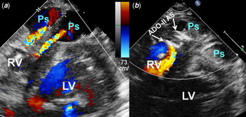
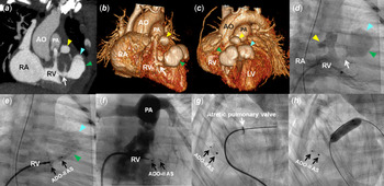
We describe a 3-day-old male newborn with 38 weeks of gestation and birth weight of 2.63 kg who underwent transcatheter intervention for pulmonary atresia with intact ventricular septum. The procedure was aborted due to right ventricular perforation with cardiac tamponade. Two weeks later, two large pseudoaneurysms (7 × 11 and 8 × 12 mm) arising from the right ventricular anterior wall were detected by echocardiography (Fig 1a). Cardiac CT and right ventricular angiography revealed three aneurysm sacs (6 × 8, 9 × 16, and 11 × 17 mm), one at the anterior region of the right ventricular outflow tract and two others arising from the anterior right ventricular free wall (Fig 2a–d). The two large false aneurysms appeared to connect to the right ventricle with a narrow neck that measured 3.8 mm in diameter and 4.9 mm in length. Because of the rapid growth of the two large pseudoaneurysms, which had a high risk of rupture, and surgical operation was not feasible because of the anterior location and concern for rupture with opening the sternum, transcatheter approach for embolisation of the pseudoaneurysms was performed at 3-week-old (body weight of 2.97 kg). A 4Fr delivery catheter (Amplatzer TorqVue LP catheter, Abbott Medical, Minnesota, United States of America) was advanced over the 0.018” Terumo guidewire into the right ventricle. This was technically challenging and successfully achieved delivery of the catheter across the narrow neck into the aneurysm. Once the sheath was in position, the guidewire was removed and the device was inserted promptly. A 5/4 Amplatzer Duct Occluder II Additional Sizes (Abbott Park, Illinois, United States of America) was selected for closure of the two large pseudoaneurysms (Fig 2e and f), as the 5 mm waist diameter and 4 mm length matched the dimension of the neck of the pseudoaneurysm and the smaller disc diameter was suitable for a small right ventricular chamber. With the left disc of the device deployed in the aneurysm, the long sheath was retracted to the right ventricle, the right disc was deployed, and then the device was released. Then, perforation of the atretic pulmonary valve was approached using a combined coronary guidewire and a microcatheter followed by sequential balloon pulmonary valvuloplasty (Fig 2g and h). There were no post-catheterisation complications. A repeat echocardiogram revealed the closure device was in a good position, and progressive thrombosis of embolised pseudoaneurysms (Fig 1b), as well as another smaller pseudoaneurysm, resolved spontaneously after right ventricular decompression. He was discharged after a 2-month hospitalisation.
Transthoracic echocardiograms. ( a ) Two large pseudoaneurysms with forward flow at colour Doppler in the short-axis view. ( b ) Two large pseudoaneurysms were embolised using a closure device (arrow) and were completely thrombosed. ADO-II AS = Amplatzer Duct Occluder II Additional Sizes; LV = left ventricle; Ps = pseudoaneurysms; RV = right ventricle.

Images of CT and interventional catheterisation. ( a–d ) Cardiac CT and right ventriculography showed three false aneurysm sacs (arrow heads). The two large aneurysms appeared as one lesion with two lobes (white arrows). ( e and f ) RVG showed an ADO-II AS (black arrows) that was placed in the narrow neck with a good position, and aneurysms regressed totally 8 months later. ( g ) Perforation of the atretic valve by the coronary guidewire. ( h ) A 6 mm Tyshak II Balloon was used for pulmonary valvuloplasty. ADO-II AS = Amplatzer Duct Occluder II Additional Sizes; AO = aorta; PA = pulmonary artery; RA = right atrium; RV = right ventricle.

Discussion
Pulmonary atresia with intact ventricular septum is a complex CHD with significant morphological variability, and transcatheter pulmonary valvotomy and sequential balloon dilatation have become established as the procedure of first choice. Reference Alwi, Geetha and Bilkis1 Although successful perforation of the atretic valve can be achieved in 80–89% of patients, this method is technically challenging and associated with severe complications, including right ventricular outflow tract perforation, stroke, sepsis, multi-organ failure, and mortality. Reference Alwi, Budi, Mood, Leong and Samion3,Reference Bakhru, Marathe and Saxena5,Reference Patil, Saxena and Gupta6 Peri-procedural complications leading to death were observed in about 10% of cases. Reference Bakhru, Marathe and Saxena5–Reference Marasini, Gorrieri and Tuo8 A major cause of early mortality is cardiac tamponade due to right ventricular outflow tract tear. Immediate pericardial drainage and surgical repair of cardiac rupture could be life-saving procedures for patients with cardiac tamponade; however, about half of the patients complicated with this complication died. Reference Bakhru, Marathe and Saxena5–Reference Marasini, Gorrieri and Tuo8 Amongst the post-procedural complications, RV pseudoaneurysm is a rare complication. A suitable approach for treating right ventricular pseudoaneurysm is surgery. For right ventricular pseudoaneurysm with the anterior location and concern for aneurysm rupture with catastrophic bleeding during opening the sternum, but our patient was too small, and therefore surgical operation was not deemed feasible. Cases of successful percutaneous closure of right ventricular pseudoaneurysm have been reported in the literature, but they are rare in childhood, and this treatment may be considered in patients who are poor candidates for surgery. Reference Herbert, Ikemba and Nugent9,Reference Alkhouli, Waits, Chaturvedi and Ling10 Because of the high risk of rupture due to rapid growth of the large false aneurysms with high right ventricular pressure, in this case, interventional catheterisation for embolisation of the pseudoaneurysms was required.
The Amplatzer Duct Occluder II is a self-expanding occlusion device with a central waist plug and retention discs deployed on both ends. This low-profile device is implanted forward from a 4Fr delivery catheter, and is feasible in right ventricular pseudoaneurysm patients weighing less than 5 kg. Despite the small size of the patient and challenging catheter course, the intervention was tolerated by minimising the time for delivery of the catheter into position in the false aneurysm. Safety and device stability was demonstrated over 1 year of follow-up in this patient.
Conclusion
Perforation of the atretic pulmonary valve in selected patients with pulmonary atresia with intact ventricular septum is effective, but right ventricular perforation is a severe complication that frequently leads to a fatal outcome. Post-procedural right ventricular pseudoaneurysm is relatively rare but life-threatening complication in patients with pulmonary atresia with intact ventricular septum who underwent interventional catheterisation. The results of the present case demonstrate that transcatheter intervention for embolisation of a right ventricular pseudoaneurysm can be a safe and effective treatment, even in a relatively small patient.
Acknowledgements
No acknowledgement was received for this study.
Financial support
This research received no specific grant from any funding agency, commercial, or not-for-profit sectors.
Conflict of interest
None.
Ethical statement
This article does not contain any studies with human participants performed by any of the authors.