Structural protein modules with specific functions, termed domains, are used and reused in evolution, with mixing and matching of various domains achieving combinations of functions. Successful elements, such as the kinase domain or immunoglobulin fold, are present in a surprising variety of proteins. The module composed of seven transmembrane α-helices (heptahelical domain; HD) is one of the greatest successes of evolutionary engineering: it constitutes the core of all G-protein-coupled receptors (GPCRs) – the most numerous protein family in the animal kingdom, with ~1000 members in most mammals (Refs Reference Bockaert and Pin1, Reference Rompler2). Structurally, GPCRs are very diverse, ranging from the virtually HD-only light receptor rhodopsin (RHO), a prototype of the largest class A, to constitutively dimeric class C receptors, where each HD is fused to the Venus flytrap domain, or the enormous (>6000 amino acid long) ‘very large GPCR’ (GPR98), with a motley collection of domains attached to its HD (Ref. Reference Fredriksson3). Different GPCRs respond to incredibly diverse stimuli, from photons and small molecules to large proteins, metal ions, and extracellular protease activity (Refs Reference Bockaert and Pin1, Reference Fredriksson3). Not surprisingly, GPCRs are targeted by about half of clinically used drugs (Ref. Reference Rompler2).
The key mechanisms of the activation of the HD core of GPCRs and signal transduction across the membrane appear to be fairly well conserved (Refs Reference Rosenbaum, Rasmussen and Kobilka4, Reference Hofmann5). Receptor transition into an active conformation triggered by agonist binding involves the movement of the transmembrane helices relative to each other (Refs Reference Hubbell6, Reference Scheerer7), which significantly changes the cytoplasmic tip of the receptor. This conformational rearrangement dramatically increases its affinity for several intracellular signalling partners. Upon activation, the great majority of GPCRs bind heterotrimeric G proteins, catalysing the release of bound GDP and its replacement with GTP, the prevalent guanyl nucleotide in the cytoplasm. GTP-liganded Gα subunit loses its affinity for the Gβγ dimer, so that both parts of the G protein dissociate from the receptor and activate or inhibit various effectors. While it remains active, a single receptor can activate dozens of G protein molecules (Ref. Reference Burns and Pugh8), amplifying the signal to ensure high sensitivity.
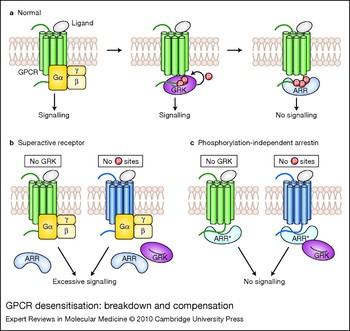
In most cases, signalling is terminated by a two-step mechanism: active receptor is selectively phosphorylated by one or more GPCR kinases (GRKs), whereupon a member of the arrestin family of proteins binds with high affinity to the cytoplasmic tip of the active phosphoreceptor (Ref. Reference Gurevich and Gurevich9), sterically precluding further G-protein interactions (Ref. Reference Krupnick, Gurevich and Benovic10) (Fig. 1a). Receptor-bound arrestin recruits GPCRs to coated pits for internalisation via its interactions with clathrin (Ref. Reference Goodman11) and the AP2 adaptor complex (Ref. Reference Laporte12). The arrestin–receptor complex often initiates another round of signalling via arrestin-mediated recruitment of mitogen-activated protein kinases (MAPKs) and other proteins (Refs Reference DeWire13, Reference Kendall and Luttrell14). Although the timing of action and relative contribution of individual members of arrestin and GRK families vary (Refs Reference Gurevich and Gurevich15, Reference Tobin, Butcher and Kong16), the general mechanism is conserved (Ref. Reference Gurevich and Gurevich9). The ability of a single GPCR upon binding different ligands to assume distinct active conformations (Ref. Reference Kobilka and Deupi17) preferentially interacting with different G proteins and other signal transducers apparently underlies biased agonism (Refs Reference Kenakin18, Reference Drake19).
GPCR desensitisation: breakdown and compensation. (a) Ligand-activated GPCRs bind heterotrimeric G proteins, catalysing exchange of GDP for GTP, and subsequent dissociation of Gα and Gβγ subunits (dissociation not shown), which regulate downstream signalling proteins. In normal situations, active GPCRs are phosphorylated by GPCR kinases (GRKs). Binding of wild-type arrestin (ARR; blue) to active phosphoreceptor stops the signalling in a timely fashion. (b) Normal GPCR (green) in the absence of GRK, or a mutant GPCR (dark blue) that does not have GRK phosphorylation sites, cannot be shut off by wild-type arrestins, which only bind phosphorylated GPCRs. In both cases, excessive receptor signalling results in pathology. (c) Expression of phosphorylation-independent arrestin (ARR*; lighter blue) compensates for the defects of receptor phosphorylation by binding unphosphorylated wild-type or mutant receptors, thereby reining in excessive signalling.

Diseases associated with excessive GPCR signalling
More than 30 human diseases have been directly linked to mutations in GPCRs (reviewed in Ref. Reference Schöneberg20). The majority of identified mutations are inactivating, and therefore recessive: disease phenotype develops only in individuals that are homozygous or compound heterozygous for the affected allele. However, activating mutations in different GPCRs underlie about a dozen disorders, ranging from dwarfism and hypo- and hyperthyroidism to several forms of cancer (Ref. Reference Schöneberg20). In some cases, constitutive activity of the receptor results in its excessive phosphorylation and association with arrestin. Constitutive desensitisation of a human vasopressin receptor mutant underlies nephrogenic diabetes insipidus (Ref. Reference Barak21), whereas in the case of rhodopsin it results in night blindness or retinal degeneration (Ref. Reference Rim and Oprian22).
At the molecular level, receptor mutations are not the only cause of excessive GPCR signalling. For example, uncontrolled rhodopsin activity can be produced by activating mutations (Ref. Reference Rao, Cohen and Oprian23), as well as desensitisation deficits due to absence of phosphorylation sites in mutant rhodopsin (Refs Reference Apfelstedt-Sylla24, Reference Kim25, Reference Restagno26), and null mutations in rhodopsin kinase (Ref. Reference Cideciyan27) or arrestin (Ref. Reference Fuchs28). All of these defects cause visual disorders ranging from night blindness to photoreceptor degeneration, which have been reproduced in genetically modified mice (Refs Reference Xu29, Reference Chen30, Reference Chen31, Reference Mendez32). A growing body of evidence suggests that GPCR genes are proto-oncogenes (Ref. Reference Allen33). Uncontrolled activation of wild-type M1, M3 and M5 muscarinic acetylcholine receptors (mAChRs; CHRM1, 3 and 5) by constant presence of an agonist (Ref. Reference Gutkind34), ectopic expression of serotonin 5HT1C receptor (HTR2C) (Ref. Reference Julius35), or expression of a constitutively active α1B-adrenergic receptor (ADRA1B) (Ref. Reference Allen33) have all been shown to induce malignant transformation. The gene encoding the angiotensin receptor was first cloned as the mas oncogene (MAS1) (Ref. Reference Young36, Reference Young37) even before its pharmacological nature was established (Ref. Reference Jackson38). Signalling by wild-type GPCRs pushes the cell out of balance in most of these cases. Another example of excessive activity of a perfectly normal receptor in cells that apparently express all relevant GRKs and arrestins is supersensitivity of dopamine receptors associated with dopamine depletion in Parkinson disease or dyskinesia, developing upon prolonged treatment of parkinsonian patients with l-DOPA (Ref. Reference Guigoni39).
One possible strategy to counteract excessive GPCR signalling is to boost the desensitisation mechanisms that normally keep it in check. Increased expression of GRKs and/or arrestins is likely to decrease the signalling to normal levels where wild-type receptors are involved, but different approaches are needed in the case of mutant receptors that cannot be phosphorylated properly.
Molecular mechanics of arrestin proteins
The first arrestin was discovered in the visual system as a negative regulator of rhodopsin signalling. As arrestin nomenclature is rather confusing, we use systematic names in this article: arrestin1 (historic names S-antigen, 48 kDa protein, visual or rod arrestin; SAG in HUGO Gene Nomeclature Committee database), arrestin2 (β-arrestin or β-arrestin1; ARRB1), arrestin3 (β-arrestin2; ARRB2), and arrestin4 (cone or X-arrestin; ARR3). Arrestin1 was shown to bind active phosphorylated rhodopsin and suppress (arrest) further G-protein activation (Refs Reference Kuhn, Hall and Wilden40, Reference Pfister41). Subsequent cloning of additional family members (Refs Reference Lohse42, Reference Attramadal43, Reference Sterne-Marr44, Reference Craft, Whitmore and Wiechmann45, Reference Murakami46) and elegant demonstration that arrestin subtypes preferentially regulate different GPCRs (Ref. Reference Lohse47) showed that the same mechanism is involved in the regulation of numerous receptors in this superfamily (reviewed in Refs Reference Gurevich and Gurevich9, Reference Carman and Benovic48) (Fig. 1a).
Three families of cytoplasmic proteins preferentially bind active GPCRs: G proteins, GRKs and arrestins. In contrast to G proteins and GRKs, arrestins have no lipid modifications or other means of recruitment to the membrane except through receptor binding. Out of four functional forms of rhodopsin – inactive (Rh), active (Rh*), inactive phosphorylated (P-Rh), and active phosphorylated (P-Rh*) – arrestins bind with high affinity only one: P-Rh*. This raises an interesting question: how do these soluble proteins, diffusing in the cytoplasm and randomly encountering GPCRs in various functional states, specifically bind only P-Rh*? What is the molecular mechanism that translates the encounter with P-Rh* into high-affinity binding? The development of a direct binding assay with femtomolar sensitivity provided vital clues: arrestins were found to specifically bind to Rh* and inactive P-Rh, albeit with much lower affinity than to P-Rh* (Ref. Reference Gurevich and Benovic49). Importantly, a C-terminal truncation generates a mutant that still binds P-Rh* best, but shows greatly enhanced affinity for Rh* and inactive P-Rh, but not for Rh (Ref. Reference Gurevich and Benovic49). These data suggested that arrestin recognises receptor activation and phosphorylation states independently, but has internal restraints precluding high-affinity binding to anything except P-Rh*. GPCR activation involves a significant conformational rearrangement of the receptor, exposing elements that were inaccessible in the basal state (Refs Reference Hubbell6, Reference Scheerer7, Reference Li50). Arrestin must interact with these newly exposed elements to distinguish between active and inactive receptor. It also has to bind receptor-attached phosphates to discriminate between phosphorylated and unphosphorylated forms.
A model of sequential multisite binding (Ref. Reference Gurevich and Benovic51) explains arrestin selectivity for P-Rh*, as follows. Arrestin has separate sites interacting with elements in Rh* exposed by its activation and with receptor-attached phosphates. Individually these interactions are relatively weak, allowing arrestin to ‘probe’ the functional state of the receptor it encounters and dissociate quickly if it turns out to be an unpreferred one. Only when arrestin encounters P-Rh* are both of the primary sites simultaneously engaged. This releases internal constraints, allowing the arrestin molecule to undergo a conformational change of its own, which brings additional secondary binding sites into contact with the receptor, greatly increasing the energy of the interaction and hence the affinity (reviewed in Ref. Reference Gurevich and Gurevich52). This model predicts that primary binding sites serve as sensors, and that the molecule works as a coincidence detector, swinging into action only when both sensors are activated simultaneously.
Obviously, a sensor in a protein can be artificially turned on by mutagenesis. One arginine in the first set of identified phosphate-binding residues behaved in this fashion: charge neutralisation generated an arrestin mutant with high binding to Rh* (Ref. Reference Gurevich and Benovic53). Subsequent demonstration that charge reversal increases Rh* binding even more, whereas Arg to Lys mutation preserving the charge does not, led to the idea that this arginine interacts with a negatively charged residue within arrestin, and that neutralisation of its charge either by receptor-attached phosphates or by mutagenesis breaks the salt bridge, thereby turning arrestin on (Refs Reference Gurevich and Benovic53, Reference Gurevich and Benovic54). Indeed, crystal structures of arrestin1 (Ref. Reference Hirsch55), 2 (Refs Reference Han56, Reference Milano57) and 4 (Ref. Reference Sutton58) revealed that between the two arrestin domains there is an arrangement of five charged residues that are largely solvent-excluded (unusual for a soluble protein), which was termed the polar core. Extensive mutagenesis proved that the salt bridge between an arginine and an aspartic acid in the polar core serves as the main phosphate sensor in all arrestins (Refs Reference Vishnivetskiy59, Reference Vishnivetskiy60, Reference Kovoor61, Reference Celver62, Reference Gray-Keller63, Reference Pan, Gurevich and Gurevich64, Reference Song65). Subsequent studies showed that the interaction between the β-strand I, α-helix I, and the arrestin C-tail serves as an auxiliary phosphate sensor and is also disrupted by receptor-attached phosphates (Refs Reference Vishnivetskiy60, Reference Gurevich66, Reference Hanson67, Reference Vishnivetskiy68). Manipulation of this interaction yielded structurally distinct mutants with high affinity for Rh*. These gain-of-function mutants that bind either active form of their cognate receptors – P-Rh* and Rh* – with comparable affinity (Refs Reference Kovoor61, Reference Celver62, Reference Gray-Keller63, Reference Gurevich69) were termed phosphorylation-independent or enhanced arrestins (Ref. Reference Song65).
Phosphorylation-independent arrestins as tools for gene therapy
The strategy for gene therapy in the case of loss-of-function mutations is conceptually straightforward: expression of a missing functional protein is likely to solve the problem. With the exception of rhodopsin expressed in photoreceptors in enormous quantities, the levels of most GPCRs in other cells are relatively low, so that even a receptor mutant with severe folding defects is unlikely to cause additional trouble by overloading the cellular protein-degradation machinery consisting of proteasomes and lysosomes.
In contrast, dealing with gain-of-function mutations is much harder. These mutations are dominant, because normal protein encoded by the second allele cannot prevent excessive signalling. Traditionally, GPCR signalling is reduced by appropriate small-molecule antagonists (blockers), which are widely used for therapeutic purposes. Antagonists targeting normal GPCRs attenuate the amplitude of signalling, while leaving the regulation of the time course of signal transduction to cellular desensitisation mechanisms mediated by GRKs and arrestins. In the case of phosphorylation-deficient GPCR mutants this mechanism does not work, so an antagonist can decrease signal intensity, but cannot restore its normal time course, which is equally important biologically. Theoretically, mutant mRNA could be eliminated with an appropriately designed ribozyme. However, since even very low expression of constitutively active or desensitisation-deficient receptor is harmful (Ref. Reference Chen30), the efficiency of this process must be extremely high. As mutant and normal mRNA encoded by the other allele could differ by as little as a single base (in the case of point and frameshift mutations), the required combination of very high efficiency and perfect selectivity is hardly realistic. Conceivably, one could deliver a ribozyme that indiscriminately destroys all endogenous mRNA for the receptor, along with the gene encoding normal protein in which the sequence is modified by silent mutations to make it resistant to this ribozyme. This places additional demands on the vector, which has to deliver two functional coding sequences instead of one. Thus, alternative strategies to dampen the signalling by gain-of-function receptor mutants are needed.
Logically, since excessive receptor signalling is at the root of the problem, increased activity of components of the desensitisation machinery could bring the system back into balance. For normal (nonmutant) GPCRs, GRK availability is rate-limiting, although extra arrestin further facilitates desensitisation in the presence of added GRK (Refs Reference Krasel70, Reference Moore, Milano and Benovic71). Obviously, where the mutation has eliminated or significantly reduced the number of GRK phosphorylation sites, an increase in the availability of wild-type GRKs and arrestins cannot achieve this goal (Fig. 1b). Theoretically, a small molecule that promotes the binding of wild-type arrestin to unphosphorylated mutant receptor in an activation-dependent manner could solve this problem. Indeed, two compounds that promote arrestin1 binding to light-activated unphosphorylated rhodopsin have been described: heparin (Ref. Reference Gurevich72) and a synthetic phosphopeptide mimicking the fully phosphorylated rhodopsin C-terminus (Ref. Reference Puig73). Although neither study directly addresses this possibility, both illustrate the same difficulties in adapting this approach for therapy. First, neither effective molecule is particularly small. Second, both carry multiple negative charges, which preclude crossing of the membrane to get inside the cell where they are expected to work. Third, both are effective at fairly high concentrations (high micromolar to millimolar). And last, but not least, both have bell-shaped dose–response curves: they promote arrestin binding to unphosphorylated Rh* only in a very narrow concentration range, and actually inhibit it above and below the optimum. While it could be possible to design smaller molecules enhancing arrestin binding to unphosphorylated receptor, a high concentration of negative charges is a must, as this molecule has to substitute for missing receptor-attached phosphates, activating the arrestin phosphate sensor. In fact, the great majority of clinically used drugs act on the extracellular side of the membrane, where the issue of membrane crossing does not arise. Still, some are hydrophobic enough to cross the membrane unaided or rely on transporters to get inside cells. Small molecules inhibiting or enhancing intracellular protein–protein interactions can and should be developed, although one has to keep in mind that this approach is not free of significant limitations. The ultimate solution might require the same type of delivery vehicle as gene transfer: one that targets specific tissues or cell types and delivers the cargo across the plasma membrane. Considering that drugs must be delivered repeatedly, whereas genes need to be delivered only once, the latter may turn out to be more feasible and economical.
Phosphorylation-independent arrestin mutants described above are promising tools for controlling excessive GPCR signalling (Fig. 1c). Their ability to blunt the signalling of normal GPCRs in the absence of receptor phosphorylation and that of phosphorylation-deficient mutants of rhodopsin (Ref. Reference Gray-Keller63), μ- (Refs Reference Celver74, Reference Macey, Lowe and Chavkin75) and δ-opioid (OPRM1, OPRD1) (Refs Reference Kovoor61, Reference Celver62), and β2-adrenergic (Refs Reference Kovoor61, Reference Celver62) (ADRB2) receptors has been convincingly demonstrated in vitro (Ref. Reference Gray-Keller63), in exogenous expression systems (Refs Reference Kovoor61, Reference Celver62, Reference Pan, Gurevich and Gurevich64, Reference Celver74), and in primary cultures of GRK-deficient neurons (Ref. Reference Macey, Lowe and Chavkin75), paving the way for in vivo experiments. This compensational strategy was recently tested in the visual system of living animals (Ref. Reference Song65).
Testing the compensational approach in the visual system
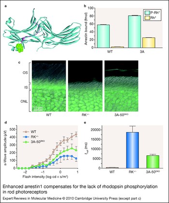
Rod photoreceptors are neurons, in which the molecular machinery responsible for light-initiated signalling is localised in a specialised compartment, termed the outer segment, separated from the rest of the cell by a narrow cilium (Ref. Reference Gurevich, Fliesler and Kisselev76). In a healthy retina, elongated outer segments of all photoreceptors are parallel and neatly arranged (Fig. 2c). Normal length of the outer segments is an indication of photoreceptor health; in visual disorders they often become shorter and visibly disorganised (Refs Reference Xu29, Reference Chen30, Reference Chen31, Reference Song65). Photoreceptor cell bodies and nuclei make up the outer nuclear layer. The thickness of this layer is a good measure of the number of surviving photoreceptors; its thinning is a clear indication of disease-associated photoreceptor loss (Refs Reference Xu29, Reference Chen30, Reference Chen31).
Rhodopsin kinase (systematic name GRK1) knockout mice (RK−/−) (Ref. Reference Chen31) display multiple deficits in the rod photoreceptors associated with excessive rhodopsin signalling. These animals express wild-type rhodopsin along with the normal complement of wild-type arrestin1, which cannot quench the signalling in the absence of rhodopsin phosphorylation (Fig. 2). Rod photoreceptors in vertebrates have extremely high light sensitivity (Ref. Reference Baylor, Lamb and Yau77). To maintain it, rods are capable of shutting off rhodopsin signalling with rapid subsecond kinetics (Refs Reference Mendez32, Reference Song65, Reference Krispel78), which requires the concerted action of rhodopsin kinase phosphorylating rhodopsin at multiple sites (Refs Reference Mendez32, Reference Doan79, Reference Vishnivetskiy80) and arrestin1, which binds light-activated phosphorhodopsin (P-Rh*) with high affinity (Refs Reference Gurevich and Benovic51, Reference Gurevich, Fliesler and Kisselev76). Very slow dissociation of arrestin1 from P-Rh* (Refs Reference Gurevich and Benovic51, Reference Vishnivetskiy80) ensures high fidelity of the shut-off and rapid recovery (Refs Reference Mendez32, Reference Doan79), whereas low arrestin1 affinity for light-activated unphosphorylated rhodopsin (Rh*) and a consequent much faster off-rate (Refs Reference Gurevich and Benovic51, Reference Vishnivetskiy80) prevents reliable shut-off in the absence of phosphorylation and slows down rod recovery by two orders of magnitude (Refs Reference Song65, Reference Burns81) (Fig. 2f,g). Therefore, even dim light completely desensitises rods of RK−/− mice due to prolonged rhodopsin signalling, causing night blindness (Ref. Reference Chen31) similar to that observed in human patients with the same genetic defect (Ref. Reference Cideciyan27). Moreover, rods in RK−/− mice degenerate within a few months (Ref. Reference Chen31). The fact that excessive signalling is at the root of all problems in RK−/− mice was convincingly demonstrated by dark-rearing of these animals, which effectively prevented photoreceptor degeneration (Ref. Reference Chen31). Thus, RK−/− mice represent a perfect model to test the compensational potential of phosphorylation-independent arrestin mutants.
Enhanced arrestin1 compensates for the lack of rhodopsin phosphorylation in rod photoreceptors. (a) Arrestin1 is an elongated two-domain molecule (crystal structure from Ref. Reference Hirsch55, PDB 1CF1). Interdomain interactions and the anchoring of the C-tail (magenta) to the body of the molecule by bulky hydrophobic residues (side chains are shown in green) stabilise its basal (inactive) conformation. (b) Wild-type (WT) arrestin1 binds light-activated phosphorylated rhodopsin (P-Rh*) much better than active unphosphorylated rhodopsin (Rh*). Alanine substitution of the residues anchoring the C-tail (3A) yields an enhanced mutant with greatly increased affinity for Rh*. (c) Rhodopsin-containing outer segments (OSs) of healthy rod photoreceptors of WT animals are long and neatly organised. Despite normal expression of WT arrestin1, the loss of rhodopsin kinase (RK−/−) results in a dramatic shortening and dishevelled appearance of the outer segments. The expression of phosphorylation-independent arrestin1 in RK-deficient animals 3A-50RK−/−Arr1−/− [3A-50DKO (double knockout) on figure] increases the length of the outer segments, indicative of the improved overall health of the photoreceptors. (d) Photoreceptor response to light stimuli in vivo is measured by electroretinography. A typical electroretinogram begins with the negative a-wave, reflecting direct response of photoreceptor cells. The amplitude of a-waves robustly increases with flash intensity in WT animals (brown curve). It is greatly diminished in RK−/− mice expressing WT arrestin1 (blue curve). However, substitution with phosphorylation-independent arrestin1-3A in the double knockout line improves light sensitivity in the absence of rhodopsin phosphorylation (green curve). (e) The recovery of the photoreceptors is measured in a double-flash protocol. A desensitising flash is delivered first, and the response to the second flash delivered at variable time intervals after the desensitising flash is compared with the control response to the flash of the same intensity delivered without the preceding desensitising flash. The time of half recovery (t half) is the interval between the two flashes allowing for half-maximum recovery of the response. Replacement of WT arrestin1 with arrestin1-3A facilitates the recovery about threefold. However, the recovery rate remains significantly slower than in WT animals, indicating that there is a lot of room for further improvement. The complete set of data was published in Ref. Reference Song65: part c is reproduced from Ref. Reference Song65, with permission from Elsevier (© 2009 Elsevier Ltd); parts b, d and e are compiled from data in Ref. Reference Song65. Abbreviations: IS, inner segment; ONL, outer nuclear layer.

Arrestins are elongated two-domain molecules, with the C-tail anchored to the body of the N-domain (Fig. 2a) (Refs Reference Hirsch55, Reference Han56, Reference Milano57, Reference Sutton58). Receptor binding induces the release of the C-tail (Refs Reference Hanson67, Reference Vishnivetskiy68, Reference Palczewski82). The deletion of the arrestin C-tail (Refs Reference Gurevich and Benovic49, Reference Pulvermuller83) or its detachment by alanine substitutions of three anchoring hydrophobic residues (3A mutation) (Ref. Reference Gurevich66) facilitates arrestin transition into a high-affinity receptor-binding state, thereby generating mutants with greatly enhanced affinity for unphosphorylated active receptor (Fig. 2b). The functional capability of enhanced arrestin1-3A was directly compared with the wild-type arrestin1 in live mice (Fig. 2). To this end, the amplitude of the light response, the recovery kinetics, and photoreceptor survival in RK−/− mouse lines that have the normal complement of wild-type arrestin1 (RK−/−Arr1+/+) or express only the enhanced 3A mutant (3A-50RK−/−Arr1−/−) (Refs Reference Song65, Reference Nair84) were compared. In phosphorylation-deficient animals, enhanced arrestin1 increases the length of the outer segments, improves rod survival, and improves the ability of rods to respond to repeated light stimulation, facilitating photoresponse recovery about threefold (Ref. Reference Song65) (Fig. 2).
These results clearly indicate that this novel compensational approach to the therapy of gain-of-function genetic disorders is feasible. However, the engineered single-step shut-off of Rh* by enhanced arrestin1-3A yields a time of half-recovery of ~6 s – that is, significantly slower than in wild-type animals (~0.4 s) (Ref. Reference Song65). Thus, additional re-engineering of the arrestin1 molecule involving the receptor-binding surface is necessary to achieve better compensation. Nonetheless, the fact that this strategy works in rod photoreceptors, equipped with the most sensitive GPCR-driven signalling system with the fastest shut-off on record, suggests that it can fairly efficiently rein in excessive signalling by other GPCRs that normally desensitise much more slowly. The difficulty of testing this idea in other disease models in vivo lies in the relative promiscuity of the two nonvisual arrestins, which bind hundreds of different GPCR subtypes (Ref. Reference Gurevich and Gurevich52).
Can we construct arrestin mutants with high receptor specificity?
Rod photoreceptors are arguably the most specialised cells, expressing millimolar concentrations of rhodopsin and its cognate arrestin1 (Refs Reference Gurevich, Fliesler and Kisselev76, Reference Hanson85, Reference Strissel86), which makes targeted compensation of rhodopsin shut-off defects by phosphorylation-independent arrestin1 fairly straightforward. In contrast, most neurons and other cells express 5–25 different GPCRs (Refs Reference Penn87, Reference Gurevich and Gurevich88) along with arrestin2 and arrestin3 (Refs Reference Gurevich and Gurevich15, Reference Gurevich, Benovic and Gurevich89, Reference Gurevich, Benovic and Gurevich90, Reference Luo, Busillo and Benovic91). Many activating mutations of nonvisual arrestins have been described (Refs Reference Kovoor61, Reference Celver62, Reference Pan, Gurevich and Gurevich64, Reference Gurevich69, Reference Celver74, Reference Macey, Lowe and Chavkin75, Reference Gurevich92, Reference Hanson93, Reference Hanson and Gurevich94, Reference Song95, Reference Carter96), but the usability of their phosphorylation-independent versions for therapeutic purposes could be questionable, because both interact with multiple receptors (Refs Reference Gurevich69, Reference Gurevich92, Reference Barak97). Thus, while an enhanced version of arrestin2/3 would likely dampen the signalling by an overactive GPCR mutant, it will also blunt the signalling by all the normal GPCRs in the same cell, likely producing unwanted side effects. Therefore, to achieve full therapeutic potential, phosphorylation-independent nonvisual arrestins may need to be rendered specific for the particular receptor they are meant to target. However, practical use of phosphorylation-independent nonvisual arrestins does not have to be entirely contingent on successful narrowing of their receptor specificity. Considering the devastating consequences of many activating GPCR mutations (Ref. Reference Schöneberg20) and the lack of viable therapeutic alternatives, existing mutants with broad receptor specificity, although imperfect, will be in many cases better than nothing. Selective expression in affected cell types can be achieved by targeted delivery and cell-type-specific promoters, both of which are being actively developed (for representative reviews see Refs Reference Miller and Dean98, Reference Beck99, Reference Sadeghi and Hitt100, Reference Lundberg101, Reference Baum102). This will limit off-target effects to the cells where the overactive mutant receptor produces the most damage, so that the overall impact of the expression of enhanced arrestin will likely be positive.
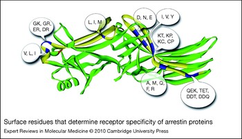
Evolution made arrestin1 quite specific for rhodopsin, with only marginal affinity for nonvisual GPCRs (Refs Reference Sutton58, Reference Gurevich92, Reference Barak97), demonstrating that arrestin protein specific for a single receptor subtype can be designed. Although several research groups using a variety of methods identified a fairly extensive receptor-binding surface on arrestins (Refs Reference Hanson67, Reference Gurevich92, Reference Hanson and Gurevich94, Reference Ohguro103, Reference Pulvermuller104, Reference Dinculescu105), much of it engages non-subtype-specific elements, such as receptor-attached phosphates (Refs Reference Gurevich and Benovic53, Reference Gurevich and Benovic54, Reference Hirsch55, Reference Sutton58, Reference Vishnivetskiy59, Reference Vishnivetskiy60, Reference Hanson and Gurevich94), with only a few elements playing a key role in defining receptor preference (Ref. Reference Vishnivetskiy106). Arrestin1 shows particularly low binding to the M2 mAChR, whereas arrestin2 binds this receptor fairly well (Refs Reference Gurevich69, Reference Gurevich92, Reference Gurevich107). Relative specificity for rhodopsin and M2 mAChR of a series of arrestin1–arrestin2 chimeras with different parts of the molecule swapped between the two subtypes identified key elements whose exchange completely reversed receptor preference of these arrestins (Ref. Reference Vishnivetskiy106). They turned out to be surprisingly small, localised on the concave sides of the two arrestin domains (Fig. 3). As a result of fairly high homology within the arrestin family (Ref. Reference Gurevich and Gurevich108), only about a dozen residues in these regions are different, with even fewer nonconservative substitutions. Since these residues must be primarily responsible for receptor specificity, judicious manipulation of these amino acids by mutagenesis appears to be a natural approach to the construction of arrestin proteins with narrowed receptor specificity. Theoretically, random mutagenesis of even five positions yields 205, or >3000 000, combinations – that is, orders of magnitude more than can be tested by any assay within a reasonable time. However, the analysis of known arrestin sequences (Ref. Reference Gurevich and Gurevich108) shows that in ~600 million years of arrestin evolution there were only a few different residues in each of the key positions (Fig. 3). Mammalian arrestins show even less variability, which brings the number of combinations to be tested down to a manageable ~30–240. This approach to designing arrestins with significantly enhanced receptor specificity looks promising, although it needs to be tested experimentally. Although mammalian arrestin2 and arrestin3 are generally regarded as promiscuous, and many GPCRs apparently bind both with the same or very similar affinity (Refs Reference Gurevich92, Reference Barak97), certain receptors demonstrate clear preference for arrestin3 (Refs Reference Oakley109, Reference DeGraff, Gurevich and Benovic110), whereas others prefer arrestin2 (Refs Reference Macey, Gurevich and Neve111, Reference Dale112). Even when a GPCR binds either nonvisual arrestin, its interaction with arrestin2 and 3 subtypes results in distinct trafficking patterns (Ref. Reference Mundell113) and signalling outcomes (Ref. Reference Ahn114). The construction of receptor-specific mutants can begin by increasing rudimentary receptor specificity already built into the two nonvisual subtypes. Obviously, engineered arrestins targeting particular receptors should be constructed on the basis of the native subtype they preferentially bind.
Surface residues that determine receptor specificity of arrestin proteins. Swapping of two elements [shown in yellow on the crystal structure of arrestin2 (Ref. Reference Han56; PDB 1G4M)] localised on the concave sides of the two domains completely reverses receptor preference of arrestin1 and arrestin2 for rhodopsin and the M2 muscarinic acetylcholine receptor, respectively (Ref. Reference Vishnivetskiy106). The positions of nonconservative substitutions in these elements are shown in blue, with alternative residues found in each position from Caenorhabditis elegans to mammals (Ref. Reference Gurevich and Gurevich108) indicated in a single-letter code. The variability in the subset of mammalian arrestin proteins is significantly smaller. The position where visual arrestins with narrow receptor specificity carry a bulky hydrophobic residue that makes the N-domain more rigid is shown in red.

Manipulation of arrestin interactions with nonreceptor partners
In addition to GPCRs, arrestins bind dozens, if not hundreds, of surprisingly diverse proteins (Refs Reference DeWire13, Reference Kendall and Luttrell14). The view that only nonvisual arrestin2 and 3, and only in their receptor-bound conformation, engage these partners (Refs Reference DeWire13, Reference Gurevich and Gurevich115) has been recently challenged by experimental evidence to the contrary (Refs Reference Hanson93, Reference Song95, Reference Song, Gurevich and Gurevich116, Reference Song117, Reference Hanson118, Reference Wu119, Reference Meng120). The emerging picture is that the three known conformational states of arrestins – free, receptor-bound, and microtubule-associated – play distinct roles in the regulation of cell signalling via direct interactions with numerous other proteins (Refs Reference DeWire13, Reference Gurevich, Gurevich and Cleghorn121). Just as modifications of receptor-binding elements generated arrestin mutants with unusual functional capabilities, re-engineering of parts mediating arrestin interactions with other partners is a viable strategy for construction of custom-designed arrestins with unique signalling properties that can be used for therapeutic purposes. However, the binding sites of very few non-GPCR partners have been elucidated with any degree of precision. Rare exceptions include clathrin (Refs Reference Goodman11, Reference Kim and Benovic122, Reference Kang123), clathrin adaptor AP2 (Refs Reference Laporte12, Reference Kim and Benovic122), microtubules and unpolymerised tubulin αβ dimers (Refs Reference Hanson93, Reference Hanson118), cAMP phosphodiesterase PDE4D5 (Ref. Reference Baillie124), and calmodulin (Ref. Reference Wu119).
Among arrestin binding partners, MAP kinases have attracted particular attention, as these proteins play a key role in life-or-death decisions in the cell. Arrestins facilitate the activation of ERK1/2 (MAPK3/MAPK1) (Ref. Reference Luttrell125), which in simplistic terms are prosurvival and proproliferation kinases, as well as activation of JNK3 (MAPK10) (Ref. Reference McDonald126), which is largely antiproliferative and/or proapoptotic. Thus, judicious rechannelling of arrestin activity towards ERK1/2 would have therapeutic potential in neurodegenerative Alzheimer and Parkinson diseases and retinal degenerations, whereas shifting it towards JNK3 may counteract excessive proliferation in cancer. Originally, receptor-bound arrestin3 was shown to scaffold the ASK1–MKK4–JNK3 (MAP3K–MAP2K4–MAPK10) cascade (Ref. Reference McDonald126). Follow-up study by the same group found that in the absence of GPCR stimulation, arrestin3 promotes JNK3 activation in cells overexpressing ASK1 (Ref. Reference Miller127), strongly implicating free arrestin3 in this process. Indeed, JNK3 was shown to bind free arrestin3 by three other research groups (Refs Reference Song95, Reference Scott128, Reference Wang129), and receptor-binding-deficient arrestin3 mutant ‘frozen’ in the basal conformation (Ref. Reference Hanson93) was found to promote JNK3 activation at least as well as wild-type arrestin3 (Ref. Reference Song117).
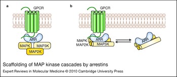
Two models of the organisation of a complex of arrestin and MAP kinases were proposed. The first, based on the original discoveries of arrestin-assisted signalling in ASK1–MKK4–JNK3 (Ref. Reference McDonald126) and c-Raf–MEK1–ERK1/2 (RAF1–MAP2K1–MAPK3/1) (Ref. Reference Luttrell125) cascades, posits that upstream MAP3Ks (c-Raf or ASK1) bind to the N-domain and downstream MAPKs (ERK1/2 or JNK3) bind to the C-domain, whereas MAP2Ks (MEK1 or MKK4) do not bind arrestins directly, but interact with the complex via cognate MAP3K and MAPK (Refs Reference Kendall and Luttrell14, Reference Miller and Lefkowitz130) (Fig. 4a). Recently, both MAP2Ks – MKK4 (Ref. Reference Song117) and MEK1 (Refs Reference Song117, Reference Meng120) – were shown to bind arrestins directly. The two arrestin domains are independent folding units that can be expressed separately and retain certain functions (Refs Reference Gurevich and Benovic49, Reference Gurevich and Benovic51, Reference Gurevich and Benovic53, Reference Gurevich92, Reference Hanson93). The demonstration that individual N- and C-domains bind JNK3, its upstream kinases ASK1 and MKK4, as well as ERK2, MEK1 and c-Raf, suggested a very different arrangement of the MAPK modules on the arrestin scaffold (Ref. Reference Song117). Recently, ASK1 interaction with sites in both arrestin domains were independently shown using peptide arrays (Ref. Reference Li131). The interaction of each kinase with both domains suggested an alternative model, where kinases are arranged side-by-side with their long axes parallel to the long axis of the arrestin, like three hotdogs on a single bun (Fig. 4b). Although several arrestin elements were recently implicated in the binding of MEK1 (Ref. Reference Meng120) and components of the ASK1–MKK4–JNK3 cascade (Ref. Reference Li131), precise footprints of each kinase on arrestin remain to be elucidated.
Scaffolding of MAP kinase cascades by arrestins. (a) The original model of mitogen-activated protein kinase (MAPK) scaffolding by receptor-bound arrestin (Refs Reference Luttrell125, Reference McDonald126, Reference Miller127). The model posits that MAPK (ERK1/2 or JNK3) and MAPK kinase kinase (MAP3K; c-Raf-1 or ASK1) directly bind arrestin, whereas MAPK kinase (MAP2K; MEK1 or MKK4) is brought to the complex via interactions with MAPK and MAP3K. (b) An alternative model posits that all three kinases in the cascade directly interact with arrestins, and each kinase binds both arrestin domains (Ref. Reference Song117). Based on the ability of receptor-binding-deficient arrestin3 to facilitate ASK1-dependent JNK3 activation (Ref. Reference Song117), this model shows that both receptor-bound and free arrestins can scaffold MAPK cascades.

The same is true for the majority of arrestin binding partners, although in many cases we have some clues. First, any protein that binds the receptor–arrestin complex must interact with the elements that are not engaged by the receptor. Indeed, when the binding site of a protein on arrestin overlaps with the receptor-interaction surface, as was shown for microtubules (Refs Reference Hanson93, Reference Hanson118) and calmodulin (Ref. Reference Wu119), as a result of higher affinity the receptor invariably outcompetes the other protein (Refs Reference Nair84, Reference Wu119). As the receptor-binding surface of arrestins is fairly well established, this narrows the field down to the sides of the molecule and the convex surfaces of the two domains (Refs Reference Gurevich and Gurevich9, Reference Gurevich, Gurevich and Cleghorn121). Second, upon GPCR binding, arrestins undergo a significant conformational rearrangement (Refs Reference Gurevich and Benovic51, Reference Carter96, Reference Schleicher, Kuhn and Hofmann132, Reference Xiao133, Reference Nobles134), which involves the release of the C-tail and a movement of the two domains relative to each other (Refs Reference Hanson67, Reference Vishnivetskiy68, Reference Vishnivetskiy135). Recent comparison of wild-type, conformationally loose 3A mutants mimicking the receptor-bound state, and receptor-binding-deficient forms frozen in the basal state showed that arrestins in different conformations bind JNK3 with similar affinity (Refs Reference Song95, Reference Song, Gurevich and Gurevich116). The partners that show similar affinity for free and receptor-bound arrestin likely engage the elements equally exposed in both states. In contrast, proteins that preferentially bind receptor-associated arrestin [e.g. clathrin and AP2 (Refs Reference Goodman11, Reference Kim and Benovic122, Reference Xiao133), ubiquitin ligase AIP4 (ITCH) (Ref. Reference Bhandari136)] must engage elements that become more exposed upon arrestin activation, whereas partners that prefer arrestin in the basal state [e.g. ubiquitin ligase MDM2 (Refs Reference Song95, Reference Song, Gurevich and Gurevich116)] either bind elements less exposed in the active conformation or require the exact spacing between the binding sites in the two domains that is only achieved in the basal conformation. Comparative studies of different arrestin subtypes provide additional information. Studies using arrestin2/3 shuttling between the nucleus and cytoplasm (Refs Reference Song95, Reference Scott128, Reference Wang129), as well as peptide arrays (Ref. Reference Li131), showed that arrestins 2 and 3 bind JNK3 equally well. Visual arrestins 1 (rod arrestin) and 4 (cone arrestin) were also shown to bind JNK3 (Refs Reference Song95, Reference Song, Gurevich and Gurevich116). Obviously, partners that bind all four vertebrate arrestins likely engage conserved residues, whereas arrestin-subtype-specific functions [e.g. JNK3 activation by arrestin3 (Refs Reference Song117, Reference McDonald126, Reference Miller127)] must be mediated by residues absent in other arrestins. These considerations reduce the number of arrestin residues to be tested for their role in binding each partner, but there is still a lot of work to be done.
Although the task of creating arrestin mutants in which individual functions are disrupted or enhanced by targeted mutagenesis appears daunting, quite a few successes in this direction have already been achieved. Many of the relevant mutants were tested in cultured cells, and some even in live animals.
Components of the internalisation machinery: clathrin and AP2
In the process of identification of compact clathrin- and AP2-binding sites in the arrestin C-tail, several mutants were created with one or both of these sites disrupted (Refs Reference Goodman11, Reference Laporte12, Reference Kim and Benovic122). The expression of these mutants, which bind receptors normally but cannot recruit the receptor–arrestin complex to the coated pit, considerably reduced GPCR internalisation (Refs Reference Goodman11, Reference Laporte12, Reference Kim and Benovic122). Interestingly, expression of mutant arrestins with a disrupted phosphoinositide-binding site (Ref. Reference Gaidarov and Keen137) and the expression of the arrestin C-tail that carries clathrin- and AP2-interaction sites but does not bind receptors (Ref. Reference Krupnick138) produces similar effects via different mechanisms: the former weakens the arrestin–receptor interaction, and the latter occupies arrestin-binding elements on clathrin and AP2 and outcompetes the arrestin–receptor complexes.
cAMP-specific phosphodiesterase PDE4D
Arrestins recruit two isoforms of cAMP-specific PDE4D to the activated β2-adrenergic receptor, thereby facilitating cAMP breakdown and reducing protein kinase A activity in the vicinity of the membrane (Ref. Reference Perry139). Arrestin3 mutants in which the binding to PDE4D was disrupted by targeted mutagenesis bind the receptor normally, but do not recruit PDE4D and therefore, in contrast to wild-type arrestin3, do not affect PKA phosphorylation of the receptor (Ref. Reference Baillie124). Moreover, a recent in vivo study showed that arrestin3–PDE4D interaction in the amygdala is important for fear conditioning (Ref. Reference Li140). The authors found that the expression of a PDE4D-binding-deficient mutant does not rescue fear conditioning in arrestin3-knockout mice, whereas the expression of wild-type arrestin3 does (Ref. Reference Li140).
MEK1 and other MAP kinases
Arrestin2 is constitutively phosphorylated at the C-terminus by ERK2, and this phosphorylation reduces its affinity for clathrin, so that receptor internalisation requires dephosphorylation of arrestin2 (Ref. Reference Lin141). An arrestin2 mutant deficient in MEK1 binding was recently constructed on the basis of identification of two key residues required for arrestin–MEK1 interaction (Ref. Reference Meng120). In cells, this mutant is not phosphorylated by ERK2, demonstrating that arrestin2 is phosphorylated by ERK2 scaffolded and activated on it. As could be expected, the peptide disrupting arrestin2 association with MEK1 blocked receptor-activation-dependent ERK2 activation in cells, while promoting arrestin-dependent receptor internalisation (Ref. Reference Meng120). Following recent identification of arrestin elements mediating the interactions with ASK1, MKK4 and JNK3 (Ref. Reference Li131), corresponding binding-deficient mutants need to be constructed and functionally tested.
Ubiquitination sites on arrestins
Activation of the β2-adrenergic receptor and arrestin binding were shown to induce the ubiquitination of both proteins, which plays distinct roles in receptor trafficking (Ref. Reference Shenoy142). Arrestins and the receptor are ubiquitinated by two different E3 ligases – MDM2 (Ref. Reference Shenoy142) and NEDD4 (Ref. Reference Shenoy143), respectively – both of which are recruited to the complex via their interactions with arrestins (Refs Reference Shenoy142, Reference Shenoy143). Subsequent studies showed that MDM2 interacts with free arrestins (Ref. Reference Wang129), and preferentially binds arrestins in their basal conformation (Refs Reference Song95, Reference Song, Gurevich and Gurevich116), suggesting that the receptor recruits a preformed arrestin–MDM2 complex; MDM2 then has a short time window to ubiquitinate receptor-bound arrestin, which assumes the active conformation, before MDM2 is released due to reduced affinity for this form (Ref. Reference Song95). In contrast, the activation of the chemokine receptor CXCR4 promotes arrestin2 interaction with E3 ubiquitin ligase AIP4 (Ref. Reference Bhandari136). In this case, depletion of arrestin2 by small interfering RNA (siRNA) does not affect CXCR4 internalisation and initial ubiquitination, but prevents its sorting from endosomes to lysosomes and subsequent degradation (Ref. Reference Bhandari136). Arrestins also recruit deubiquitinating enzymes USP33 and USP20, important for recycling and resensitisation to the β2-adrenergic receptor (Ref. Reference Berthouze144).
Interestingly, particular lysines that are ubiquitinated in the same arrestin3 depend on the receptor it binds to: interaction with the angiotensin II type 1a receptor (AGTR1) promotes the modification of lysines 11 and 12, whereas binding to the vasopressin V2 receptor (AVPR2) directs ubiquitination to lysines 18, 107, 108, 207 and 296 (Ref. Reference Shenoy and Lefkowitz145). Intriguingly, the elimination of individual sets of two (11 + 12) or five (18 + 107 + 108 + 207 + 296) lysines (by conservative Lys to Arg substitutions) selectively reduces arrestin3 affinity for the angiotensin or vasopressin receptor, respectively, and prevents arrestin-dependent ERK activation only by that receptor (Ref. Reference Shenoy and Lefkowitz145). Thus, the two mutants described in this study selectively lost the ability to promote internalisation and postendocytic signalling by one specific GPCR, but not by another (Ref. Reference Shenoy and Lefkowitz145). The effects of the same quintuple Lys to Arg arrestin3 mutant on M1 and M2 mAChRs were also receptor-specific: the mutation reduced stable arrestin–M2 association and M2 downregulation, while increasing downregulation of M1 (Ref. Reference Mosser146). Interestingly, the rate of M1 or M2 internalisation was not affected (Ref. Reference Mosser146). However, one should keep in mind that the M2 receptor belongs to the group of GPCRs that avidly bind and require arrestins for desensitisation, but normally internalise via arrestin-independent pathway(s) (Refs Reference Gurevich69, Reference Gurevich92, Reference Gurevich107, Reference Pals-Rylaarsdam147, Reference Lee148). An arrestin3 mutant where all 31 lysines that could be ubiquitinated were replaced with arginines was found to interact with AP2 and components of the ERK cascade normally, but displayed reduced affinity for the β2-adrenergic receptor and clathrin (Ref. Reference Shenoy149). This mutant failed to enhance receptor internalisation and formation of stable signalosomes that retain activated ERK, whereas arrestin3–ubiquitin fusion showed the opposite phenotype (Ref. Reference Shenoy149). Thus, manipulation of the ubiquitination sites on nonvisual arrestins changes their functional characteristics in a receptor-specific manner.
Interactions mediating arrestin oligomerisation
Arrestin1 self-associates, cooperatively forming dimers and tetramers (Refs Reference Schubert150, Reference Imamoto151, Reference Hanson152). Interestingly, although arrestin1 invariably crystallises as a tetramer (Refs Reference Hirsch55, Reference Granzin153), the biologically relevant solution tetramer was shown to be different from that observed in the crystal (Ref. Reference Hanson152), and considerable effort was required to elucidate its shape and the interaction interfaces between protomers (Ref. Reference Hanson154). Based on this information, two self-association-deficient arrestin1 mutants were constructed (Ref. Reference Hanson154), both of which retained normal ability to bind rhodopsin and microtubules. The receptor-binding surface of arrestin1 monomers within the tetramer and both possible dimers are shielded by sister subunits (Ref. Reference Hanson154), so it is not surprising that only monomeric arrestin1 can bind rhodopsin (Ref. Reference Hanson152). These data suggest that oligomers are a storage form, but it is unclear why arrestin1 robustly self-associates, whereas cone-specific arrestin4 does not (Ref. Reference Hanson155). The exact role of oligomerisation in the biology of arrestin1 remains to be elucidated, although the molecular tools necessary to establish it are ready (Ref. Reference Hanson154).
Arrestin2 forms dimers of different configuration in several crystal forms (Refs Reference Han56, Reference Milano57). In some cases, the dimerisation interface appears to be significantly strengthened by inositol hexakisphosphate (InsP 6) diffused into preformed crystals (Ref. Reference Milano156). The ability of InsP 6 to promote arrestin2 dimerisation was confirmed in solution (Refs Reference Hanson155, Reference Milano156). In contrast, InsP 6 reduces self-association of arrestin1 (Ref. Reference Hanson155), which could be interpreted as an indication that the shapes of arrestin1 and arrestin2 oligomers are different. Homo- and hetero-oligomers of both nonvisual arrestins were observed in overexpressing cells, suggesting that this process occurs in vivo (Ref. Reference Storez157). Arrestin3 has a nuclear export signal (NES) in its C-terminus, whereas arrestin2 does not, so that arrestin3 is predominantly cytoplasmic, while arrestin2 is also present in the nucleus (Refs Reference Song95, Reference Scott128, Reference Wang129). Coexpression of arrestin3 suppresses nuclear localisation of arrestin2, suggesting that hetero-oligomerisation regulates subcellular localisation of arrestins (Ref. Reference Storez157). Indeed, targeted disruption of arrestin2/3 oligomerisation increased nuclear localisation of arrestin2 without affecting its interactions with clathrin, AP2 or ERK2 (Ref. Reference Milano156). Considering that arrestin2 plays a role in regulation of gene expression (Ref. Reference Kang158), this may have important biological implications. Subsequent studies showed that disruption of arrestin3 oligomerisation by mutagenesis reduced its interactions with MDM2 and inhibited p53-dependent antiproliferative effects of arrestin3 (Ref. Reference Boularan159), as well as arrestin3 interaction with ERK1/2 (Ref. Reference Xu160), identifying additional functions of arrestin3 oligomers. These clues suggest several possible biological functions of arrestin2/3 oligomerisation, although many mechanistic questions remain unanswered (see Ref. Reference DeFea161 for an excellent review).
Thus, a number of arrestin mutants with selectively disrupted individual functions have already been created and successfully used as research tools. Although many more kinds of designer arrestins are needed to explore and manipulate other facets of their function, some of the existing ones might well have therapeutic potential that needs to be explored.
Conclusions and future directions
Traditionally, medicine largely relies on small molecules that act as long as they are in the body. The main advantage of conventional drugs is the ease of delivery and straightforward ways of structure optimisation for maximum effect and acceptable pharmacokinetics. As far as killing invading bacteria or viruses with distinct biochemistry goes, where strong constant action is an advantage, small molecules are perfect therapeutic tools. However, problems arise when we need to suppress (or boost) the activity of endogenous enzymes and receptors. The key drawback is that enzyme inhibitors and receptor blockers affect a single process in the extremely complex network regardless of the functional state of the system. This single-mindedness of conventional therapies becomes a problem in many disorders, where hitting a single target too hard often results in drug-induced imbalances as bad as the disease itself. That is why therapeutic approaches are shifting towards types of drugs that act in a context-dependent manner: partial agonists, which increase or decrease signalling when the level of the endogenous agonist is low or high, respectively, and allosteric modulators, the effect of which is contingent on the presence of endogenous agonists (reviewed in Refs Reference Conn, Christopoulos and Lindsley162, Reference Kenakin163). Proteins are the ultimate context-dependent signalling machines perfected over millions of years of rigorous evolutionary experimentation. Each function of every protein is controlled by sophisticated regulatory mechanisms that are already in place in the body and will likely prevent introduced protein from overdoing whatever we made it to do.
Mechanistic understanding of the inner workings of a protein paves the way to subtle manipulation of its individual functional characteristics for therapeutic purposes without destroying its biologically advantageous complexity and regulation. Recent findings show that re-engineering of arrestin proteins to create optimised versions with therapeutic potential is feasible (Ref. Reference Song65). Although this approach is still in its infancy, the ability of phosphorylation-independent arrestins to compensate for defects in receptor phosphorylation, as well as construction of other arrestin mutants with changed individual functional modalities, is very encouraging.
Potential benefits from the elucidation of molecular details of arrestin interactions with MAP kinases, ubiquitin ligases, and other signalling proteins are great enough to make this work worth doing: it will enable the engineering of mutants that channel the signalling to the pathways of choice. Arrestins designed to facilitate prosurvival signalling via ERK, MDM2 and/or other interaction partners could help the neurons affected by degenerative disorders to survive. By the same token, mutant arrestins channeling signalling to proapoptotic or antiproliferative pathways could be of interest in cancer, where rapid proliferation is the key problem. It is important to keep in mind that to achieve a therapeutic effect it is not necessary to kill malignant cells. Numerous precancerous cells emerge in the body every day, and our immune system disposes of them quite successfully. Only the ones that proliferate so fast that they outrun the immune response create problems. Thus, simply slowing down proliferation would likely make these cells vulnerable enough for the immune system to eliminate them.
Structurally, arrestins are inviting targets for creative protein engineering. One side of the molecule, where the concave surfaces of the two domains are localised, is responsible for receptor and microtubule binding. The opposite side carries the interaction sites for the majority of nonreceptor binding partners, which do not overlap with the receptor footprint. The third element, the detachable arrestin C-tail, carries the sites for the key components of the internalisation machinery of the coated pit: clathrin and AP2. Each of these elements can be manipulated without affecting the other two. Thus, necessary structural information will enable us to construct arrestins that connect the receptor of interest to the signalling pathway of our choosing. Although targeted re-engineering of all arrestin functions is still years away, several arrestin mutants with special functional characteristics have already been constructed and tested. Phosphorylation-independent mutants with high affinity for active unphosphorylated receptors were shown to normalise signalling in the absence of GPCR phosphorylation in vitro (Ref. Reference Gray-Keller63), in model systems such as Xenopus oocytes (Refs Reference Kovoor61, Reference Celver62) or HEK293 cells (Ref. Reference Pan, Gurevich and Gurevich64), in more challenging primary neuronal culture (Ref. Reference Macey, Lowe and Chavkin75), and even in rod photoreceptors of living mice (Ref. Reference Song65). We can also make arrestins that do not bind GPCRs very well but have increased affinity for microtubules instead (Ref. Reference Hanson93), which will be useful to direct specific signalling proteins to the cytoskeleton. The possibilities of further engineering are limited only by the available structural information.
Arrestins represent just four out of >20 000 proteins we have, and their versatility is unlikely to be unique. Virtually every protein consists of several structural elements, has multiple functions, and interacts with many binding partners. Thus, most proteins are amenable to subtle functional adjustments by targeted mutagenesis, as described here for arrestins. The difficulty of this approach is that a lot of precise structural and functional information about the protein of interest is necessary to make it feasible. In contrast, small molecules producing desired effects can often be identified by high-throughput screening of huge chemical libraries without the least knowledge of any molecular mechanisms. One only needs to know the desired target (e.g. a particular GPCR subtype or enzyme isoform) and proteins that should not be affected (e.g. highly homologous other GPCRs, or closely related other enzymes) to make a potential drug highly specific. After that, many well-established methods of therapeutic delivery of small molecules can be tested and the best one found. However, the very specificity so much sought after usually underlies the downside of small molecules: their single-mindedness makes it too easy to overdo whatever they are meant to do, causing side effects.
Intracellular signalling proteins, native or engineered, can be delivered only by gene transfer, which means that necessary methods are much more complicated and still need further development. This is well worth the effort: in contrast to commonly used single-purpose small molecules, which cannot be regulated in any way except via patient intake, proteins are sophisticated multifunctional machines, with their lifespan and every aspect of the function subject to complex regulation in the cell. Subtle changes in signalling pathways by an increase in expression of selected protein or by introducing its functionally modified version will remain under control of normal cellular mechanisms, minimising the chances of excesses. This approach will likely be developed first for congenital disorders, primarily those with molecular errors caused by gain-of-function mutations. However, when technical hurdles of targeting gene transfer to a particular tissue or cell type are overcome, one can envision many potential uses of these sophisticated molecular tools in a variety of disorders where normal cellular functions get out of hand. The most likely targets include diseases requiring adjustment of cell signalling, such as neurodegenerative disorders (caused by runaway neuronal death), cancer (runaway proliferation), and allergies and autoimmune disorders (runaway immune response).
Acknowledgements and funding
This work was supported in part by NIH grants EY011500, GM077561, GM081756 (V.V.G), and NS065868 (E.V.G). The authors thank Dr Robert J. Lefkowitz for critical reading of the manuscript, as well as the reviewers for constructive suggestions.