I. INTRODUCTION
Since Li et al. Reference Li, Cai, An, Kim, Nah, Yang, Piner, Velamakanni, Jung, Tutuc, Banerjee, Colombo and Ruoff1 published the growth of graphene on copper via chemical vapor deposition (CVD), there has been extensive research in the understanding of the basic growth mechanism of graphene and to extending the two dimensional growth observed on copper to other metal surfaces, as well as to dielectric surfaces. Reference Batzill2 While much progress has been made in growing large-area graphene films, Reference Yan, Lin, Peng, Sun, Zhu, Li, Xiang, Samuel, Kittrell and Tour3–Reference Iwasaki, Park, Konuma, Lee, Smet and Starke12 more is required before wafer-scale single-crystal graphene can be achieved. One bottleneck in synthesizing large-area graphene grains is that it has been difficult to decrease the number of nucleation sites. Ideally, as in the classical case of bulk single crystal growth, a single nucleation site is all that is needed to initiate growth of single crystal graphene. This assumption is valid as long as the interaction between the substrate and graphene is weak, such as the case for graphene on copper. Another approach for producing large area crystalline graphene is to grow the graphene epitaxially. However, the high cost of the single crystal metal substrates makes this technique prohibitively expensive for most applications. A number of studies have been performed to attempt to understand the nucleation of graphene on Cu; however, there has not been wide application of this understanding, and there are many aspects that could influence nucleation that have not been evaluated. Reference Yan, Lin, Peng, Sun, Zhu, Li, Xiang, Samuel, Kittrell and Tour3,Reference Yu, Jauregui, Wu, Colby, Tian, Su, Cao, Liu, Pandey, Wei, Chung, Peng, Guisinger, Stach, Bao, Pei and Chen4,Reference Wang, Wang, Bao, Yang, Zhu, Xie and Zhang8,Reference Luo, Lu, Singer, Berck, Somers, Goldsmith and Johnson13–Reference Wood, Schmucker, Lyons, Pop and Lyding15 Some techniques have been used since the discovery of graphene growth on Cu, e.g., hydrogen annealing, electrochemical polishing, etching in acid solutions etc., to create a clean copper surface with some success. It is found however that even long high temperature annealing under hydrogen still leaves carbon on the copper surface. Reference Kim, Mattevi, Calvo, Oberg, Artiglia, Agnoli, Hirjibehedin, Chhowalla and Saiz16 In addition, there is a recent report that graphene etching in hydrogen is caused by trace amounts of oxygen in the hydrogen source gas rather than the hydrogen itself. Reference Choubak, Biron, Levesque, Martel and Desjardins17 Kim et al. Reference Kim, Mattevi, Calvo, Oberg, Artiglia, Agnoli, Hirjibehedin, Chhowalla and Saiz16 suggest that “we should consider that this residual carbon might perturb growth.” We find that the residual carbon not only perturbs growth but promotes graphene nucleation as well.
The challenge of getting low nucleation density, and thus large graphene grains, relies on reproducibly obtaining a clean copper surface free of the adventitious carbon, or other carbon sources before methane is introduced into the CVD system. Obtaining a perfectly clean surface ex situ in a perfect clean room environment, free of any dust or hydrocarbons, still would not prevent carbon from being on the surface before growth since carbon monoxide is reported to form a carbon layer on copper even at room temperature (which becomes graphitic at 300 °C). Reference Santoni and Urban18 To eliminate any preformed graphene nucleation sites, the substrate must be cleaned in situ just before graphene growth. Some other metals used as substrates for growing graphene, like iridium and ruthenium, are already commonly cleaned in situ at high temperatures by introducing a small amount of oxygen, which “burns off” any remaining carbon contaminants. Reference Loginova, Bartelt, Feibelman and McCarty19
We report here that copper with an oxidized surface, including cuprous oxide (Cu2O) and cupric oxide (CuO), can act as a self-cleaning substrate for graphene growth by CVD. Both Cu2O and CuO thermally decompose into metallic copper below copper's melting point at low oxygen partial pressures and release oxygen from the substrate surface. With the assistance of a residual gas analyzer (RGA), we found that the released oxygen is free to react with the carbon residues on the copper surface and forms carbon monoxide and carbon dioxide, leaving a clean copper surface apparently free of carbon for large-area graphene growth.
II. EXPERIMENTAL
Normal copper foil cleaning method: after rinsing with acetone, copper foils were dipped into acetic acid for 2 h and then dried with nitrogen. Electropolishing was done in a homemade electrochemistry cell as reported previously Reference Zhang, Lee, Piner, Kholmanov, Wu, Li, Ji and Ruoff20 with the acetic acid cleaned Cu foil [25-μm-thick Cu foil, 99.8%, Alfa Aesar No. 13382; 125-μm-thick Cu foil, oxygen-free-high-conductivity (OFHC), Eagle Brass, C10200, Leesport, PA] as the anode and a large Cu plate as the cathode.
Copper oxidation was performed in a Ney Vulcan 3-550 box furnace (Vernon Hills, IL). Cu foils were heated to 250 or 300 °C (heating the substrates much above 250 °C heavily oxidizes the copper, which then takes a long time to fully reduce in the CVD system) under ambient atmosphere at 1° per minute and immediately allowed to cool, at 1° per minute in the box furnace, to room temperature.
All residual gas analysis experiments were conducted in a high-vacuum CVD system with a 10 cm diameter quartz tube chamber equipped with a turbopump to reach vacuum levels compatible with the RGA (a vacuum pressure of 10−8 Torr was reached at 100 °C before heating the system further). The entire CVD system has a 10 cm inner diameter and is 2.5 m long. Gas analysis was performed using a Stanford Research Systems Quadrupole Residual Gas Analyzer (SRS RGA100), which is located next to the turbopump inlet 1.5 m from the furnace center.
All experiments comparing nucleation densities among OFHC copper substrates with different pregrowth cleaning, oxidizing, and/or electropolishing were conducted in a vacuum CVD system with a 25 mm diameter chamber equipped with only a rotary vane pump (Edwards RV8, Sanborn, NY). A vacuum level of about 10 mTorr was reached in the chamber before gas introduction. The CVD growth was carried out with a relatively lower methane (Air Gas, 99.999%, Austin, TX) flow rate (0.1–0.2 sccm) and a hydrogen (Air Gas, 99.999%) flow rate of 10 sccm at 1040 °C, following a 1 h hydrogen anneal (10 sccm, 60 mTorr).
Scanning electron microscopy (SEM) images were acquired with an FEI Quanta-600 FEG environmental SEM (Houston, TX) using an acceleration voltage of 30 kV. Raman spectra (WITec Alpha 300 micro-Raman imaging system) were obtained using a 488 nm wave length incident laser.
III. RESULTS AND DISCUSSION
A. Comparison of graphene nucleation density on different copper substrates
We first studied the effect of the normal cleaning methods and the resulting graphene nucleation density and domain size. We chose two kinds of commonly used commercial Cu foils, the nonoxygen-free copper [hereafter referred to as oxygen-rich (OR) copper, 25-μm thick, 99.8%, Alfa Aesar No. 13382] and the OFHC copper (125-μm thick, Eagle Brass, C10200) for comparison. Both copper foils were thoroughly cleaned with acetone and acetic acid shortly before a high temperature annealing with hydrogen. Figures 1(a) and 1(b) show that indeed residues—likely carbon since Raman spectra taken from the residue on the OFHC copper had both D and G peaks—remained on both copper surfaces after annealing at 1040 °C for 1 h with hydrogen flowing (10 sccm), although much less is on the OR copper compared to on the OFHC copper. This is attributed to the oxygen present in OR copper.
SEM images of (a) 99.8% Cu foil and (b) OFHC Cu foil surfaces after cleaning with acetone, acetic acid, and annealing at 1040 °C for 1 h under 10 sccm H2 showing differing amounts of surviving carbon residue. Four of the small remaining residue spots have been circled in Fig. 1(a). Maps of the Raman G-peak position for graphene grown on (c) 99.8% copper and (d) OFHC copper.

To measure and compare the graphene nucleation density and the resulting domain size on the two kinds of Cu foils after normal cleaning and hydrogen protected annealing, 13C methane was used for the first 5 min of growth, followed by growth with normal methane, which is composed predominately of 12C. The as-grown graphene islands then have 13C at their centers with a Raman G-peak position of ∼1525 cm−1 and normal graphene surrounding the 13C region with a G-peak at ∼1585 cm−1. Reference Ferrari and Basko21–Reference Li, Cai, Colombo and Ruoff23 Raman maps of graphene grown in the same system at the same time following the same cleaning process show that the graphene islands on the commonly used 99.8% OR copper foil are ∼20 µm across, compared to the ∼2 µm graphene domains on the OFHC copper foil [Figs. 1(c) and 1(d)]. Since the main difference between the two copper foil substrates is that one is oxygen free while the other is not, we used copper oxides to mimic the 99.8% copper and found that this yielded still lower nucleation densities. Nucleation density on copper oxides is dramatically decreased and millimeter-sized graphene islands are readily achieved on the thermally reduced preoxidized copper surface.
B. Thermal reduction of copper oxide
Copper can be readily oxidized and forms Cu2O and CuO after heating in air. Reference Chen, Brown, Levendorf, Cai, Ju, Edgeworth, Li, Magnuson, Velamakanni, Piner, Kang, Park and Ruoff24–Reference Dubé, Workie, Kounaves, Robbat, Aksub and Davies26 The copper substrates were oxidized in a box furnace under ambient atmosphere. The surface of the oxidized copper foil becomes dark brown (cuprous oxide) when heated to 250 °C and then black (cupric oxide) when heated to 300 °C, for the heating time we used.
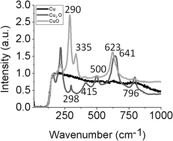
CuO is reported to thermally decompose to Cu2O under vacuum around 350 °C. Reference Goswami and Trehan27 Cupric oxide and cuprous oxide can be distinguished from each other not only by color but also by Raman spectroscopy. Reference Gan, Yu, Tay, Tan, Zhao and Fu28,Reference Gong, Lee and Yang29 Figure 2 shows the Raman spectra from the surface of OFHC copper foil after having been oxidized at 300 °C to form cupric oxide and then after being thermally reduced to cuprous oxide under vacuum at 600 °C. Although Cu2O is stable and solid at atmospheric pressure up to 1230 °C, well above the melting point of copper (1085 °C), it will thermally decompose into metallic copper below copper's melting point at low oxygen partial pressures (below ∼10−6 Torr at 1000 °C based on our experimental data). Figure 2 also shows the Raman spectra of the cuprous oxide sample after being thermally reduced to metallic copper under vacuum at 1000 °C.
Raman spectra from the same copper sample after being oxidized at 300 °C (CuO), vacuum annealed at 600 °C (Cu2O), and vacuum annealed at 1000 °C (Cu).

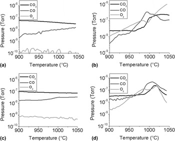
To show the relationship between the oxygen partial pressure and temperature, a 50 mm square piece of the as-oxidized 125-µm-thick OFHC Cu foil was placed in the CVD system and the system was pumped down to ∼10−8 Torr. The system with the preoxidized copper oxide substrate was heated to 1080 °C and then immediately allowed to cool. While heating and cooling, the composition of the CVD chamber atmosphere was measured downstream of the sample using an RGA. Figure 3 shows oxygen partial pressure versus temperature as the sample was allowed to cool. At higher temperature, the cuprous oxide will thermally decompose even at higher oxygen pressure. This temperature/pressure profile was quite reproducible between copper preoxidation levels and cooling rates. The logarithmic relationship between the oxygen partial pressure and temperature (as shown in Fig. 3) fits well with previously published results. Reference Kodera, Kusunoki and Shimizu30 (We note that our data fit almost exactly with that of Kodera et al. but only if °C is used instead of K. More recent work has shown that the earlier reports including that of Kodera et al. drastically overestimated the oxygen solubility in copper. Reference Hallstedt, Risold and Gauckler31 In fact, Kodera et al. overestimated the oxygen pressure for the other metal oxides measured in their work—which they also mention in their publication.) However, if the substrate is too lightly oxidized, the oxygen pressure will drop prematurely due to the substrate becoming completely reduced (this is the case of the 99.8% Cu foil from Alfa Aesar, Ward Hill, MA), which reaches a maximum oxygen pressure around 1020 °C with no detectable oxygen in the system by ∼1050 °C [Fig. 4(d)]. While the oxygen pressure is dependent on temperature, when the oxygen pressure starts to decrease again depends on the amount of original cuprous oxide in the substrate and the temperature at which it is being reduced; thus, the needed anneal temperature and time to fully reduce all the cuprous oxide is dependent on the substrate and can vary from substrate to substrate. As expected, heating OFHC copper does not result in an increase in oxygen partial pressure as shown in Fig. 3(c). To be mentioned, ex situ oxidation followed by vacuum thermal reduction does not noticeably affect the texture or grain size distribution of the copper substrate.
Log plot of the partial pressure of oxygen evolving from copper oxide versus inverse temperature while cooling the sample.

The oxygen, carbon monoxide, and carbon dioxide partial pressures in the CVD system versus the furnace temperature on initial heating of (a) just the CVD chamber and sample holder, (b) oxidized OFHC copper, (c) OFHC copper, and (d) 99.8% copper.

During this thermal decomposition, not only is a metallic copper surface obtained, but oxygen is also released from the substrate surface, which raises the possibility of copper oxides as a “self-cleaning” substrate for graphene growth. Indeed, the released oxygen does react with any adventitious carbon on the copper surface, forming carbon monoxide and carbon dioxide. Fig. 4(b) shows the CO and CO2 partial pressure, along with the O2 partial pressure, while heating the oxidized copper foil for the first time. [The background gas partial pressures from the system chamber have been subtracted so that only the gasses evolving from the substrate are shown in Fig. 4(b)]. The background pressures remain below 5 × 10⁻⁸ Torr and are shown in Fig. 4(a). As the oxygen evolves from the copper oxide, it reacts with the surface carbon forming both a peak in the CO pressure and a corresponding dip in the O2 pressure around 995 °C. At higher temperature, the CO2 pressure increases until the surface carbon has been depleted, indicated by a decrease in the CO2 pressure. How much CO and CO2 are generated depends on the quantity of carbon residues present on the substrate and varies between different substrates. To obtain a surface free of carbon, the CO and CO2 pressures must decrease (indicating that carbon is depleted) before the O2 pressure decreases due to the copper becoming fully reduced. This is the case for the partial pressures while heating the 99.8% foil shown in Fig. 4(d), where some (but not all) of the carbon is burned off and why there is still some of the likely carbon residue visible in Fig. 1(a). If the substrate is not sufficiently oxidized to provide enough oxygen to fully burn off any surface carbon, then the surface will not be completely clean for graphene growth.
C. Large-area graphene growth on preoxidized copper foils
To show the high effect of copper oxidation before graphene growth on reducing the nucleation density, we grew sub-monolayer graphene on OFHC copper cleaned with pure acetic acid to remove any native surface oxide layer Reference Chavez and Hess32 and as-oxidized OFHC copper under the same growth conditions (CH4 flow = 0.2 sccm, t = 25 min) in a vacuum CVD system with a 25 mm diameter chamber equipped with a rotary vane pump (Edwards RV8). Before growth, a vacuum level of about 10 mTorr was reached in the chamber before a 1-h hydrogen anneal (10 sccm, 60 mTorr) at 1040 °C. We then examined the nucleation density via SEM. As can be seen in Figs. 5(a) and 5(c), the nucleation density is over 50 times lower on the preoxidized substrate than on the Cu foil that was not oxidized before growth. When the methane flow rate is decreased to 0.1 sccm, there are even fewer nucleation sites on the preoxidized Cu foil, as shown in Fig. 5(e), where there are only 2 graphene islands detected in the whole image area (∼5 mm2). Since it is known that electro-polishing can also reduce nucleation density by decreasing the pregrowth surface roughness at room temperature before inserting the substrate into the CVD chamber, Reference Zhang, Lee, Piner, Kholmanov, Wu, Li, Ji and Ruoff20 both electro-polishing and preoxidation were used in conjunction. While electro-polishing alone [Fig. 5(b)] yields 3 times higher graphene nucleation than preoxidation [Fig. 5(c)], electro-polishing and then oxidizing the copper before growth yields very sparse graphene islands [Fig. 5(e)]. In fact, the nucleation density of electro-polished and oxidized copper is over 20 times lower than that on just oxidized copper, which is compared in detail in the table of Fig. 5. So by using both techniques of electro-polishing and oxidizing the copper before growth, nucleation density can be suppressed by over 1000 times compared to using simply acetic acid cleaned OFHC copper. With the decreased methane flow rate of 0.1 sccm, there were no graphene flakes found after 25 min of growth over a fairly large area on the oxidized plus electro-polished Cu foil [Fig. 5(f)], thus opening the possibility of synthesizing extremely large-area graphene islands on the Cu surface, as shown in Fig. 5(g). Individual graphene islands with lateral size larger than 1 mm can be readily achieved on the as-oxidized Cu foil after 5 h total growth time with 0.1 sccm methane. The Raman spectrum [inset of Fig. 5(g)] from the large-area islands confirms that it is high-quality monolayer graphene. Between the large islands that are over 1 mm across, some smaller graphene islands were obtained on the same sample due to new nucleation sites formed during the long graphene growth process, which ideally should be avoided to get large-area individual graphene islands.
SEM images of sub-monolayer graphene on (a) OFHC copper, CH4 = 0.2 sccm; (b) electro-polished OFHC copper, CH4 = 0.2 sccm; (c) preoxidized OFHC copper, CH4 = 0.2 sccm; (d) electro-polished and preoxidized OFHC copper, CH4 = 0.2 sccm; (e) preoxidized OFHC copper, CH4 = 0.1 sccm; (f) electro-polished and preoxidized OFHC copper, CH4 = 0.1 sccm. The comparison of the nucleation sites on the Cu foils with the four different treatments [Figs. 5(a)–5(d)] at the same methane flow rate (0.2 sccm) are summarized in the right table. (g) SEM image of graphene domains, with a Raman spectra insert from one, with lateral size larger than 1 mm on electro-polished and preoxidized OFHC copper foil with 0.1 sccm CH4 for 5 h at 1040 °C.

D. Comparison with previous studies of oxygen mediated growth
There have been a couple of recently published papers on the influence of oxygen on the growth of graphene on copper substrates. For instance, Hao et al. Reference Hao, Bharathi, Wang, Liu, Chen, Nie, Wang, Chou, Tan, Fallahazad, Ramanarayan, Magnuson, Tutuc, Yakobson, McCarty, Zhang, Kim, Hone, Colombo and Ruoff33 studied the influence of oxygen on the growth of graphene in copper enclosures. By introducing oxygen into the CVD chamber right before the introduction of methane, they found that the graphene nucleation rate was reduced. Our study shows that large graphene domains can be obtained without using an enclosure if the copper surface is sufficiently clean before growth is initiated. Another recent paper published by Robinson et al. Reference Robinson, Ong, Mowll, Tyagi, Gaskill, Geisler and Ventrice34 studied the influence of a chemisorbed oxygen layer on the growth of graphene on the Cu(100) surface. This study also found that the nucleation rate on the surface with a chemisorbed oxygen layer was lower than when the growth was initiated on the Cu(100) surface that was not pre-dosed with oxygen. These studies clearly show that oxygen plays an important role in the initial nucleation rate of graphene on copper surfaces.
IV. CONCLUSIONS
In summary, adventitious carbon or other carbonaceous compounds on the surface of Cu can increase graphene nucleation density. Copper oxidation with subsequent reduction at temperatures above 900 °C was found to be an effective way to clean the copper surface before graphene growth. This process was used to grow large-area individual graphene islands of over a millimeter across.
ACKNOWLEDGMENTS
Carl W. Magnuson gratefully appreciates support from Sandia National Labs LDRD fellowship program. We thank the Keck Foundation (749046) and ONR (N00014-10-1-0254) for support.