Introduction
Embryonic stem (ES) cells are derived from inner cell mass/epiblast of the blastocyst. They have distinct properties, such as long-term self-renewal and expression of transcription factor Oct-4 for maintenance of undifferentiated status. Under certain conditions, they can give rise to three primary germ layers as well as germ cells. Also, genetically modified cell clones can be created by gene targeting and clonal selection. For such reasons, ES cells have been a great tool for studying early mammalian development and transgenesis. Since the derivation of ES cells from mouse blastocysts (Evans & Kaufman, Reference Evans and Kaufman1981; Martin et al., Reference Martin1981), many researchers have reported successful derivation of ES cells or ES-like cells in hamster (Doestshman et al., Reference Doetshman, Williams and Maeda1988), rat (Ouhibi et al., Reference Ouhibi, Sullivan, English, Colledge, Evans and Clarke1995), cat (Yu et al., Reference Yu, Jin, Yin, Cho, Jeon, Lee and Kong2008), cattle (Strelchenko, Reference Strelchenko1996), pig (Li et al., Reference Li, Li, Hou, Sun, Sun and Wang2004), rhesus monkey (Thomson et al., Reference Thomson, Kalishman, Golos, Durning, Harris, Becker and Hearn1995) and human (Thomson et al., Reference Thomson, Itskovitz-Eldor, Shapiro, Waknitz, Swiergiel, Marshall and Jones1998). Recently, ES cells or ES-like cells have also been established from somatic cell nuclear transfer (SCNT) preimplantation embryos in mouse (Wakayama et al., Reference Wakayama, Tabar, Rodriguez, Perry, Studer and Mombaerts2001), bovine (Wang et al., Reference Wang, Duan, Sung, Jeong, Yang and Tian2005) and primate (Byrne et al., Reference Byrne, Pedersen, Clepper, Nelson, Sanger, Gokhale, Wolf and Mitalipov2007). In addition to their importance for studies of mammalian embryogenesis, porcine embryonic stem cells (ESCs) can be exploited to produce transgenic animals for use as xenotransplantation donors. Most notably in the pig, the combination of SCNT and stem cell establishment technology has proven to be of great value for clinical applications of xenotransplantation, which makes it possible to bulk-produce immunosuppressive organs for xenotransplantation. Porcine ESCs are derived from early embryos, but lose their pluripotency over time in culture (Moore & Piedrahita, Reference Moore and Piedrahita1997). Attempts to generate pluripotent ES-like cells from porcine in vitro fertilized or in vivo produced embryos have been reported in only a few studies (Miyoshi et al., Reference Miyoshi, Taguchi, Sendai, Hoshi and Sato2000; Li et al., Reference Li, Zhang, Hou, Jiao, Zheng and Wang2003). Most of these ES-like cells lacked definitive markers for the characterization of ES cells and failed to maintain their undifferentiated status. This deficiency may have been due to suboptimal culture conditions, as the majority of the cells failed to survive expansion over long periods of time. Moreover, successful establishment of ESCs derived from SCNT and the production of chimeras have been reported only for murine and bovine models (Cibelli et al., Reference Cibelli, Stice, Golueke, Kane, Jerry, Blackwell, Ponce de León and Robl1998; Wakayama et al., Reference Wakayama, Tabar, Rodriguez, Perry, Studer and Mombaerts2001). In this study, we established porcine ES-like cells from blastocysts derived from SCNT of fetal and neonatal fibroblasts into enucleated oocytes.
Materials and Methods
Oocyte collection and culture of cumulus–oocyte complexes
Ovaries were retrieved from prepubertal gilts at a local slaughterhouse and transported to the laboratory in physiological saline at 30–35°C within 2 h. Follicular fluid and cumulus–oocyte complexes (COCs) from follicles (3–6 mm in diameter) were aspirated using an 18-gauge needle attached to a 10-ml disposable syringe. Compact COCs were selected and cultured in tissue culture medium (TCM)-199 (Life Technologies) that was supplemented with 10 ng/ml epidermal growth factor, 4 IU/ml pregnant mare serum gonadotropin (PMSG) (Intervet, Boxmeer) and human chorionic gonadotropin (hCG) (Intervet) and 10% (v/v) porcine follicular fluid (pFF). The pFF was aspirated from the follicles (3–7 mm) of the prepubertal gilt ovaries. After centrifugation at 500 g for 30 min, supernatants were collected and filtered sequentially through 1.2-μm and 0.45-μm syringe filters (Gelman Sciences). Prepared pFF was stored at −20°C until use. Each group of 50 COCs was cultured in 500 μl TCM-199 at 39°C in a humidified atmosphere of 5% CO2 and 95% air. After culturing for 22 h, the COCs were washed three times and cultured in PMSG- and hCG-free TCM-199 medium for an additional 22 h. At the end of culture maturation, the COCs were transferred to HEPES-buffered NCSU-23 medium that contained 0.5 mg/ml hyaluronidase (Sigma-Aldrich Corp.) for 1 min and the cumulus cells were subsequently removed by gentle pipetting.
Preparation of miniature pig fibroblasts for SCNT
Miniature pig fibroblasts for use as donor cells were isolated from cloned fetus (CF1) on day 30 of gestation and tail fibroblasts were isolated from a newborn cloned piglet (CP1). The fetal head was removed using iris scissors and the soft tissues, which included the liver and intestines, were discarded by scooping out with two watchmaker's forceps. To isolate tail fibroblasts, the external surface of the tail was shaved and then washed several times. After washing twice with PBS (Life Technologies), the carcass was minced with a surgical blade on a 100-mm culture dish (Becton Dickinson). The minced fetal and tail tissues were dissociated for 1–2 h in DMEM (Life Technologies) that was supplemented with 0.1% (w/v) trypsin and 1 mM EDTA (Life Technologies). Trypsinized cells were washed once by centrifugation at 300 g for 10 min and subsequently seeded into 100-mm plastic culture dishes. Seeded cells were cultured for 6–8 days in DMEM that was supplemented with 10% (v/v) FBS (Life Technologies), 1 mM sodium pyruvate, 1% (v/v) non-essential amino acids (Life Technologies) and 10 μg/ml penicillin–streptomycin solution, at 39°C in a humidified atmosphere of 5% CO2 and 95% air. After removal of unattached clumps of cells or explants, the attached cells were further cultured until confluent, subcultured at intervals of 5–7 days by trypsinization for 5 min in 0.1% trypsin and 0.02% EDTA and stored after two passages in freezing medium in liquid nitrogen at–196°C. The freezing medium consisted of 80% (v/v) DMEM, 10% (v/v) DMSO and 10% (v/v) FBS. Prior to SCNT, cells were thawed, cultured for 3–4 days until 80% confluent and then subjected to serum starvation. For serum starvation, cells were cultured for 3 days in DMEM that was supplemented with 0.5% FBS. Individual cells were retrieved from the monolayer by trypsinization for 30 s and subsequently used for SCNT.
SCNT and embryo culture
After 42 h of in vitro maturation (IVM), a cumulus-free oocyte was held with a holding micropipette and the zona pellucida was partially dissected with a fine glass needle so as to make a slit near the first polar body. The first polar body and adjacent cytoplasm that, presumably, contained the metaphase II chromosomes were extruded by squeezing the oocytes with the same needle. Oocytes were enucleated in HEPES-buffered NCSU-23 that was supplemented with 0.3% BSA. After enucleation, the oocytes were stained with 5 μg/ml bisbenzimide (Hoechst 33342; Sigma) for 5 min and observed under an inverted microscope that was equipped with epifluorescence to visualize the residual DNA. Oocytes that harboured DNA were excluded from the experiments. Using a fine pipette, a trypsinized fetal fibroblast with a smooth cell surface was transferred into the perivitelline space of an enucleated oocyte. Couplets were equilibrated for 4 min with 0.26 M mannitol solution that contained 0.5 mM HEPES, 0.1 mM CaCl2 and 0.1 mM MgCl2 and transferred to a chamber that contained two electrodes overlaid with mannitol solution. The couplets were simultaneously fused and activated with a single DC pulse of 2.0 kV/cm for 30 s delivered by the BTX Electro-Cell Manipulator 2001 (BTX Inc.). Activated oocytes were washed three times with NCSU-23 that was supplemented with 4 mg/ml fatty acid-free BSA and placed in 25-μl microdrops (10 oocytes per drop) of NCSU-23 under mineral oil and cultured at 39°C, in 5% CO2, 5% O2 and 90% N2. The reconstructed embryos were cultured for 168 h after activation; cleavage and blastocyst formation were monitored under a stereomicroscope at 48 h and 168 h, respectively.
Preparation of feeder cells
Mouse embryonic fetal fibroblasts (MEFs) were isolated from day-13.5 postcoitum fetuses of CF1 mice (Thomson et al., Reference Thomson, Itskovitz-Eldor, Shapiro, Waknitz, Swiergiel, Marshall and Jones1998). The head was removed using iris scissors and soft tissues, such as the liver and intestines, were discarded by scooping out with two watchmaker's forceps. After washing twice with PBS (Life Technologies), the carcass was minced with a surgical blade on a 60-mm culture dish (Becton Dickinson). The minced fetal tissues were pipetted several times, washed once by centrifugation at 300 g for 10 min and subsequently seeded into 100-mm plastic culture dishes. Seeded cells were cultured for 5–7 days in DMEM that was supplemented with 10% (v/v) FBS (Life Technologies), 0.1 mM β-mercaptoethanol, 1% (v/v) non-essential amino acids (Life Technologies) and 10 μg/ml penicillin–streptomycin, at 37°C, in a humidified atmosphere of 5% CO2 in air. Feeder cells were cultured in DMEM (Life Technologies) that was supplemented with 10% FBS (Life Technologies), 0.1 mM β-mercaptoethanol, 1% (v/v) non-essential amino acids (Life Technologies) and 10 μg/ml penicillin–streptomycin. MEFs at 80% confluence were treated with 10 μg/ml mitomycin C (Sigma) for 2 h to arrest mitosis and then washed several times in PBS and replated at a concentration of 0.8 × 10⁵ cells/well in gelatin-coated, 4-well cell culture dishes.
ICM isolation and maintenance of embryonic stem-like cells
After the zona pellucidae from SCNT blastocysts were removed with 0.5% pronase E (Sigma), the zona-free SCNT blastocysts were washed with DMEM/F12 (Life Technologies) that was supplemented with 20% knockout serum replacement (Life Technologies). ICMs were isolated from blastocysts by mechanical removal of the trophectoderm using a narrow glass pipette. They were transferred onto the MEF feeder layer (Thomson et al., Reference Thomson, Itskovitz-Eldor, Shapiro, Waknitz, Swiergiel, Marshall and Jones1998) and ICM outgrowths were passaged to plates with fresh medium and new MEFs by mechanical dissection. To maintain porcine ES-like cells, about 200 undifferentiated ES-like cell colonies were mechanically isolated under a dissecting microscope and the clumps were propagated on a new feeder layer every 5–7 days. The ES–like cells were cultured in DMEM/F12 that was supplemented with 20% Knockout serum replacement, 0.1 mM β-mercaptoethanol, 0.1 mM non-essential amino acids, 1% (v/v) penicillin–streptomycin solution and 20 ng/ml human recombination basic FGF (Invitrogen).
Marker assay
The pNTES-1 (embryonic stem-like cells derived from SCNT blastocyst with CF1) and pNTES-2 (embryonic stem-like cells derived from cloned blastocyst with CP1) cells collected from passages 28 and 24, respectively, were washed with PBS, fixed in 4% (v/v) formaldehyde at room temperature for 10 min, washed twice with PBS and then stained with NBT/BCIP solution (Roche Molecular Biochemicals). The colour reaction was stopped with PBS. For immunostaining of surface markers, ES-like cells were washed with PBS and fixed in 4% paraformaldehyde at 4°C for 30 min. Anti-Oct-4 antibody (SC-9081) was purchased from Santa Cruz Biotechnology and antibodies against SSEA-1 (MAB4301), SSEA-4 (MAB4304), TRA-1–60 (MAB4360) and TRA-1–81 (MAB4381) were purchased from Chemicon. All antibodies were diluted 1:100 in PBS and added for 3 h. After washing with PBS three times, FITC-conjugated IgM and IgG (SSEA-4) (Chemicon) were used as secondary antibodies.
Karyotyping
The chromosomes of established pNTES-1 and pNTES-2 cells were analyzed at passages 28 and 24, respectively. pNTES-1 and pNTES-2 cells were incubated in medium that was supplemented with 0.02 μg/ml colcemide (Sigma) for 1 h at 37°C in an atmosphere of 5% CO2 in air. After trypsinization and treatment with hypotonic KCl (0.56%) for 20 min, the cells were placed in a hypotonic solution, fixed in a 3:1 (v/v) mixture of methanol and acetic acid and spread on clean microscopic slides by gentle dropping. After staining with Giemsa (1:20 dilution; Sigma) for 20 min, the chromosomes were examined at ×1000 magnification.
Microsatellite analysis
Genomic DNA samples from pNTES-1 and pNTES-2 cells and their donor fibroblasts cells were extracted using the standard phenol extraction method. Briefly, mechanically dissected pNTES-1 and pNTES-2 colonies and trypsinized donor cells were incubated overnight in lysis buffer [0.05 M Tris (pH 8.0), 0.05 M EDTA, 0.5% SDS] that contained proteinase K (Intron). Proteins were removed by phenol extraction and DNA was precipitated by the addition of ethanol. Genomic DNA samples were subjected to microsatellite analyses using nine porcine DNA microsatellite markers (S0005, S0007, S0086, S0230, SW61, SW902, SW980, SW1661 and SWR 1004), each of which was labelled with one of the fluorescent dyes: FAM, TET and HEX. Length variations were detected by PCR amplification with fluorescently labelled locus-specific primers and PAGE on an automated DNA sequencer (ABI 373; Applied Biosystems). Proprietary software (GeneScan and Genotyper; Applied Biosystems) was used to estimate the PCR product size in nucleotides.
Reverse transcription-polymerase chain reaction
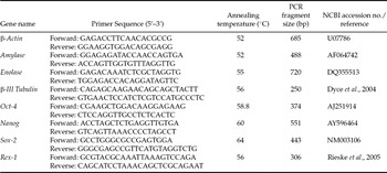
Total RNA samples from embryoid bodies of cultured pNTES-1 and pNTES-2 cells were extracted using Trizol (Invitrogen) and dissolved in 20 μl RNase-free water, as described by Kim et al. (Reference Kim, Lee, Lee, Kim, Jeong, Kim, Kang, Lee and Hwang2005). Total RNA was subjected to reverse transcription-polymerase chain reaction (RT-PCR). The reverse transcription step was carried out at 37°C for 60 min. Individual RT reactions (33 μl each) were performed in 5 mM MgCl2, 1× RT buffer, 2.5 mM oligo(dT), 1 mM dNTP and 50 IU of murine leukemia virus reverse transcriptase (MoMLV; Amersham Pharmacia Biotechnologies). The expression of porcine β-actin was used as a control for RT-PCR amplification. The sequences of the primers used are listed in Table 1. The cDNA (2 μl) was amplified in a 50-μl PCR reaction that contained 250 U Ex-Taq polymerase (Takara, Shiga, Japan), 5 μl of 10× buffer, 2.5 mM deoxy-NTPs and 50 pmol of specific primers. Amplified PCR products were visualized under UV light on a 1.5% agarose gel in 1× TAE buffer that contained 1 μg/ml ethidium bromide. The image of each gel was recorded using the Gel Doc 2000 Gel Documentation System (Bio-Rad Laboratories).
Primers used for PCR of gene transcripts from porcine SCNT ES-like cell lines

Results
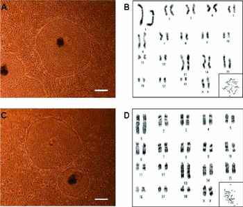
In total, 46 and 14 SCNT blastocysts were produced from day-30 fetal fibroblasts (CF1) and newborn piglet fibroblasts (CP1), respectively. As shown in Table 2, no significant differences between the two groups were noted with respect to reconstruction, cleavage and blastocyst formation rates. ICMs from zona pellucida-free blastocysts were mechanically dissected, washed several times and plated on a mitotically inactivated MEF feeder layer. Colony formation was detected 5–8 days after initial seeding. The porcine ES-like cells showed typical ES cell morphology, with compact shape and distinct borders. Under high-magnification phase-contrast microscopy, ES-like cells were found to have prominent nucleoli and a high ratio of nucleus to cytoplasm with ribosomes (Fig. 1A, C). However, some of the colonies maintained their ESC morphology for only 2–5 passages and then differentiated or degenerated. Only two lines were successfully cultured without differentiation or degeneration. Each of these lines was propagated continuously for more than 52 (pNTES-1) or 48 (pNTES-2) passages. The rate of ES-like cell establishment using CF1 was lower than that obtained using CP1 (2.2% vs. 7.1%; Table 2).
Establishment of porcine SCNT embryonic stem-like cells derived from cloned blastocysts reconstructed with fetal (F1) and newborn (P1) fibroblasts of cloned pigs

aCF1 denotes fetal fibroblasts isolated from cloned fetus on day 30 of gestation.
bCP1 denotes piglet fibroblasts isolated from cloned newborn piglets.
cPercentage of enucleated oocytes.
dPercentage of reconstructed oocytes.
ePercentage of reconstructed oocytes that developed to 2-cell embryos.
fPercentage of established ES-like cells from used blastocysts.
Phase-contrast images and karyotypes of pNTES-1 and pNTES-2 cells. Morphologies of pNTES-1 (A) and pNTES-2 (C) cells at 28 and 24 passages, respectively. The pNTES-1 and pNTES-2 cells show typical ESC morphology and have a distinct border. Scale bar = 200 μm. G-banding karyotypes of pNTES-1 (B) and pNTES-2 (D) cells at 28 and 24 passages, respectively. Both cell types have 18 pairs of autosomes and normal XX karyotypes.

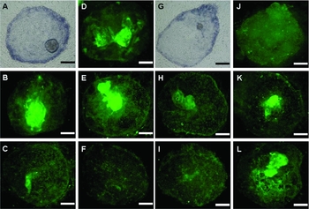
The karyotype of pNTES-1 and pNTES-2 cells was normal, with 38 chromosomes and XX sex chromosomes (Fig. 1B, D). As in the mouse and human ESCs, pNTES-1 and pNTES-2 cells showed a high level of AP activity and Oct-4 expression. Immunostaining of the pNTES-1 and pNTES-2 cells were performed using several antibodies against cell surface carbohydrates, including anti-SSEA-1, anti-SSEA-4, anti-TRA-1–60 and anti-TRA-1–81. Similar expression patterns were observed for pNTES-1 and pNTES-2 cells (Fig. 2).
Characterization of pNTES-1 and pNTES-2 cells. pNTES-1 and pNTES-2 cells were characterized using six stem-cell-specific markers. pNTES-1 (28 passages) shows high levels of alkaline phosphatase (AP) (A), stage-specific embryonic antigen (SSEA)-4 (B), TRA-1–60 (C), SSEA-1 (D), Oct-4 (E) and TRA-1–81 (F). pNTES-2 (24 passages) cells were characterized using the same markers: AP (G), SSEA-4 (H), TRA-1–60 (I), SSEA-1 (J), Oct-4 (K) and TRA-1–81 (L).

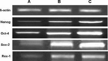
The expression levels of Oct-4, Nanog, Sox-2 and Rex-1 in pNTES-1 and pNTES-2 cells were measured by RT-PCR. The levels of these markers were higher in pNTES-1 and pNTES-2 cells than in their respective blastocysts (Fig. 3).
Expression of Nanog, Oct-4, Sox-2 and Rex-1 genes in NT blastocysts (A), pNTES-1 (B) and pNTES-2 (C) cells, respectively. Scale bar = 200 μm.

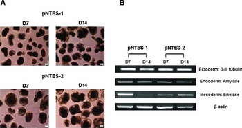
When pNTES-1 and pNTES-2 cells were cultured for 14 days on plastic petri dishes, cystic embryoid bodies formed (Fig. 4). The expression levels of several cell specific genes of the three embryonic germ layer cells were assessed in day-7 and day-14 embryoid bodies. The mRNA transcripts of β-III tubulin (ectoderm, neuron), amylase (endoderm, pancrease) and enolase (mesoderm, muscle) were detected by RT-PCR.
Light microscopic images of embryoid bodies and RT-PCR detection of genes typical for three germ layers. (A) Light microscopic images of embryoid bodies cultured for 7 days or 14 days. (B) RT-PCR detection of the expression levels of genes characteristic of the three germ layers in embryoid bodies derived from pNTES-1 and pNTES-2 cells. Scale bars = 100 μm.

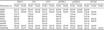
Microsatellite analysis of pNTES-1 and pNTES-2 cells and its donor cells confirmed that the pNTES-1 and pNTES-2 cells were genetically identical to their donor cells (CF1 and CP1) (Table 3).
Microsatellite analysis of pNTES-1, pNTES-2 and respective donor cellsa

aFor each microsatellite marker, the genotype was determined by size (bp).
bMiniature pig fetal fibroblasts used as donor cells for CF1c and CP1d.
cDay 30 cloned fetal fibroblasts and donor cell used for pNTES-1
dNewborn cloned piglet fibroblasts and donor cell used for pNTES-2.
eNegative control: ESCs derived from IVF blastocysts.
Discussion
In the present study, two porcine ES-like cell lines were established from SCNT blastocysts produced via the recloning of fetal or neonatal fibroblast of cloned pigs. These cells have typical ESC morphology and AP activity, express surface marker proteins (SSEA-1, SSEA-4, Oct-4, TRA-1–60 and TRA-1–81) and readily formed embryoid bodies in suspension culture. In addition, microsatellite analyses revealed genetic identity between the pNTES-1, pNTES-2 and donor cells.
The successful establishment of ES cells derived from nuclear transferred embryos is of potential importance for many research areas, such as cell therapy, the production of biopharmaceuticals and studies of mammalian development. Among the many animal models used for these purposes, genetically modified pig models have proven to be invaluable tools, owing to the physiological similarities between pigs and humans. This advantage may be an important consideration in the production of porcine xenogeneic organs for the treatment of human diseases through genetic engineering.
Although several attempts on establishing ES cells in pig have been reported so far, characterization results have not been definitive as those from mice. Strojek et al. (Reference Strojek, Reed, Hoover and Wagner1990) reported that DMEM with 10% FCS yielded best results on establishing ES cells, however, they were not able to subculture more than 3 to 5 passages layers. Wianny et al. (Reference Wianny, Perreau and Hochereau de Reviers1997) also investigated the proliferation rate and differentiation state in porcine ICMs and epiblasts by analyzing the percentages of cells in S-phase and they found that ICMs from day-7 blastocysts remained undifferentiated in vitro, whereas those from day-11 epiblasts differentiated rapidly. Chen et al. (Reference Chen, Shiue, Bertolini, Medrano, BonDurant and Anderson1999) isolated pluripotent embryo-derived cell lines with suitable undifferentiated morphology only from day-7 and day-8 early hatched blastocysts. Li et al. (Reference Li, Zhang, Hou, Jiao, Zheng and Wang2003) used ICMs from blastocysts directly flushed from uterus for derivation of ES cell lines. Although cells initially grew on mitotically inactive MEFs, the outgrowth was not vigorous and stopped at passage 6. Such limitation might be due to suboptimal culture conditions, as the majority of the cells failed to expand over extended period.
The ESCs culture medium is usually supplemented with various components, e.g., cytokines and growth factors, for the maintenance of cell pluripotency and self-renewal activity. Of these factors, leukemia inhibitory factor (LIF) and basic fibroblast growth factor (bFGF) are essential elements. It has been reported that LIF inhibits the differentiation of ESCs and that bFGF stimulates ESC proliferation (Lee & Piedrahita, Reference Lee and Piedrahita2000). However, some researchers have reported that culturing ESCs on a layer of feeder cells without the addition of cytokines or growth factors is sufficient for the maintenance of ESCs (Chen et al., Reference Chen, Shiue, Bertolini, Medrano, BonDurant and Anderson1999). Based on these previous reports, the culture medium for pNTES-1 and pNTES-2 cells were refined in the present study. It was found that the addition of bFGF (20 ng/ml) without LIF could support the growth of porcine NTES-like cells while maintaining its pluripotency. No morphological difference was noticed when LIF was omitted from the culture medium.
Teratoma formation has been an essential characteristic of ESCs in humans and mice. We attempted to generate teratoma in SCID mice, but it was unsuccessful (data not shown). Similarly Anderson et al. (Reference Anderson, BonDurant, Goff, Groff and Moyer1996) failed to induce teratoma formation after transplantation of bovine and porcine ICMs into the kidney capsules of athymic mice. One possible explanation for these failures might be the incompatibility and immune rejection of cells derived from different species.
In conclusion, we have established and maintained successfully embryonic stem-like cell lines (pNTES-1 and pNTES-2 cells) using the ICMs of blastocysts produced by SCNT with fetal or neonatal fibroblasts of cloned pigs. The pNTES-1 and pNTES-2 cells displayed typical ESC surface markers and showed high levels of AP activity and Oct-4 level. Microsatellite analyses demonstrated genetical identity between the pNTES-1 and pNTES-2 and their donor cells and the pNTES-1 and pNTES-2 cells expressed transcripts those are specific for the three germ layers of embryoid bodies obtained by suspension culture. Our results will contribute to establishing transgenic technology in livestock and an efficient model system for studying early mammalian development.