INTRODUCTION
The rare pilumnoid genus Dentoxanthus Stephensen, Reference Stephensen, Jessen and Spärck1946, is monotypic. Dentoxanthus iranicus Stephensen, Reference Stephensen, Jessen and Spärck1946, was described from the Persian Gulf on the basis of one small female specimen. Its systematic position has been debated at length since its original description, and was questionably referred to as belonging to the families Xanthidae or Parthenopidae sensu lato, even after males were subsequently found in Pakistan (Stephensen, Reference Stephensen, Jessen and Spärck1946; Serène et al., Reference Serène, van Luc and Van Luom1958; Serène, Reference Serène1968; Tirmizi & Serène, Reference Tirmizi and Serène1971; Tirmizi & Kazmi, Reference Tirmizi and Kazmi1982).
A second species, Denthoxanthus komodoensis, was described by Serène (Reference Serène1971) from Komodo Island in Indonesia on the basis of a small female measuring 8.2 × 7.6 mm. In the 1980s, the consensus was that Denthoxanthus was a member of the subfamily Eumedoninae Dana, 1852, and part of the family Parthenopidae (Tirmizi & Kazmi, Reference Tirmizi and Kazmi1982). Števčić & Ng (Reference Števčić and Ng1988) reviewed the literature and argued that Dentoxanthus iranicus had pilumnoid affinities, and suggested it should be referred to as belonging to the Pilumnidae family, within its own tribe. Ng & Števčić (Reference Ng and Števčić1993) later described a new genus, Otognathon for Denthoxanthus komodoensis.
Števčić (Reference Stevčić2005) formally established the tribe Dentoxanthini for Dentoxanthus, but did not indicate if Otognathon also belonged to this tribe. Ng et al. (Reference Ng, Guinot and Davie2008) discussed the taxonomy of Dentoxanthus and the systematics of the Pilumnoidea at length, and recognized Galenidae Alcock, 1898, within Pilumnoidea. This family, comprising four subfamilies – Galeninae Alcock, 1898; Halimedinae Alcock, 1898; Dentoxanthinae Števčić, 2005; and Parapanopinae Števčic, 2005 – contained most of the free-living pilumnoids, which have a ‘xanthid’-like appearance and differ from typical pilumnids, which are usually distinctly setose. They also placed and redefined Eumedoninaes sensu stricto in the Pilumnidae family (see discussion in Ng & Clark, Reference Ng and Clark2000). Ng et al. (Reference Ng, Guinot and Davie2008) also argued that Otognathon was not a dentoxanthine but more likely a varunid (Grapsoidea) instead. This was subsequently confirmed by Ng et al. (Reference Ng, Komai and Ng2009) when they described a second Otognathon species from Japan from material that included male specimens.
The recent collection of a male specimen from Gujarat in India (a first record for India) allowed additional taxonomic observations to be made for the species. The specimens examined have been deposited in the Zoology Museum of The M.S. University of Baroda (ZL), Gujarat, India; and the Zoological Reference Collection (ZRC) of the Lee Kong Chian Natural History Museum (previously Raffles Museum of Biodiversity Research), National University of Singapore. The measurements provided (in millimetres) are of the maximum carapace width and lengths, respectively. The abbreviations G1 and G2 are used for the male first and second gonopods, respectively.
RESULTS
SYSTEMATICS
Family PILUMNIDAE Samoeuelle, 1819
Subfamily DENTHOXANTHINAE Števčić, Reference Stevčić2005
Dentoxanthus iranicus Stephensen, Reference Stephensen, Jessen and Spärck1946
(Figures 1–3)
Dentoxanthus iranicus Stephensen, Reference Stephensen, Jessen and Spärck1946, male (18.8 × 14.9 mm) (ZL-AR-CR-83), Gujarat, India. Overall habitus, colour fresh after collection.

Dentoxanthus iranicus Stephensen, Reference Stephensen, Jessen and Spärck1946: 164, Figure 43; Tirmizi & Serène, Reference Tirmizi and Serène1971: 27, Figure 3A–D; Tirmizi & Kazmi, Reference Tirmizi and Kazmi1982: 310, Figures 2 & 3; Števčić & Ng, Reference Števčić and Ng1988: 1; Ng et al., Reference Ng, Guinot and Davie2008: 137.
MATERIAL EXAMINED
One male (18.8 × 15.0 mm) (ZL-AR-CR-83), Jakhau fishing harbour, Gulf of Kachchh, 22°13′48″N 68°36′44″E, Gujarat, India, from muddy bottoms, 20–25 m water depth, trawl, coll. by J. Trivedi and K.D. Vachhrajani, 27 March 2015; one male (23.7 × 18.3 mm) (ZRC 1999.1104), Karachi Fish Harbour, Pakistan, coll. by University of Karachi, 1990s.
DIAGNOSIS
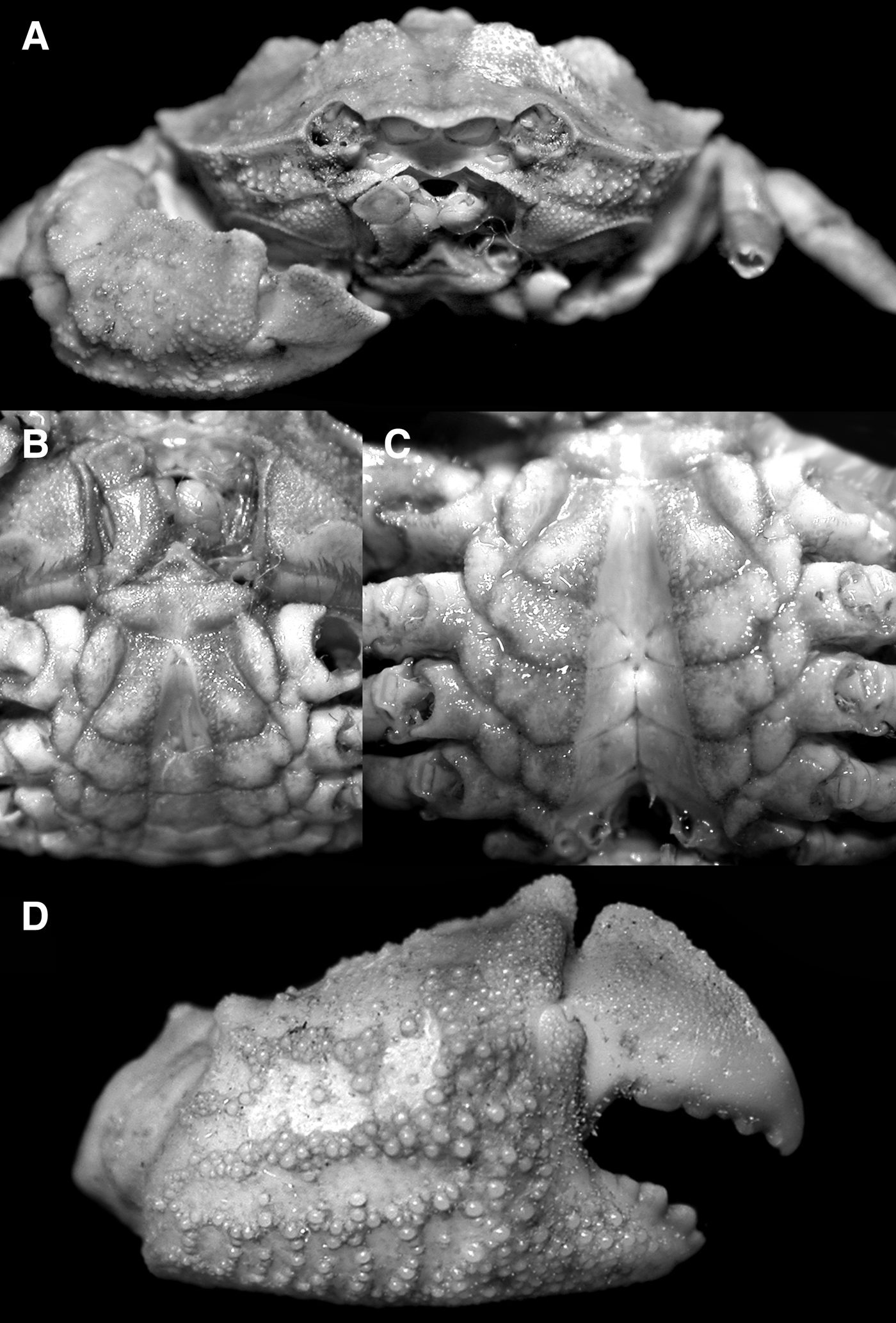
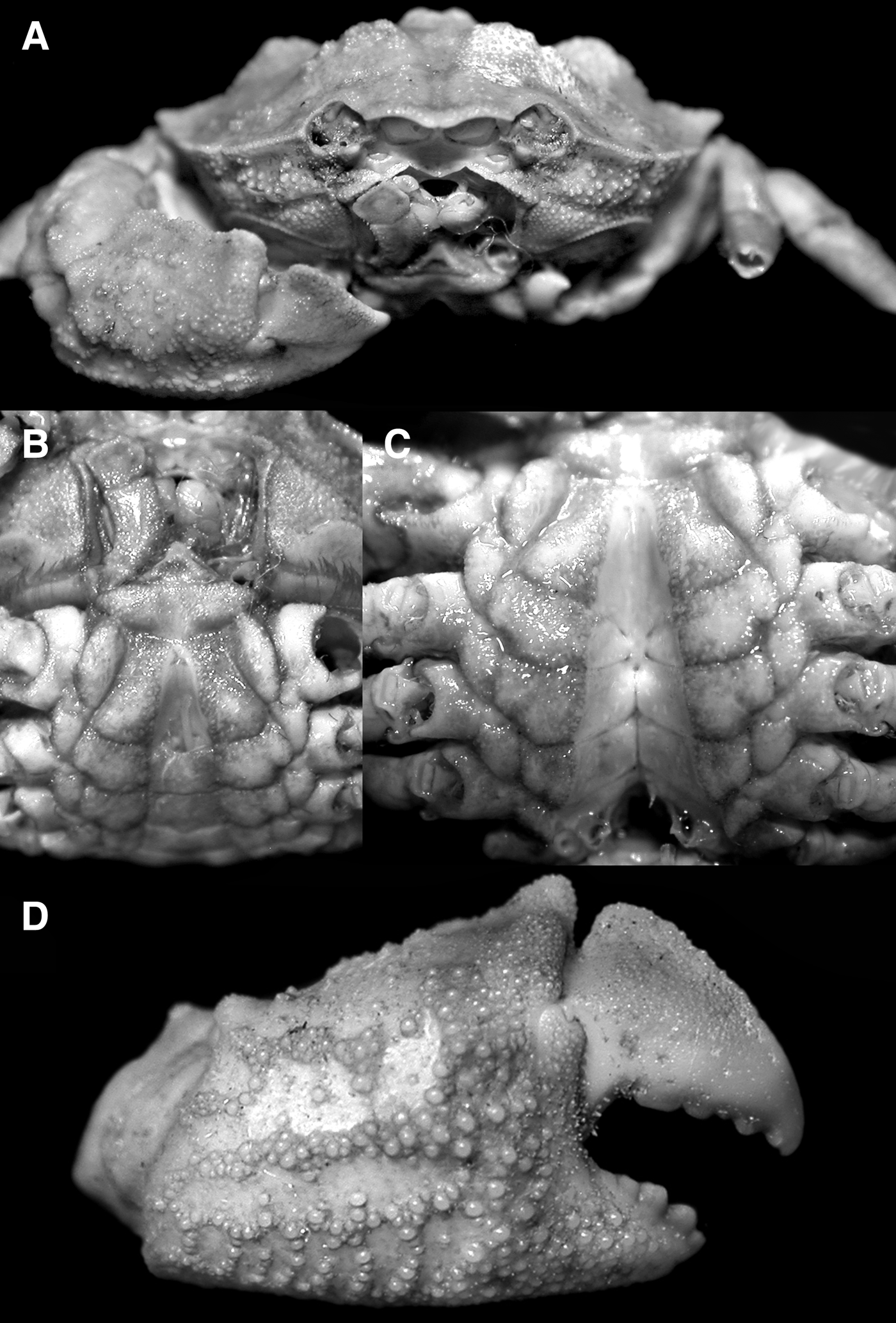
Carapace hexagonal; regions clearly demarcated, protogastric, mesogastric, metagastric, cardiac and mesobranchial regions swollen, granulated; epibranchial and metabranchial surfaces raised, covered with granules (Figures 1, 3A & 4A); front prominent, lamelliform, margin weakly bilobate, contiguous with supraorbital margin (Figures 1, 3A & 4A); anterolateral margin with wide external orbital angle and two lobiform teeth with subcristate margins and large lateral tooth (Figures 1, 3A & 4A); suborbital margin with two large teeth, separated by gap or cleft (Figures 2A & 4B); posterior margin of epistome with prominent median triangle, with median longitudinal fissure (Figures 2A & 4B); endostome without ridges; third maxilliped with merus quadrate, anteroexternal angle not distinctly auriculiform (Figures 2A & 4B); chelipeds closely opposed to carapace sides when retracted (Figures 1 & 4A); chelae short, stout, fingers shorter than palm, not pigmented black or dark brown, dorsal margin strongly cristate, dactylus curved, with prominent crista on dorsal two-thirds of margin (Figure 4A); ambulatory legs, short, stout, meri with prominent dorsal cristae, outer surface of carpus bicristate, with prominent dactylo-propodal lock (Figures 1 & 4A); thoracic sternum transversely narrow, sternites 2 and 3 separated by distinct transverse suture; sternite 4 with prominent lateral swelling, separated from median part by deep oblique groove, sternite 8 not visible when abdomen closed (Figures 2B, C & 4C, D); sutures between sternites 4/5 and 5/6 medially interrupted; sutures for sternites 6–8 complete, joining medially to form longitudinal groove (Figure 2C); sternoabdominal cavity deep, reaching to median part of fused sternites 3 and 4 (Figures 2B & 4C); male abdominal locking mechanism on sternite 5 peg-like, positioned just before suture between sternites 4 and 5 (Figure 2C); male abdomen longitudinally narrow, somites 1 and 3 completely covering surfaces of thoracic sternum, reaching bases of coxae of last pair of ambulatory legs, sternite 8 completely covered (Figures 2B, 3B & 4C); G1 relatively stout, strongly sinuous, distal part recurved, hook-like (Figure 3C, D).
Dentoxanthus iranicus Stephensen, Reference Stephensen, Jessen and Spärck1946, male (18.8 × 14.9 mm) (ZL-AR-CR-83), Gujarat, India. A, frontal view of cephalothorax; B, anterior thoracic sternum and male abdomen; C, sternoabdominal cavity; D, outer view of right chela.
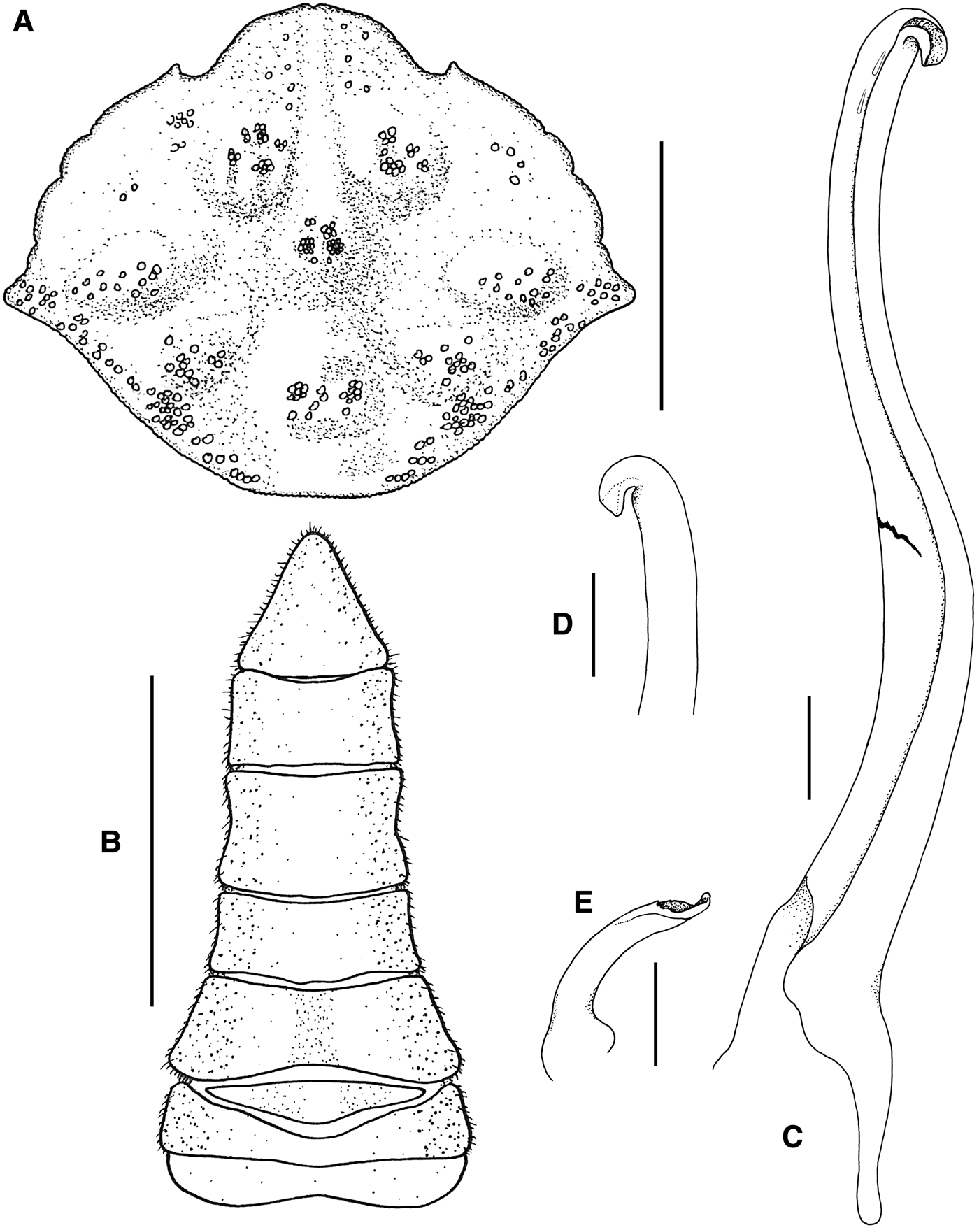
Dentoxanthus iranicus Stephensen, Reference Stephensen, Jessen and Spärck1946, male (18.8 × 14.9 mm) (ZL-AR-CR-83), Gujarat, India. A, dorsal view of carapace; B, abdomen; C, left G1 (ventral view, with break medially); D, distal part of left G1 (ventral view); E, left G2. Scale bars: A = 10 mm; B = 5 mm; C–E = 0.5 mm.

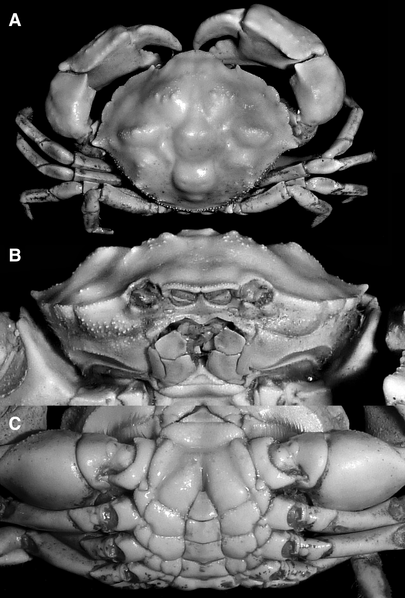
Dentoxanthus iranicus Stephensen, Reference Stephensen, Jessen and Spärck1946, male (23.7 × 18.3 mm) (ZRC 1999.1104), Karachi, Pakistan. A, overall habitus; B, frontal view of cephalothorax; C, thoracic sternum and abdomen.
DESCRIPTION OF MALE
Carapace hexagonal, wider than long; regions clearly demarcated, separated by wide grooves; epigastric region not inflated, relatively flat, with longitudinal row of small granules; protogastric region swollen, surface with numerous granules; mesogastric region gently convex, not swollen, smooth; metagastric region small, gently swollen, covered with granules; cardiac region prominently swollen, covered with granules; intestinal region not prominently swollen, smooth; epibranchial region with postero-median ovate part swollen, covered with granules; mesobranchial region with median swelling covered with granules; posterolateral and metabranchial regions covered with numerous granules (Figures 1, 3A & 4A). Front prominent, projecting anteriorly, lamelliform; margin granulated, contiguous with supraorbital margin, margin weakly bilobate, each lobe with margin sinuous, lobes separated by shallow indentation, sometimes almost undiscernible (Figures 1, 3A & 4A). Supraorbital margin short, concave, with two long, narrow fissures, external angle with sharp tooth; externalorbital angle wide, margin gently convex to sinuous, subcristate, lined with small granules, separated from rest of margin by distinct cleft; rest of anterolateral margin with two lobiform teeth, margins subcristate, lined with small granules; second lobe larger than first lobe; with large lateral tooth, dorsal margin with longitudinal row of granules (Figures 1, 3A & 4A). Orbit short, round; eye completely filling orbit; ocular peduncle short, stout, smooth; cornea large, pigmented (Figures 1, 2A & 4A). Posterolateral margin gently concave, margin curving inwards towards intestinal region just before posterior carapace margin; posterolateral surfaces covered with distinct granules (Figures 1, 3A & 4A). Posterior carapace gently convex, subcristate, lined with small granules (Figures 1, 3A & 4A). Suborbital margin with two large teeth, separated by gap or cleft, separated from external orbital angle by notch; suborbital and subhepatic regions covered with distinct granules; pterygostomian region covered with small granules (Figures 2A & 4B). Antennule large, fossa large, deep, flagellum folding obliquely; proepistome distinct (Figures 2A & 4B). Antenna with rectangular basal article; flagellum short, lodged inside orbit (Figures 2A & 4B). Epistome transversely rectangular; posterior margin with prominent median triangle, with long median longitudinal fissure, separated from lateral parts by short fissure, lateral margins almost straight (Figures 2A & 4B).
Endostome without ridges. Third maxilliped with outer surfaces granulated; ischium subrectangular, with distinct submedian oblique sulcus, inner surface strongly granulated, inner margin dentate; merus quadrate, anteroexternal angle not distinctly auriculiform, median part depressed; exopod stout, distal end reaching to edge of merus, flagellum elongate (Figures 2A & 4B).
Chelipeds slightly asymmetrical; outer surfaces with numerous granules, inner surfaces smooth; closely opposed to carapace sides when retracted (Figures 1 & 4A). Basis-ischium fused, short, anterior margin subcristate, granulated, smoother and more rounded in large males (Figure 4C). Merus short, subtriangular; anterior margin distinctly granulated, inner distal dorsal tooth sharp, directed upwards, with subdistal dorsal, upwardly directed tooth. Carpus subovate, surface with rows of granules, inner angle with low, upwardly-directed tooth and large, broad basal tubercle; outer margin subcristate, margin uneven, surface behind margin may be gently depressed, forming sublobiform structure; subdistal part of outer margin of carpus with low subdorsal swelling or large rounded tubercle (Figures 1 & 4A). Chelae short, stout, fingers shorter than palm, not pigmented black or dark brown, outer surface with longitudinal rows of granules (may be eroded), surfaces between rows depressed; dorsal margin strongly cristate, proximal part projects laterally and posteriorly forming lobiform structure, distal part vertically oriented, dentiform (Figures 1, 2D & 4A). Pollex short, covered with numerous granules, cutting edge with three teeth (Figures 1, 2A, D & 4A). Dactylus curved, with prominent crista on dorsal two-thirds of margin, cutting edge with five teeth, basal 2 submolariform (Figure 4A).
Ambulatory legs short, stout; second pair longest; fourth pair shortest (Figures 1 & 4A). Basis-ischium fused, covered with small granules. Outer surfaces of merus and carpus covered with small granules, rough. Merus with prominent dorsal crista (relatively lower in large specimens), proximal third of crista granulated, rest of crista smooth (Figures 1 & 4A). Outer surface of carpus bicristate, with surface between cristae gently concave; propodus subovate, margins covered with dense pubescence, distal margin with prominent dactylo-propodal lock (Figures 1 & 4A). Dactylus unarmed, with surfaces pubescent except for pectinated tip (Figure 4A).
Thoracic sternum transversely narrow, surfaces covered with small granules, denser along margins and along sternoabdominal cavity; covered with short setae which partially obscures surface in smaller specimens (Figures 2B, C & 4C). Sternites 1 and 2 completely fused forming triangular structure, median surface depressed; sternites 2 and 3 separated by distinct transverse suture; sternites 3 and 4 fused except for lateral cleft and median transverse groove; sternite 4 with prominent lateral swelling, separated from median part by deep oblique groove, longitudinal median groove present in large male, reaching to end of sternoabdominal cavity, less distinct in smaller male (Figures 2B & 4C). Sternites 5–7 progressively more narrow; sternite 8 not visible when abdomen closed (Figures 2C & 4D). Sutures between sternites 4/5 and 5/6 medially interrupted; sutures for sternites 6–8 complete, joining medially to form longitudinal groove (Figure 2C). Penis extending out of inner surface of condyle of coxa of last ambulatory leg, gonopore large, penis short. Sternoabdominal cavity deep, narrow, reaching to median part of fused sternites 3 and 4 (Figures 2B & 4C). Male abdominal locking mechanism short, peg-like process on anterior part of sternite 5, just before suture between sternites 4 and 5 (Figure 2C).
Male abdomen longitudinally narrow; all somites and telson freely articulating; surfaces smooth or with scattered granules except for somite 1(Figures 2B, 3B & 4C). Somite 1 wide, trapezoidal, surface distinctly granulated, completely covering surfaces of thoracic sternum, reaching bases of coxae of last pair of ambulatory legs, sternite 8 completely covered; somite 2 less wide than somite 1; somite 3 widest, trapezoidal, reaching anterior base of coxae of last pair of ambulatory legs, completely covering somite 8; somite 4 trapezoidal lateral margins gently concave; somites 5 and 6 quadrate, lateral margins gently concave; telson elongate, triangular, lateral margins gently concave, tip rounded (Figures 2B, 3B & 4C).
G1 relatively stout, strongly sinuous, distal part recurved, hook-like, tip subtruncate with opening wide, subdistal surface with row of strong setae (Figure 3C, D). G2 short, sigmoid; distal part spatuliform with tip upcurved as small flap, margins with small spines (Figure 3E).
COLOUR
The colour of the freshly collected Indian specimen was yellowish-orange on dorsal surfaces of the carapace and chelipeds, with the ambulatory legs reddish-brown (Figure 1).
DISTRIBUTION
The species was described from the Gulf of Oman (Stephensen, Reference Stephensen, Jessen and Spärck1946), and has since been reported from off Karachi in Pakistan (Tirmizi & Serène, Reference Tirmizi and Serène1971; Tirmizi & Kazmi (Reference Tirmizi and Kazmi1982) and now also in Gujarat State in western India.
REMARKS
There is considerable variation in the degree of granulation on the dorsal surface of the carapace and chelipeds in Dentoxanthus iranicus, not all of which can be explained by size differences alone. The holotype specimen from the Gulf of Oman is a small subadult female 10.5 × 7.0 mm and has the front relatively broad, the lateral carapace teeth proportionately larger and more strongly directed laterally, and the subdistal margin of the outer margin of the carpus has a lobiform tubercle with a subdistal tooth also present (Stephensen Reference Stephensen, Jessen and Spärck1946: Figure 43A). The larger adult female 17.0 × 13.0 mm subsequently reported by Tirmizi & Serène (Reference Tirmizi and Serène1971) from Pakistan has a proportionately narrower front and much shorter lateral carapace teeth, although the carpus also has a lateral tooth on the outer margin as well as a subdistal tubercle (Tirmizi & Serène, Reference Tirmizi and Serène1971: Figure 3A). The granulation on the chela of their specimen, however, appears to be much stronger (Tirmizi & Serène, Reference Tirmizi and Serène1971: Figure 3C) than in the specimen of Stephensen (Reference Stephensen, Jessen and Spärck1946: Figure 43D). The female of Tirmizi & Serène's (Reference Tirmizi and Serène1971) approaches the size of the present Indian male (18.8 × 15.0 mm) and agrees in the carapace shape as well as stronger granulation on the carapace, third maxillipeds and chelae (Figures 1 & 2A, D; Tirmizi & Serène, Reference Tirmizi and Serène1971: Figure 3A–C). The lateral margins of the carpus of the cheliped, however, are uneven but not dentate and the subdistal edge only has a low tubercle (Figure 1). Tirmizi & Kazmi (Reference Tirmizi and Kazmi1982) reported on a small male 17.2 × 13.0 mm from Pakistan, and while it is similar in size to the present Indian male (18.8 × 15.0 mm), the dorsal surface of the carapace and outer surface of chelae are distinctively less granular (Tirmizi & Kazmi, Reference Tirmizi and Kazmi1982: Figure 2A–C), with the lateral margin of the cheliped carpus uneven but not dentate, and the subdistal surface possessing a large tubercle (Tirmizi & Kazmi, Reference Tirmizi and Kazmi1982: Figure 2A, C). The larger male reported by Tirmizi & Kazmi (Reference Tirmizi and Kazmi1982) measures 24.5 × 19.5 mm (similar to the present Pakistan male 23.7 × 18.3 mm) and at this size, the dorsal surface of the carapace and surfaces of the chelae and carpus are much smoother, with the granulation mostly reduced and the cristae generally lower (Figure 4A, B; Tirmizi & Kazmi, Reference Tirmizi and Kazmi1982: Figure 3A–D). The outer margin of the cheliped carpus in these larger specimens is almost entire, and the subdistal tubercle is low (Figure 4A; Tirmizi & Kazmi, Reference Tirmizi and Kazmi1982: Figure 3C, D). Despite these observations, there is little doubt that all the material now referring to Dentoxanthus iranicus belongs to this one species, with the differences due mainly to variation.
The male from India is probably not fully adult as the G1 is rather weakly chitinized and the longitudinal median groove on thoracic sternite 4 is shallow and not prominent. The distal part of the G1 has only a few strong setae (Figure 3D). In any case, the G1 of the Indian male (Figure 3D, E) agrees very well in shape and general structure with that from Pakistan (see Tirmizi & Kazmi, Reference Tirmizi and Kazmi1982: Figures 2E & 3F).
It has been suggested that Dentoxanthus is close to Parapanope De Man, 1895 (see Stephensen, Reference Stephensen, Jessen and Spärck1946), and there are indeed many similarities, notably in the shape and form of the carapace (see Guinot, Reference Guinot1985). However, unlike Dentoxanthus, the ambulatory meri and carpi of Parapanope species are not cristate, and the chelae do not have the distinctive dorsal cristae on the palm and dactylus (Guinot, Reference Guinot1985; Guinot & Ng, Reference Guinot and Ng1988). The G1 structures are similar, both being distinctively hooked at the distal part. The G1 of Dentoxanthus, however, is distinctive in that it is relatively much stouter than those of Parapanope species (cf. Guinot, Reference Guinot1969, Reference Guinot1985). Ng et al. (Reference Ng, Guinot and Davie2008: 137) noted the two genera were probably closely related but retained them in different subfamilies in the Galenidae for the time being.
ACKNOWLEDGEMENTS
The authors are grateful for the financial assistance from Gujarat State Biodiversity Board, Government of Gujarat under the research project ‘Documentation of crustacean biodiversity of Gujarat’ and to the Ministry of Earth Sciences, New Delhi under the network program of ESTC cell for the research project on ‘Studies on brachyuran crabs of Saurashtra coast.’ J.N. Trivedi is thankful to his university, UGC, for the award of a meritorious fellowship. We thank Mr Gunjan Soni and Ms Barkha Purohit for their help during field surveys and Ms Dhruva Trivedi for some of the drawings of the specimen.