

Case Presentation
A 61-year-old male with a pertinent medical history of stage IV diffuse large B-cell lymphoma (treated with rituximab, cyclophosphamide, doxorubicin hydroxide and vincristine sulphate [R-CHOP] 10 years prior, in remission, no exposure to radiation and no known chemotherapy-related sensory deficits after treatment), clonal cytopenia of undetermined significance (CCUS) (following R-CHOP treatment) with a TET2 mutation, hypertension and hyperlipidaemia, presented for evaluation of sensory symptoms.
Two months prior to presentation, he developed acute-onset numbness and tingling in the entire right leg. The numbness and tingling progressed over the next few weeks to the entire right arm and later involved patchy areas of the left arm and left leg. He also developed numbness on the right side of the face in the perioral area, roof of the mouth and tongue and subsequently decreased taste. He also noted difficulty with ambulation 1–2 weeks prior to this clinic encounter, which he attributed to right leg weakness (proximal, asymmetric with right-sided predominance). The leg weakness had been present for years and deteriorated after a skiing injury and fall about 10 months prior to this visit.
Neurological examination showed decreased sensation to pinprick in the right lower face (V2–V3), mild muscle weakness of right hip flexion and foot dorsiflexion (4/5 Medical Research Council grading), absent deep tendon reflexes in the upper and lower extremities, decreased sensation to pinprick in the medial right foot and calf, markedly decreased vibratory and proprioceptive sensation even to large excursions of both feet at the ankle, a positive Romberg test and wide-based gait. Mental status and other cranial nerves were normal, and there were no other signs of ataxia aside from gait. As stated above, the weakness in the right leg was present for years per patient.
Discussion: Localisation
A profound symmetric decrease in vibration and proprioception in the feet (accompanied by a positive Romberg sign) with relatively spared motor function (except for chronic right leg motor deficits) suggested involvement of large sensory nerve fibres, dorsal root ganglia, sensory root fibres and/or the dorsal columns. The presence of ataxia in the absence of eye movement abnormalities, dysarthria, hypotonia, dysdiadochokinesia or vestibular symptoms argues against cerebellar or vestibular ataxia and is suggestive of sensory ataxia. Moreover, dermatomal pinprick alteration and trigeminal involvement favoured a lesion that was patchy/segmental and non-length-dependent (excluding the most common sensory polyneuropathies). Absent deep tendon reflexes favoured a lesion in the reflex arc, more likely in the peripheral nervous system. Therefore, a problem within the sensory root fibres or dorsal root ganglia is more likely.
Discussion: Differential Diagnosis
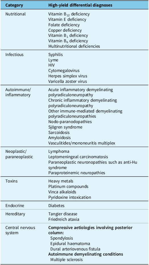
The differential diagnoses for this case are wide and summarised in Table 1. They include nutritional (either single or combined) deficiency polyneuropathies and myeloneuropathies, toxic sensory neuronopathies (i.e. B6 toxicity), infections of dorsal columns or sensory roots, paraproteinemic neuropathies, infiltrative/neoplastic processes, autoimmune/inflammatory conditions, primary immune-mediated demyelinating radiculoneuropathies and mononeuritis multiplex. Moreover, certain spinal cord-related disorders such as posterior compressive aetiologies and central demyelinating conditions are still considered, although are less likely in the absence of bowel/bladder symptoms and with persistent areflexia.
Summary of differential diagnoses
The presence of cranial neuropathy narrows the differential somewhat; the list includes Lyme disease, sarcoidosis, autoimmune disorders including Sjögren syndrome and scleroderma, paraneoplastic or immune-mediated radiculoneuropathies, leptomeningeal carcinomatosis and neurolymphomatosis, new-onset diabetes with amyotrophy with concurrent cranial neuropathy and Tangier disease.Reference Russell1 Guillain–Barré syndrome is excluded given the time course. Given the unusual distribution of symptoms, combined central and peripheral demyelination also needs to be considered, such as what is described in association with antibodies against paranodal proteins.Reference Fehmi, Scherer, Willison and Rinaldi2
Case Presentation: Initial Workup (Laboratory Tests and Electrodiagnostic Studies)
The workup should evaluate for the more likely diagnoses first, including screening blood tests and electromyography (EMG), along with neuroimaging, given the facial symptoms. In this particular patient, malignancy and paraneoplastic conditions are higher in the differential given the prior history of lymphoma. Laboratory workup included nutritional labs (vitamins B6, B12 and E, methylmalonic acid, copper, zinc), serum protein electrophoresis, anti-antineutrophil cytoplasmic antibodies, anti-double-stranded DNA antibodies, an extractable nuclear antigen panel, a paraneoplastic panel (including anti-CRMP5 and anti-Hu) and ganglioside antibodies, which were all normal. The antinuclear antibody titre was elevated at 1:160, which was also positive nine months prior. Initial CBC on admission showed haemoglobin of 176 g/L (17.6 g/dL), mean corpuscular volume of 90.1 fL and WBC of 4.8 cells/L. The patient had chronic leukopoenia in the context of his CCUS as stated above. Testing for Lyme disease and syphilis was not done in this patient.
Two months into symptom onset, the patient underwent an EMG of the right upper and lower extremities that showed only minor non-specific changes (absent H-reflex and prolonged ulnar F-wave latency), with no evidence of acquired segmental demyelination or sensory or motor fibre loss.
Discussion: Updated Differential Diagnosis
The normal sensory nerve conduction responses in the setting of prominent clinical large fibresensory loss imply a pre-ganglionic lesion. The prolonged F-wave and H-reflex latencies with normal distal motor conduction responses should also prompt a differential diagnosis shift towards a root-level process.
Case Presentation: Imaging Studies
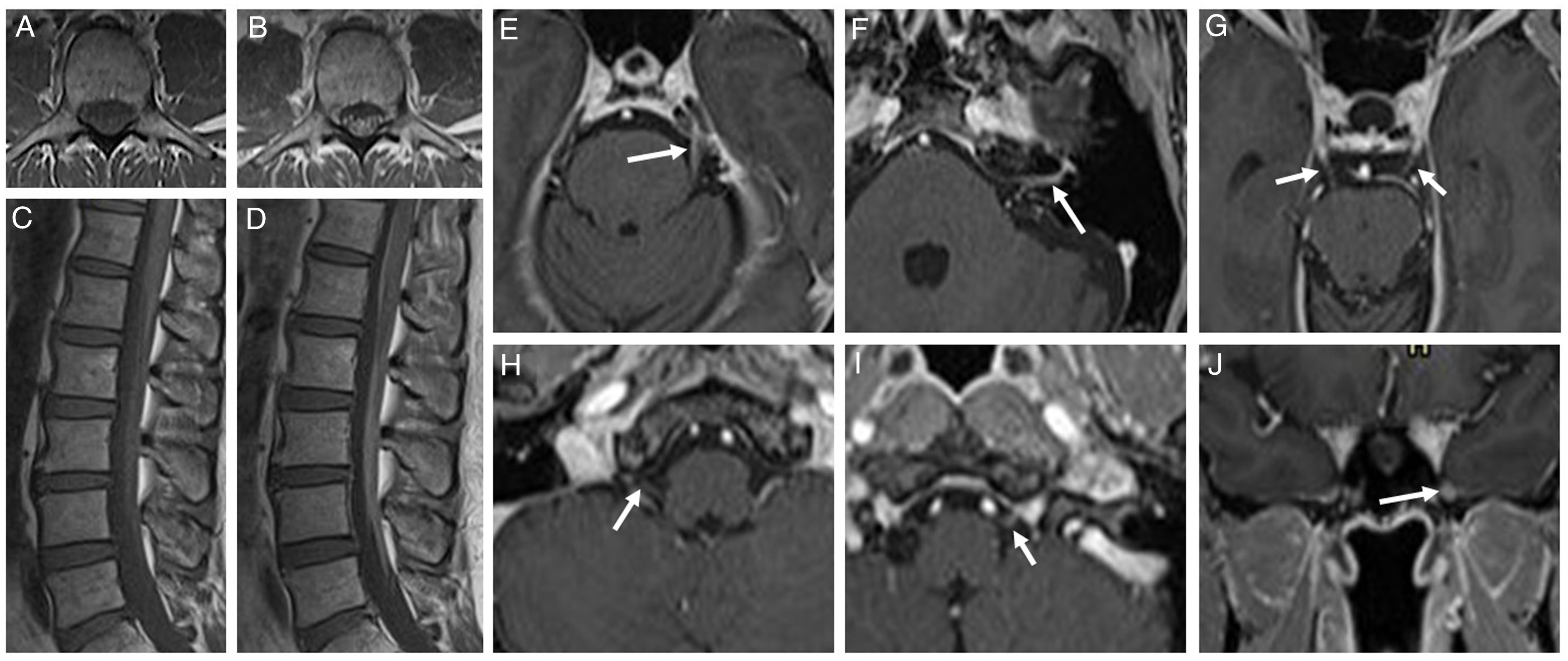
Now three months into symptom onset, an MRI of the neuroaxis was completed and revealed mild contrast enhancement along the bilateral 3rd, left 5th, left 7th/8th complex, right 10th, left 12th and left maxillary (V2) cranial nerves, smooth enhancement of the cauda equina nerve roots and leptomeningeal enhancement along the conus medullaris (Figure 1). There was no enhancement of the right 5th nerve and no examination findings indicative of the 3rd nerve deficit.
T1-weighted MRI of the thoracic and lumbar spinal cord and brain, without and with gadolinium enhancement. Axial view of cauda equina, pre-contrast (A) and post-contrast (B). Sagittal view of lumbar spine and cauda equina pre-contrast (C) and post-contrast (D). Enhancement of cranial nerves: left 5th nerve (E), left 7th and 8th nerve complex in internal acoustic canal (F), bilateral 3rd nerve (G), right 10th nerve (H), left 12th nerve (I) and left maxillary nerve (V2) (J).
Discussion: Updated Differential Diagnosis
The differential diagnoses are further narrowed down to sarcoidosis, a paraneoplastic or immune-mediated radiculoneuropathy and nodo-paranodopathies. The imaging findings warrant further workup with CSF studies.
Case Presentation: CSF Studies and Further Evaluations
Lumbar puncture was performed, with CSF studies showing one total nucleated cell, zero red blood cells, protein 2.06 mg/mL (206 mg/dL), glucose 3.72 mmol/L (67 mg/dL) and an elevated immunoglobulin index of 0.70 (normal range 0–0.61). This was consistent with cytoalbuminologic dissociation. CSF tests performed for infection (culture and stain, meningitis/encephalitis panel), inflammation (IgG index, oligoclonal bands) and malignancy (cytology, flow cytometry, antineuronal antibodies) were unremarkable.
Given his history of malignancy, cancer screening needs to be done. Therefore, CT scan of the chest, abdomen and pelvis was performed and showed no evidence of new or recurrent malignancy. Whole-body positron emission tomography (PET) scan also showed no evidence of malignancy. The lack of perihilar lymphadenopathy on CT/PET or other systemic features to suggest sarcoidosis made this an unlikely diagnosis.
EMG can be repeated to evaluate for new findings, evidence of demyelination or new motor involvement. Somatosensory evoked potential (SSEP) may show delayed or absent response in certain autoimmune polyradiculopathies. Repeat EMG of the right upper and lower extremities one month after the prior study showed similar findings with unremarkable nerve conduction studies and no evidence of sensory or motor fibre loss. Unexpectedly, the ulnar F response had normalised. He also underwent median and posterior tibial SSEPs that showed increased latency of the posterior tibial SSEP; however, the results were affected by poor signal-to-noise ratio.
Case Presentation: Diagnosis and Management
This patient was diagnosed with chronic immune sensory polyradiculopathy (CISP) and underwent treatment with intravenous immunoglobulin (IVIG) 2 g/kg over five days. Subjectively, he reported improved tingling in his face and slight improvement with gait. His neurological examination immediately before discharge showed a return of the brachioradialis (1+) and patellar reflexes (1+), but otherwise remained unchanged.
Discussion: Chronic Inflammatory Demyelinating Polyradiculoneuropathy (CIDP) and Other Sensory Polyradiculoneuropathies
CIDP comprises a group of immune-mediated radiculoneuropathies that affect peripheral roots with or without the peripheral nerves.Reference Köller, Kieseier, Jander and Hartung3 The classic form involves dorsal (sensory) and ventral (motor) nerve roots and peripheral nerves and presents with a relapsing-remitting or progressive course of sensory and motor deficits in distal and proximal extremities.Reference Köller, Kieseier, Jander and Hartung3 EMG typically shows evidence of acquired segmental demyelination in the form of conduction velocity slowing, prolonged distal motor latency, conduction block and/or temporal dispersion.Reference Köller, Kieseier, Jander and Hartung3 The sensory variant of CIDP predominantly involves dorsal roots and sensory nerve fibres and can present with sensory ataxia.
CISP, a pure sensory root polyradiculopathy, can mimic pure sensory CIDP. It tends to involve the dorsal nerve roots only and spares peripheral nerves distal to and including the dorsal root ganglion, resulting in normal sensory and motor nerve conduction studies.Reference Sinnreich, Klein, Daube, Engelstad, Spinner and Dyck4 Normal sensory conduction studies are the primary differentiation point from pure sensory CIDP. In these cases, neuroimaging is of diagnostic value and may provide evidence of contrast enhancement in the nerve roots. Elevated protein in the CSF and abnormal SSEPs are also described.Reference Sinnreich, Klein, Daube, Engelstad, Spinner and Dyck4 Regarding the presentation, patients with CISP tend to have more involvement in the lower extremities, with decreased/absent reflexes.Reference Sinnreich, Klein, Daube, Engelstad, Spinner and Dyck4 Cranial nerve involvement is rare and reported once only in CISP.Reference Trip, Saifee and Honan5 In this particular patient, the prolonged ulnar F-wave is not completely compatible with CISP because it implies motor root involvement. However, the normalisation of this F response in the second study potentially suggested technical failure during the first study. In the context of otherwise normal sensory and motor conduction responses and no evidence of acquired segmental demyelination, CISP was felt to be the most accurate diagnosis at this point.
Case Presentation: Follow-up
At a follow-up visit, four months after symptom onset (and one month after IVIG treatment), the patient reported gradual subjective improvement in his sensory symptoms. He continued to have numbness over the right side of the face and tip of the tongue but with improvement in taste. He also had minimal residual subjective numbness/tingling in the fingertips with partial improvement of the sensory deficit in the legs. However, objectively, his ambulation remained impaired and possibly deteriorated, requiring the use of a cane.
The partial response of sensory symptoms and worsening of ataxia and gait warrants reconsideration of diagnosis and management. Here, the therapeutic plan was changed to high-dose intravenous methylprednisolone (1 gram/day for 3 days), followed by IVIG (1 g/kg every three weeks) as maintenance therapy. Moreover, a nodopathy antibody panel (Mayo Clinic Laboratories, test code CIDP) was sent in consideration of other differential diagnoses discussed above.
Case Presentation: New Findings and Change of Management
On the next follow-up visit, six months after symptom onset, his nodopathy antibody panel resulted positive for contactin-1 IgG and neurofascin-155 (NF-155) IgG-4 antibodies. His examination showed new mild weakness in knee extension and foot dorsiflexion bilaterally, though several reflexes were now present. This suggested a partial but incomplete response to IVIG. His ambulation was still impaired, requiring the use of a rollator, and he reported feeling weaker and more off-balance. With the development of new clinical motor symptoms, the diagnostic impression became more uncertain, and CISMP (chronic immune sensorimotor polyradiculopathy) was considered. Moreover, the presence of paranodal protein antibodies altered his therapeutic plan, which was switched to rituximab, though this couldn’t be started immediately due to insurance delays. His weakness then progressed to involve bilateral shoulder abduction, elbow extension, wrist extensor and finger abduction. He also developed a postural tremor. Subsequently, he underwent plasma exchange due to the progression of symptoms with improvement of sensory symptoms, ataxia and gait. Immediately following the plasma exchange, he was started on rituximab 1,000 mg with further improvement of gait and motor symptoms. A second dose of rituximab was given one month later with further motor improvement at last follow-up.
Discussion: Sensorimotor Polyradiculoneuropathies
CISP-plus is an extension of CISP with similar clinical manifestations, but with additional minor involvement of peripheral sensory and motor nerve fibres on EMG.Reference Shelly, Shouman and Paul6 CISMP is another type of autoimmune polyradiculoneuropathy that involves both sensory and motor roots and presents with motor and sensory findings on clinical examination.Reference Thammongkolchai, Suhaib, Termsarasab, Li and Katirji7 In electrodiagnostic studies, patients have evidence of sensory and motor root involvement, with abnormal F-waves, H-reflexes and evidence of chronic denervation. CSF studies could show elevated protein with normal total nucleated cell count.Reference Thammongkolchai, Suhaib, Termsarasab, Li and Katirji7,Reference Khadilkar, Patel, Shah, Deshmukh, Patel and Mansukhani8 CISMP appears to have more cranial nerve involvement as described in a case series where three out of nine patients had involvement of at least one cranial nerve.Reference Thammongkolchai, Suhaib, Termsarasab, Li and Katirji7
Discussion: Nodo-paranodopathies
Nodo-paranodal neuropathies are a group of immune-mediated neuropathies caused by the presence of antibodies against nodes, paranodes and juxta-nodes. These antibodies were first found in supposed variants of CIDP or Guillain–Barré syndrome and therefore classified under that umbrella, although more recent studies suggest that these may represent a distinct peripheral neuropathy syndrome.Reference Fehmi, Scherer, Willison and Rinaldi2,Reference Khadilkar, Kamat and Patel9,Reference Fehmi, Vale, Keddie and Rinaldi10 The NF-155- and contactin-1-associated variants usually present with acute/subacute onset of distal > proximal motor weakness, ataxia and tremor.Reference Fehmi, Vale, Keddie and Rinaldi10,Reference Uncini11 Both of these paranodopathies are shown to respond poorly to IVIG. Therefore, any incomplete response to standard immune therapies in a patient thought to have CIDP or one of its variants should prompt evaluation for these antibodies.
Discussion: Treatment Options
For the treatment of CIDP variants, IVIG is commonly used as first-line therapy with plasmapheresis and high-dose intravenous steroids being used less often. Prior reports have used other immunosuppressive medications such as azathioprine, mycophenolate mofetil, cyclophosphamide, methotrexate and rituximab.Reference Sinnreich, Klein, Daube, Engelstad, Spinner and Dyck4,Reference Thammongkolchai, Suhaib, Termsarasab, Li and Katirji7,Reference Rotta, Sussman, Bradley, Ayyar, Sharma and Shebert12,Reference Menon, Katzberg and Bril13 Paranodopathy antibodies like NF-155 and contactin-1 are mainly IgG-4 subtypes. IVIG acts by suppressing the complement cascade, and because IgG-4 doesn’t significantly activate complement, this likely explains the poor response to IVIG.Reference Fehmi, Vale, Keddie and Rinaldi10 In these patients, rituximab has been more effective in decreasing antibody-producing cells.Reference Fehmi, Vale, Keddie and Rinaldi10 This patient underwent treatment with IVIG and steroids with an unfavourable response. This prompted reconsidering the initial diagnostic impression and evaluating the patient for variants associated with nodo-paranodal antibodies. The presence of NF-155 and contactin-1 antibodies resulted in a change in management.
Discussion: Clinical Reasoning
In this patient, the lack of motor symptoms and benign electrodiagnostics on initial presentation led to a diagnosis of CISP. While the patient had mild weakness in right hip flexion, this started months before and appeared to be unrelated to his acute-onset neuropathic symptoms. Of note, the prolonged F-wave response may have suggested initial motor root involvement, but without weakness, it was uncertain. However, the subsequent development of clear motor symptoms changed the diagnostic impression. Initially, CISMP was considered as the possible diagnosis (as compared to CISP + given the extent of motor symptoms), but the lack of new electrodiagnostic data after the development of significant weakness makes this uncertain.
Later, the patient was found to have paranodal antibodies, suggesting a diagnosis of paranodopathy. Here, the presentation does not fully match previously reported clinical phenotypes reported in paranodopathies. Most importantly, he presented with a sensory-dominant form of disease with progression to a sensorimotor clinical picture. Of note, a CISP-like picture is previously reported in paranodopathies with contactin-1 antibodies;Reference Shelly, Klein and Dyck14 however, the presence of both NF-155 and contactin-1 antibodies along with the phenotype shift is unusual. Ultimately, a diagnosis of paranodopathy is felt to be most appropriate as he is following a typical treatment course, being somewhat refractory to IVIG and steroids, with a more robust response to rituximab.
In conclusion, the differential diagnoses for sensorimotor polyradiculoneuropathies are broad. A thorough history and neurological examination can narrow this down, but early antibody testing and electrodiagnostic data in particular may help facilitate early treatment success and avoid CIDP misdiagnoses. It is imperative to re-evaluate and reassess the initial diagnosis if there is a suboptimal response to treatment or a change in clinical phenotype.
Author contributions
SB: Data acquisition, drafting the manuscript; HM: data acquisition, drafting the manuscript; VD: data acquisition, drafting the manuscript; SM: conception, data acquisition, revising the manuscript; NT: conception, revising the manuscript; DO: conception, revising the manuscript; AS: conception, revising the manuscript; MK: conception, revising the manuscript; RM: conception, revising the manuscript.
Funding statement
None.
Competing interests
SB, HM, VD, SM and MK: nothing to disclose; DO: research support from the National Institutes of Health, National Multiple Sclerosis Society, Patient-Centered Outcomes Research Institute, Race to Erase MS Foundation, Genentech, Genzyme, Bristol Myers Squibb and Novartis. Consulting fees from Bristol Myers Squibb, Genentech/Roche, Novartis and Pipeline Therapeutics; AS: North American Neuroendocrine Tumor Society (NANETS) Clinical Investigator Scholarship, The Robert A. Winn Diversity in Clinical Trials Award, honoraria for presentations at American Society of Hematology (ASH) Meeting on Hematologic Malignancies, Aplastic Anemia and MDS International Foundation Webinar, ASH Friday Satellite Symposia, travel support from Acute Leukemia Forum, ASH Meeting on Hematologic Malignancies and ASH Friday Satellite Symposia. Leadership role on Early Career Development Committee; RM: Advisory Board for AstraZeneca.


 Open access

