Introduction
Cumulus-oocyte complexes (COCs) used for the in vitro production of bovine embryos (IVP) are commonly selected based on the morphological characteristics of cumulus cells and of the oocyte itself, particularly appearance of the ooplasm evaluated by stereomicroscopy (Leibfried and First, Reference Leibfried and First1979). Conventionally, oocytes destined for IVP are recovered from follicles with a minimum diameter of 3 mm, a diameter when the oocytes reach their full size (Hyttel et al., Reference Hyttel, Viuff, Fair, Laurincik, Thomsen, Callesen, Vos, Hendriksen, Dieleman, Schellander, Besenfelder and Greve2001) up to a maximum diameter of 6 to 8 mm in an attempt to avoid selection of oocytes retrieved from dominant follicles or follicles in the early stage of atresia, although antral atresia can occur in any size of developing follicle and specially in follicles larger than 5 mm in diameter (Rodgers and Irving-Rodgers, Reference Rodgers and Irving-Rodgers2010). Despite these criteria, the selection of oocytes based on morphological quality does not permit to assess whether they have already acquired full competence to resume and complete meiosis, as well as the competence for embryonic development after fertilization (Rizos et al., Reference Rizos, Ward, Duffy, Boland and Lonergan2002). As a consequence, many incompetent oocytes are selected for use in IVP programs, which results in lower rates of embryo production (30% to 45% of structures that reach the blastocyst stage) when compared to in vivo observations (Rizos et al., Reference Rizos, Ward, Duffy, Boland and Lonergan2002).
Previous studies have demonstrated the existence of a relationship between oocyte competence and follicular diameter (De Bem et al., Reference De Bem, Adona, Bressan, Mesquita, Chiaratti, Meirelles and Leal2014; Labrecque et al., Reference Labrecque, Fournier and Sirard2016). Although it is well established that oocytes recovered from follicles with a diameter greater than 3 mm have satisfactory developmental competence (Blondin and Sirard, Reference Blondin and Sirard1995), subsequent studies have shown linear progression of the competence of oocytes derived from follicles measuring 3 to 10 mm (Labrecque et al., Reference Labrecque, Fournier and Sirard2016; Blondin et al., Reference Blondin, Vigneault, Nivet and Sirard2012). However, the vast majority of studies describing oogenesis and folliculogenesis in the cow were conducted on the European continent or in North America, where most animals belong to the taurine group (Hyttel et al., Reference Hyttel, Viuff, Fair, Laurincik, Thomsen, Callesen, Vos, Hendriksen, Dieleman, Schellander, Besenfelder and Greve2001; Lodde et al., Reference Lodde, Modina, Galbusera, Franciosi and Luciano2007; Lodde et al., Reference Lodde, Modina, Maddox-Hyttel, Franciosi, Lauria and Luciano2008; Luciano et al., Reference Luciano, Franciosei and Modina2011; Lodde et al., Reference Lodde, Franciosi, Tessaro, Modina and Luciano2013). In contrast, few studies have investigated whether differences in metabolic and nutritional status between the Bos taurus and Bos indicus (zebu) genetic groups, which have a direct impact on ovarian physiology (Sartori et al., Reference Sartori, Gimenes, Monteiro, Melo, Baruselli and Bastos2016), may affect oocyte capacities. For example, a larger diameter of the ovulatory follicle and lower peripheral concentrations of progesterone are found in Bos taurus females, while Bos indicus females have higher peripheral concentrations of insulin-like growth factor 1, a higher antral follicle count, a greater recovery rate of oocytes by ultrasound-guided follicular aspiration, better oocyte viability, and higher rates of in vitro blastocyst production (reviewed by Sartori et al., Reference Sartori, Gimenes, Monteiro, Melo, Baruselli and Bastos2016). Considering these differences in endocrine and ovarian physiology between the two genetic groups, the relationship between follicle size and oocyte competence needs to be further investigated in zebu cattle.
Chromatin remodeling during oogenesis is an important parameter for assessing differentiation and acquisition of oocyte competence (Luciano et al., Reference Luciano, Franciosei and Modina2011; Lodde et al., Reference Lodde, Franciosi, Tessaro, Modina and Luciano2013). Studies using oocytes from the ovaries of animals slaughtered on the European continent (Lodde et al., Reference Lodde, Modina, Galbusera, Franciosi and Luciano2007; Luciano et al., Reference Luciano, Franciosei and Modina2011) have shown that oocytes in germinal vesicle (GV) recovered from early and mid-stage antral follicles exhibit different patterns of chromatin configuration, in which condensation progressively increases from GV0 (diffuse filamentous chromatin and full transcriptional activity) to GV3 (fully condensed chromatin and transcriptional inactivity). Most oocytes retrieved from early antral follicles (< 2 mm) are still growing, are in the GV0 phase, and are meiotically incompetent, i.e., their capacity to resume and complete meiosis is limited. On the other hand, most of the fully grown oocytes recovered from antral follicles measuring 2–6 mm in diameter are distributed across the GV1, GV2 and GV3 phases and are meiotically competent (Pytel et al., Reference Pytel, Zyzynska-Galenska, Gajewski and Papis2024; Lodde et al., Reference Lodde, Modina, Galbusera, Franciosi and Luciano2007). High embryonic developmental competence is acquired in the GV1 to GV2 transition, before the decrease in FSH levels and follicular dominance, whereas the GV2 to GV3 transition usually occurs following the peak and decline in FSH levels within a follicular wave and the onset of follicular atresia. Thus, although all oocyte chromatin configurations can be found at all follicular diameters, it can be assumed that most oocytes included in growing follicles are at GV1 stage and have low developmental competence; most oocytes included in follicles with low FSH are supposedly at GV2 stage (high developmental competence); and most oocytes included in atretic follicles are supposedly at GV3 stage and have intermediate developmental competence (Luciano and Sirard, Reference Luciano and Sirard2018).
From the above, it is clear that follicle size is not a reliable criterion to determine the selection of a homogeneous population of oocytes regarding the chromatin configuration. However, considering that the progressive compaction of chromatin is related to the acquisition of meiotic and developmental competence during development of the oocyte present in the antral follicle, as well as the existence of particularities in ovarian physiology of female Bos indicus, the present study was designed with the objective of evaluating the relationship between chromatin compaction and acquisition of competence by oocytes derived from antral follicles with different diameters on random days of estrous in naturally cycling Nellore females (a tropical Bos indicus breed). More specifically, we assessed the following parameters: (1) the relationship between follicle size and oocyte diameter, morphological quality, integrity of transzonal projections and chromatin compaction; (2) the relationship between follicle size and acquisition of oocyte meiotic competency, assessed by its ability to resume and complete the meiotic maturation; and (3) the relationship between follicle size and acquisition of oocyte developmental competency, assessed by its ability to reach the blastocyst stage after in vitro fertilization.
Material and methods
Experimental design
The antral follicles present in ovaries removed from the carcasses of Nellore cows (Bos indicus) slaughtered in slaughterhouses were dissected and classified according to their diameter in early antral (diameter of 0.5 to 2 mm), mid antral (diameter > 2 to 6 mm), and late antral (diameter > 6 mm; the largest follicles could reach 15 mm, but most of them were between 7 and 12 mm). Then, the oocytes were matured in vitro to assess the meiotic competence. Immature and in vitro matured oocytes were evaluated for diameter, chromatin compaction and transzonal projections (TZPs) integrity. The matured oocytes were then fertilized to assess the developmental competence.
Follicle dissection and selection of oocytes
In the present study, the oocyte donor females were not subjected to a prior protocol for follicular synchronization, since it is rarely performed in the routine for ovum pick-up/IVP for use in commercial assisted reproduction in production animals, and is also not used in the routine of scientific experiments. Therefore, our objective was to evaluate the competencies of oocytes obtained on random days of the estrous cycle in a way that is more compatible with practical reality. Only ovaries from non-pregnant females were included in the study. The ovaries were removed from the carcasses of slaughtered cows and transported to the laboratory in thermoses with saline solution at a temperature of 30 to 35oC. Follicles containing evident blood vessels and with a translucent and pearly appearance were dissected free of ovarian stromal tissues with tweezers, scissors, and a scalpel blade. The follicles were kept in Petri dishes (Corning Inc., Acton, MA, USA) containing saline solution heated to 36oC throughout the dissection procedure. After dissection, the diameter of the follicles was measured with a cutimeter for size classification and the follicles were divided into the following experimental groups: 1) early antral (diameter of 0.5 to 2 mm); 2) mid antral (diameter > 2 to 6 mm), and 3) late antral (diameter > 6 mm). The follicles of each category were carefully and individually ruptured under a stereomicroscope with the aid of two tweezers that were pulled in opposite directions. The content of the follicle was released into a Petri dish containing Dulbecco’s phosphate-buffered saline (PBS) supplemented with 1% polyvinyl alcohol (Sigma-Aldrich, St. Louis, MO, USA) and 75 μg/mL penicillin/streptomycin (Sigma) and then examined for the selection of COCs.
In all experimental groups, only grade 1 oocytes were selected, i.e., oocytes surrounded by at least four layers of compact cumulus cells and with cytoplasm containing homogeneous granulation (Aguila et al., Reference Aguila, Treulen, Therrien, Felmer, Valdivia and Smith2020).
In vitro maturation of oocytes
The selected COCs were cultured in 100-μL microdroplets of maturation medium [TCM199 (Gibco BRL, Grand Island, NY, USA) supplemented with 0.2 mM pyruvate (Sigma), 25 mM sodium bicarbonate (Sigma), 75 μg/mL amikacin (Sigma), 0.5 μg/mL follicle-stimulating hormone (Folltropin-V; Bioniche Animal Health, Ontario, Canada), 100 IU/mL human chorionic gonadotropin (Vetecor; Hertape Calier, Juatuba, MG, Brazil), and 10% fetal calf serum (Gibco)], covered with mineral oil (Sigma), in an atmosphere of 5% CO2 in air at 38.5oC and maximum humidity for 22 hours.
Measurement of oocyte diameter
Immature oocytes (immediately after removal of the follicular environment – 0 hours) and oocytes matured in vitro for 22 hours were transferred to 1.5-mL microtubes (Corning) containing 30 μL of 0.2% hyaluronidase solution (Sigma) in PBS supplemented with 1% polyvinylpyrrolidone (PBS-PVP) and denuded by vortex mixing for approximately 2 minutes. Next, the oocytes were washed twice in PBS-PVP, added to a drop of 60 μL PBS-PVP in a Petri dish, and examined under a light microscope (IX51, Olympus, Tokyo, Japan). Images of each oocyte were obtained for the measurement of diameter (excluding the zona pellucida) using the ImageJ software (1.49v).
Evaluation of nuclear maturation and transzonal projections evaluation
Immature and mature oocytes were denuded as described above. Next, the oocytes were fixed in 4% paraformaldehyde (Sigma) for 10 minutes at room temperature, followed by permeabilization with Triton X-100 (0.5%, v/v; Sigma) in 0.1% sodium citrate solution (Sigma) for 5 minutes at room temperature. The oocytes were then incubated in Hoechst 33342 (1 µg/mL; Sigma) and Alexa Fluor 488 phalloidin conjugated to FITC (1 μg/mL; Molecular Probes, Invitrogen, Eugene, OR, USA) in PBS-PVP for 15 minutes in the dark at room temperature. After being washed twice in PBS-PVP, the oocytes were placed between a slide and coverslip and evaluated immediately under an inverted epifluorescence microscope (IX51, Olympus, Tokyo, Japan) for the determination of chromatin configuration (excitation at 404 nm and emission at 526 nm) and TZPs in the zona pellucida region (excitation at 495 nm and emission at 517 nm). Immature oocytes (GV oocytes) were classified according to chromatin condensation (GV0 to GV3), as described by Lodde et al. (Reference Lodde, Modina, Galbusera, Franciosi and Luciano2007). Mature oocytes were classified according to meiosis stage as follows: GV (immature), germinal vesicle breakdown (GVBD), metaphase I (MI), and metaphase II (MII – complete maturation). Regarding the evaluation of TZPs, oocytes were analyzed as proposed by Albarracín et al. (Reference Albarracín, Morató, Izquierdo and Mogas2005) and those presenting a ring-shaped stained layer of actin immediately beneath the plasma membrane were classified as having intact TZPs. Those showing interrupted staining of the actin ring, diffuse or irregular distribution of actin filaments, or absence of actin ring or filaments were classified as having retraction of the TZPs.
In vitro embryo production
At the end of in vitro maturation, part of the oocytes were transferred to 100-μL microdroplets of fertilization medium [Tyrode’s albumin lactate pyruvate medium containing 0.2 mM sodium pyruvate, 6 mg/mL fatty acid-free bovine serum albumin (BSA), 25 mM sodium bicarbonate, 13 mM sodium lactate, 50 µg/mL amikacin sulfate, 20 µM penicillamine, 10 µM hypotaurine, 2 µM epinephrine, and 10 µg/mL heparin (all from Sigma)]. Semen from a single Nellore bull (Bos indicus) from a single batch was used. The straws were thawed in a water bath at 36oC for 40 seconds and their content was centrifuged over a discontinuous Percoll density gradient (45 and 90%; GE Healthcare, Uppsala, Sweden) for 7 minutes at 2,750 g. The retrieved sperm pellet was evaluated and sperm concentration was adjusted to 25 × 106 spermatozoa/mL of fertilization medium. Approximately 1 × 10⁵ spermatozoa were added to each fertilization drop. After fertilization (18 hours in 5% CO2 in air at 38.5oC), presumptive zygotes were washed, partially denuded by vortex mixing, and transferred to 100-µL microdroplets of modified synthetic oviduct fluid supplemented with 0.2 mM L-glutamine (Sigma), 0.34 mM sodium citrate (Sigma), 2.8 mM myo-inositol (Sigma), 2% BME essential amino acids (Sigma), 1% MEM nonessential amino acids (Sigma), 0.5% BSA, and 2.5% fetal calf serum. The embryos were cultured for 7 days in an incubator in an atmosphere of 5% CO2 in air at 38.5oC. The cleavage rate and embryo development were evaluated 72 hours (day 3) and 168 hours (day 7) post-insemination, respectively.
Statistical analysis
Each step of the experiment was repeated at least three times for each proposed evaluation in independent replicates. Within each replicate, a culture microdroplet containing 25 oocytes was considered the experimental unit of each group. Data on nuclear maturation rate, TZPs integrity rate, and the rates of formation of blastocysts were arcsines transformed and subjected to analysis of variance, and the means were compared using Tukey’s post hoc test. All analyses were performed using JMP software (SAS Institute, Cary, NC, United States). The level of statistical significance was set at P < 0.05.
Results
Recovery rates and morphological quality of oocytes derived from follicles of different sizes in Nellore cattle
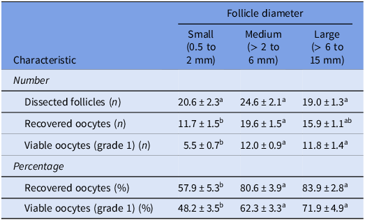
The present study evaluated oocytes recovered from ovarian follicles classified as early antral (diameter of 0.5 to 2 mm), mid antral (> 2 to 6 mm), and late antral (> 6 mm), which were obtained from Nellore females on a random day of the estrous cycle. The mean follicular diameter was 1.37 ± 0.02 mm for early antral dissected follicles (n = 365), 3.51 ± 0.04 mm for mid antral follicles (n = 303), and 9.77 ± 0.14 mm for late antral follicles (n = 293) (data not shown). The mean number of recovered oocytes in each replicate of the experiment (n = 10 replicates) was 11.7 ± 1.5 (early antral follicles), 19.6 ± 1.5 (mid antral follicles), and 15.9 ± 1.1 (late antral follicles); the mean number was lower for early antral follicles compared to mid antral follicles (P < 0.05) and both did not differ from late antral follicles. The oocyte recovery rate was lower (P < 0.05) for early antral follicles (57.9 ± 5.3%) compared to mid- (80.6 ± 3.9%) and late antral follicles (83.9 ± 2.8%). Regarding the morphological quality of recovered oocytes, the viability rate (grade 1) was lower (P < 0.05) when oocytes were recovered from early antral follicles (48.2 ± 3.5%) compared to those retrieved from mid- (62.3 ± 3.3%) or late antral follicles (71.9 ± 4.9%). These results are summarized in Table 1.
Effect of follicle diameter on oocyte characteristics in Nellore

Data are presented as mean ± standard error of the mean (SEM) of 10 replicates. Only grade 1 oocytes (Aguila et al., Reference Aguila, Treulen, Therrien, Felmer, Valdivia and Smith2020) were selected for the experiments. abDifferent letters in the same row indicate significant differences (P < 0.05).
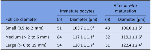
The diameter of immature oocytes was measured immediately after removal from the follicular environment and selection (Table 2). Oocytes recovered from early antral follicles had a smaller (P < 0.05) diameter (103.7 ± 1.9 µm) than those retrieved from mid- (117.1 ± 1.1 µm) and late antral follicles (120.1 ± 1.7 µm). With respect to mature oocytes, a larger diameter (P < 0.05) was observed for oocytes recovered from mid- (119.1 ± 1.6 µm) and late antral follicles (122.4 ± 2.4 µm) compared to the diameter of oocytes recovered from early antral follicles (106.0 ± 1.5 µm).
Diameter of oocytes recovered from follicles of different sizes on a random day of the estrous cycle of Nellore females

Data are presented as mean ± standard error of the mean (SEM) of 4 replicates. abMean values in the same column followed by different letters indicate significant differences (P < 0.05).
Meiotic competence and transzonal projections evaluation in oocytes derived from follicles of different sizes in Nellore cattle
Immature oocytes recovered from follicles with different diameters on a random day of the estrous cycle of Nellore females were classified according to the stage of chromatin compaction (Figure 1). A larger proportion (P < 0.05) of oocytes at the GV0 stage was found among oocytes recovered from early antral follicles (82.0 ± 4.2%) compared to mid- (9.9 ± 3.4%) and late antral (12.8 ± 3.4%). On the other hand, no difference between groups was observed for the percentage of oocytes at GV1 (15.1 ± 11.0% to 30.6 ± 8.9%), GV2 (2.9 ± 13.9% to 56.9 ± 11.4%), or GV3 (0 to 9.1 ± 6.2%).
Chromatin configuration from GV0 to GV3 [according to Lodde et al. (Reference Lodde, Modina, Galbusera, Franciosi and Luciano2007)] of immature oocytes recovered from follicles of different sizes on a random day of the estrous cycle of Nellore females. (A) The graph shows the percentage (mean ± standard error of the mean of 5 independent replicates) of distribution patterns of germinal vesicle (GV) status in the following experimental groups: Early antral = oocytes (n = 42) recovered from dissected follicles measuring 0.5 to 2 mm in diameter; Mid antral = oocytes (n = 76) recovered from dissected follicles measuring > 2 to 6 mm in diameter; Late antral = oocytes (n = 45) recovered from dissected follicles measuring > 6 mm in diameter. abDifferent letters within each pattern of GV status indicate significant differences (P < 0.05). (B) Representative images of GV stages of oocytes stained with Hoechst 33342 for the identification of nuclei (blue). Magnification: 20×.

Chromatin configuration was again evaluated after in vitro maturation to determine the acquisition of meiotic competence (Figure 2). Markedly higher (P < 0.05) percentages of GV (78.7 ± 14.1%) were observed among oocytes recovered from early antral follicles compared to those derived from mid- (5.5 ± 5.5%) and late antral (2.2 ± 2.2%) follicles. There were no differences between groups in the percentages of GVBD (0% to 11.3 ± 6.6%) or MI oocytes (10.0 ± 7.7% to 30.5 ± 2.7%). On the other hand, the percentage of oocytes that completed meiotic maturation (reached the MII stage) was higher (P < 0.05) when they were recovered from late antral follicles (84.1 ± 3.6%) compared to mid antral follicles (59.7 ± 5.0%). None of the oocytes derived from early antral follicles reached the MII stage (0%).
Meiosis progression after in vitro maturation of oocytes recovered from follicles of different sizes on a random day of the estrous cycle of Nellore females. (A) The graph shows the percentage (mean ± standard error of the mean of 4 independent replicates) of chromatin configuration in the following experimental groups: Early antral = oocytes (n = 38) recovered from dissected follicles measuring 0.5 to 2 mm in diameter; Mid antral = oocytes (n = 47) recovered from dissected follicles measuring > 2 to 6 mm in diameter; Late antral = oocytes (n = 51) recovered from dissected follicles measuring > 6 mm in diameter. Abbreviations: GV = germinal vesicle; GVBD = GV breakdown; MI = metaphase I; MII = metaphase II (complete maturation). abDifferent letters within each pattern of chromatin configuration status indicate significant differences (P < 0.05). (B) Representative images of the meiosis status of oocytes stained with Hoechst 33342 for the identification of nuclei (blue). Magnification: 20×.

Actin microfilaments were evaluated in oocytes to classify the integrity of TZPs (Figure 3). A higher percentage (P < 0.05) of intact TZPs was observed in immature oocytes recovered from early antral follicles (74.1 ± 5.4%) compared to those recovered from mid- (30.7 ± 6.9%) or late antral follicles (42.0 ± 6.0%). At the end of maturation, there was no difference between groups in the percentage of oocytes that showed TZP retraction (45.9 ± 10.7% to 65.1 ± 8.7%).
Transzonal projections (TZP) evaluation in oocytes recovered from follicles of different sizes on a random day of the estrous cycle of Nellore females. The graph shows the percentage (mean ± standard error of the mean of 5 independent replicates) of actin microfilaments configuration in the following experimental groups: Early antral = oocytes (n = 82) recovered from dissected follicles measuring 0.5 to 2 mm in diameter; Mid antral = oocytes (n = 83) recovered from dissected follicles measuring > 2 to 6 mm in diameter; Late antral = oocytes (n = 81) recovered from dissected follicles measuring > 6 mm in diameter. abDifferent letters within each pattern of TZP configuration indicate significant differences (P < 0.05).

Developmental competence of oocytes derived from follicles of different sizes in Nellore cattle
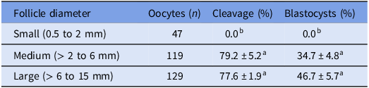
Oocytes retrieved from follicles with different diameters were fertilized at the end of in vitro maturation to assess their competence for embryonic development (Table 3). No cleavage was observed, and consequently no embryos were produced after fertilization, for oocytes recovered from early antral follicles. No difference in cleavage rates was observed for oocytes recovered from mid- (79.2 ± 5.2%) and late antral follicles (77.6 ± 1.9%). Likewise, the rate of embryonic development to the blastocyst stage was similar for oocytes derived from mid- (34.7 ± 4.8%) and late antral follicles (46.7 ± 5.7%).
Cleavage rate and embryonic development to the blastocyst stage after fertilization of oocytes recovered from follicles of different sizes on a random day of the estrous cycle of Nellore females

Data are presented as mean ± standard error of the mean (SEM) of 7 replicates. abMean values in the same column followed by different letters indicate significant differences (P < 0.05).
Discussion
Oocytes are transcriptionally active until the antral follicle reaches 2–3 mm in diameter, with a corresponding size of the oocyte of approximately 110 µm (reviewed by Fair, Reference Fair2003). From this point onwards, the follicle continues to grow while oocytes reach their final size of approximately 120 µm in diameter, acquire meiotic competence, and nuclear transcription becomes almost completely inactive (Hyttel et al., Reference Hyttel, Viuff, Fair, Laurincik, Thomsen, Callesen, Vos, Hendriksen, Dieleman, Schellander, Besenfelder and Greve2001; Lodde et al., Reference Lodde, Modina, Maddox-Hyttel, Franciosi, Lauria and Luciano2008; studies carried out in taurine cattle). Previous studies (Gimenes et al., Reference Gimenes, Sá Filho, Carvalho, Torres-Júnior, Souza, Madureira, Trinca, Sartorelli, Barros, Carvalho, Mapletoft and Baruselli2008) have shown that follicular diameter seemed to be smaller in Nellore than in Holstein cattle (Bos taurus). Despite this, our results demonstrated that immature oocytes derived from follicles smaller than 2 mm were not yet fully grown (< 110 µm), presented a low morphological viability rate (grade 1) and were predominantly at GV0 stage, agreeing with previous studies in taurine cattle that demonstrated that approximately 90% of oocytes recovered from early antral follicles smaller than 2 mm in diameter have the chromatin configuration at GV0 stage (Luciano and Sirard, Reference Luciano and Sirard2018). This finding explains why these oocytes exhibited a lower capacity to resume and complete meiotic maturation when compared to oocytes derived from follicles measuring >2–6 up to 15 mm in diameter and were also incompetent for embryonic development as they were unable to cleave after in vitro fertilization and develop to the blastocyst stage. Although we did not evaluate the transcriptional profile of oocytes and their respective cumulus cells in the present study, we can infer that the lack of competence acquisition by these oocytes may also be due to the fact that molecular maturation (biosynthesis and accumulation of key molecules and transcripts) was incomplete, as previous studies identified the mRNA expression profile as a determining factor affecting the development of oocytes derived from small follicles (Luciano and Sirard, Reference Luciano and Sirard2018; Labrecque et al., Reference Labrecque, Fournier and Sirard2016; Blondin et al., Reference Blondin, Vigneault, Nivet and Sirard2012; Racedo et al., Reference Racedo, Wrenzycki, Herrmann, Salamone and Niemann2008). Thus, since a greater proportion of oocytes derived from early antral follicles in the present study exhibited nuclear configuration in GV0 (transcriptionally active) and that such oocytes also exhibited a greater proportion of intact TZPs compared to oocytes derived from mid- and late antral follicles, it can be suggested that they are still accumulating the transcripts necessary for the synthesis of proteins that will enable the later stages of oocyte development until the maternal-to-zygotic transition (Labrecque et al., Reference Labrecque, Fournier and Sirard2016; Blondin et al., Reference Blondin, Vigneault, Nivet and Sirard2012; Racedo et al., Reference Racedo, Wrenzycki, Herrmann, Salamone and Niemann2008; Lequarre et al., Reference Lequarre, Vigneron, Ribaucour, Holm, Donnay, Tran-Dalbies, Callesen and Mermillod2005). Taken together, these data suggest that, in line with the literature for taurine cattle (Hyttel et al., Reference Hyttel, Viuff, Fair, Laurincik, Thomsen, Callesen, Vos, Hendriksen, Dieleman, Schellander, Besenfelder and Greve2001; Fair, Reference Fair2003), zebu oocytes smaller than 110 µm recovered from follicles that measure less than 2 mm in diameter are meiotically and developmentally incompetent. These oocytes exhibit inadequate rates of nuclear and, supposedly, molecular maturation after in vitro culture, a fact that makes them unsuitable for immediate use in IVP protocols.
The results of the present study also showed that GV oocytes recovered from mid (> 2 to 6 mm) and late antral follicles (> 6 mm) had a higher rate of morphological viability when compared to those derived from follicles < 2 mm in diameter. These oocytes had already reached full size and exhibited different degrees of chromatin compaction. Previous studies in taurine cattle have shown a balanced distribution in the chromatin configuration (GV1, GV2 and GV3 phases) of oocytes recovered from medium antral follicles (2–8 mm) (Lodde et al., Reference Lodde, Modina, Galbusera, Franciosi and Luciano2007; Dieci et al., Reference Dieci, Lodde, Labreque, Dufort, Tessaro, Sirard and Luciano2016; Luciano and Sirard, Reference Luciano and Sirard2018). In contrast, we found that most oocytes were at GV2 phase and only a few at GV3 phase, regardless of whether they were recovered from mid- or late antral follicles. Considering that GV2 and even GV3 oocytes without signs of atresia represent the population of oocytes that have acquired greater embryonic developmental potential (Dieci et al., Reference Dieci, Lodde, Labreque, Dufort, Tessaro, Sirard and Luciano2016; Luciano and Sirard, Reference Luciano and Sirard2018), our finding supports previous reports that have shown that Bos indicus oocytes are of better quality and may explain the improved performance in IVP programs when compared to those of Bos taurus (Sartori et al., Reference Sartori, Gimenes, Monteiro, Melo, Baruselli and Bastos2016).
The fully grown oocytes retrieved from follicles >2 mm in the present study were able to resume meiosis, corroborating reports in the literature on taurine cattle that demonstrated that fully grown oocytes with GV1 to GV3 chromatin configuration are meiotically competent (Hyttel et al., Reference Hyttel, Viuff, Fair, Laurincik, Thomsen, Callesen, Vos, Hendriksen, Dieleman, Schellander, Besenfelder and Greve2001; Lodde et al., Reference Lodde, Modina, Galbusera, Franciosi and Luciano2007; Lodde et al., Reference Lodde, Modina, Maddox-Hyttel, Franciosi, Lauria and Luciano2008; Luciano et al., Reference Luciano, Franciosei and Modina2011; Lodde et al., Reference Lodde, Franciosi, Tessaro, Modina and Luciano2013; Luciano and Sirard, Reference Luciano and Sirard2018). However, we observed that those oocytes derived from late antral follicles (> 6 mm) had a greater ability to reach metaphase II at the end of in vitro culture when compared to oocytes retrieved from mid antral follicles (> 2 to 6 mm), suggesting that the acquisition of competence to complete meiotic maturation is influenced by follicle size in Nellore. According to the literature, follicular deviation occurs at a diameter of 5.4 to 6.2 mm in Bos indicus cows versus 8.3 to 9.8 mm in Holstein cattle (Sartori et al., Reference Sartori, Gimenes, Monteiro, Melo, Baruselli and Bastos2016; Gimenes et al., Reference Gimenes, Sá Filho, Carvalho, Torres-Júnior, Souza, Madureira, Trinca, Sartorelli, Barros, Carvalho, Mapletoft and Baruselli2008). Thus, the finding that oocytes derived from late antral follicles (> 6 mm) presented greater meiotic competence in Nellore seems paradoxical, since follicular deviation is usually characterized by the fall in FSH levels and onset of atresia (Beg and Ginther, Reference Beg and Ginther2006; Luciano and Sirard, Reference Luciano and Sirard2018), which may result in a decrease in oocyte quality and viability. However, a former study in taurine cattle has shown that oocytes included in follicles in the early stages of atresia are more competent than those included in growing follicles (Blondin and Sirard, Reference Blondin and Sirard1995). In addition, it has been shown that, even in the post-mortem period (the time between slaughter and oocyte collection), oocyte competence is still modulated by cumulus cells, which release specific RNAs that are transferred to the oocyte via transzonal projections in a communication system similar to the exosome (Macaulay et al., Reference Macaulay, Gilbert, Scantland, Fournier, Ashkar, Bastien, Saadi, Gagné, Sirard, Khandjian, Richard, Hyttel and Robert2016). Based on this background, a reasonable explanation for our finding could be that most oocytes included in larger follicles have already accumulated the key molecules that characterize the acquisition of meiotic and developmental competence. Additionally, although follicular atresia was not evaluated in the present study, it can be assumed that oocytes retrieved from larger non-atretic or slightly atretic follicles of Nellore females remained for a longer period of time under the beneficial influence of follicular factors that optimize the nuclear maturation and, supposedly, also cytoplasmic and molecular maturation, enabling the acquisition of greater competences. In fact, the effect of local factors was recently reported, where the dynamics of transcripts in oocytes at three different ages post meiosis I arrest in Caenorhabditis elegans were assessed (Trimmer et al., Reference Trimmer, Zhao, Seemann, Chen, Mondal, Ben-Yakar and Arur2023). Authors found that some maternal populations of transcripts increased with oocyte age and this phenomenon was not due to de novo transcription, but as a result of transport into the oocyte from neighboring somatic cells, which were not connected to each other via gap junctions. Assuming that a similar mechanism occurs in mammalian oocytes, the acquisition of external mRNA from somatic cells may modify the acquisition of oocyte competence, and this impact may be greater the longer the period of time in which the oocyte remains included in the follicular environment.
Taken together, the results of the analysis of chromatin compaction showed that the acquisition of competence to resume and complete meiosis is highly influenced by follicle size in zebu cattle (Figure 4), with respectively low, medium, and high meiotic competence for oocytes recovered from early (< 2 mm), mid (> 2 to 6 mm), and late antral follicles (> 6 mm). The greater meiotic competence of oocytes derived from late antral follicles may explain the trend observed towards higher numerical blastocyst production when compared to those retrieved from mid antral follicles, which despite not having statistical significance, can be an interesting finding from a commercial point of view. This result may corroborate previous studies conducted on taurine cows, which demonstrated that oocytes derived from follicles > 6 mm resulted in higher blastocyst rates compared to those obtained from follicles < 4 mm (Lequarre et al, Reference Lequarre, Vigneron, Ribaucour, Holm, Donnay, Tran-Dalbies, Callesen and Mermillod2005), indicating a positive correlation between greater oocyte developmental competence and follicle size. Although oocytes included in follicles > 3 mm are transcriptionally inactive or at least present a drastic reduction in transcription capacity, they undergo a series of progressive changes in cytoplasmic, nuclear, and molecular events inside the follicle. These events culminate in a linear increase in the acquisition of competence by oocytes derived from follicles measuring 3–6 mm up to 10 mm (Labrecque et al., Reference Labrecque, Fournier and Sirard2016; Blondin et al., Reference Blondin, Vigneault, Nivet and Sirard2012) and are the result of the coordinated bidirectional communication between the oocyte and follicular cells, which provides adequate conditions for the differentiation and acquisition of oocyte capacities (Eppig et al., Reference Eppig, Wigglesworth, Pendola and Hirao1997; Simon et al., Reference Simon, Goodenough, Li and Paul1997). This bidirectional communication occurs in different ways, including the secretion of soluble molecules directly into the follicular fluid (bidirectional paracrine effect mediated by oocyte-secreted factors or factors secreted by follicular cells), the transfer of low molecular weight molecules by transzonal projections, and via extracellular vesicles that are secreted by somatic follicular cells and that can transport large molecules such as proteins, RNA, miRNA, and lipids from cumulus cells to the oocyte (Marchais et al., Reference Marchais, Gilbert, Bastien, Macaulay and Robert2022; Del Collado et al., Reference Del Collado, Andrade, Meirelles, Silveira and Perecin2018). Thus, the interaction between components of the ovarian follicular environment ensures the integrity of the processes related to the production of a competent oocyte through the coordinated communication between cells present in the follicle (Clarke, Reference Clarke2017).
Acquisition of oocyte competencies in Nellore (Bos indicus) cattle during follicular development.

In conclusion, oocytes recovered from follicles > 2 mm on random days of estrous in naturally cycling Nellore females are suitable for use in IVP, while oocytes recovered from smaller follicles are not. The morphological appearance, size and chromatin compaction configuration of immature oocytes indicated that oocytes recovered from early antral follicles (< 2 mm) are incompetent for use in IVP. Oocytes recovered from mid- (> 2 to 6 mm) or late antral follicles (diameter > 6 mm, the majority being between 7 and 12 mm) were homogeneous in terms of morphology, size and TZP integrity, in addition to exhibiting similar configurations of chromatin compaction, with most of them being at GV2 and few at GV3. The higher occurrence of these patterns of chromatin configuration may help explain the better oocyte quality and improved performance of zebu donors in IVP programs. Despite the greater meiotic competence exhibited by oocytes derived from late antral follicles, the developmental competence was similar for oocytes derived from mid- and late antral follicles, suggesting that the acquisition of developmental competence is achieved by oocytes contained in follicles with smaller diameters than those described for taurine cattle. Therefore, our data suggest that differences in ovarian physiology between bovine breeds may impact the acquisition of competences in oocytes included in antral follicles and demonstrate the need to deepen the understanding of the multiple biological processes that occur within the follicular environment to modulate the quality of an oocyte.
Acknowledgments
The authors thank Brasfrigo and JBS for donating the biological material used in the present study; Luana T. Rodrigues, Luana R. Vale, Adão A. Custodio and Alexandre J. Teixeira for technical assistance.
Author contributions
Maria Isabela Azeredo Silva: Investigation, Data curation, Writing – original draft. Beatriz Caetano da Silva Leão: Conceptualization, Investigation, Data curation, Writing – original draft. Giovana Barros Nunes: Investigation, Data curation. Cintia Rodrigues da Silva: Investigation, Data curation. Nathalia de Souza Rocha-Frigoni: Conceptualization, Investigation. Priscila Chediek Dall’Acqua: Investigation, Data curation, Writing – original draft. Gisele Zoccal Mingoti: Conceptualization, Data curation, Funding acquisition, Investigation, Project administration, Supervision, Writing – original draft, Writing – review & editing.
Funding
This research was supported by São Paulo Research Foundation – FAPESP (grant numbers #12/10083-8, #12/10084-4, #15/06733-5 and #19/11174-6) and Coordenação de Aperfeiçoamento de Pessoal de Nível Superior – CAPES (Finance Code 001). M.I.A.S. was supported by an Institutional Scholarship Program (PIBIC) from Conselho Nacional de Desenvolvimento Científico e Tecnológico – CNPq (grant number #121966/2016-0). G.Z.M. was awarded with scholarship from CNPq (grant number #307416/2015-1).
Competing interests
The authors declare that there is no conflict of interest that could be perceived as prejudicing the impartiality of the research reported.
Ethical standards
The study was approved by the Bioethical Committee of the School of Agricultural and Veterinarian Sciences, São Paulo State University (UNESP), Jaboticabal, SP, Brazil, under protocol number 008649/18. Experiments were conducted in accordance with Law 11.794 of October 8, 2008, Decree 6899 of July 15, 2009, and meeting the rules issued by the National Council for Control of Animal Experimentation (CONCEA).