Introduction
In the prenatal period, especially in the last weeks of pregnancy, the skeletal system is subject to intense changes, providing efficient functioning of the locomotor system after birth. Bone is a unique tissue, since during adult life it protects the vital organs and helps support the body. Bone as an organ also takes a part in production of blood cells. In view of rapid growth during development and in childhood, the structural and functional homeostasis of bone and its formation must be precisely regulated.Reference Stevens and Graham 1 It is known that in utero sensitivity of developing organ structures is extremely large, and even the smallest interference of pharmacological or toxic agents impairs or inhibits the normal development of the organism.Reference Śliwa and Dobrowolski 2 Glucocorticoids (GCs) are not only hormones produced naturally by the human body. Many information including elevated GCs level of mothers under stress conditions transfer the placenta and reach the fetus, changing its pattern of the development, to better adapt it for life after the birth. GCs are also therapeutic substances that are used in the treatment of many diseases. They are widely used as anti-inflammatory and immunosuppressive drugs in the regulation of stress response, inflammation and energy homeostasis.Reference Lu and Cidlowski 3 Further, GCs are also used during pregnancy and their impact on the development of fetal skeletal and possible implications for neonatal life are intensively investigated.Reference Crowther, McKinlay, Middleton and Harding 4 , Reference Śliwa, Dobrowolski and Tatara 5
Acomys cahirinus is an unusual animal model characterized by rapid development and substantial autonomy shortly after birth, which makes it an extremely valuable research object. Moreover, the offspring are furred, have open eyes, and sophisticated locomotor capabilities, and organogenesis is largely complete by the end of gestation. This animal model was chosen also because of the relatively long period of pregnancy, comparing to other small rodents, and due to the fact that spiny mouse is a precocial species characterized by rapid development of the fetus, the young are relatively mature and mobile from the moment of birth, ensuring the offspring considerable autonomy shortly after birth. This kind of fast development facilitates the analysis of bone development. Therefore, in the present study, we choose the spiny mouse as a most suitable among mice to skeletal maternal programming.
The adverse effects of glucocorticoids on the skeleton are diverse and may lead to a decrease in bone mass and many disorders of its structure. In vitro studies demonstrate that doses of GCs higher than the physiological range, observed for example during childbirth stress, inhibit the osteoblastic function and have a negative impact on bone mass.Reference Śliwa, Dobrowolski and Tatara 5 , Reference Katta, Jin and Ingham 6 GCs can injure fetal skeletal growth and development of the whole body including the intestinal tract, and is one of the causes of delayed postnatal development.Reference Śliwa and Dobrowolski 2 , Reference Śliwa, Tatara and Kowalik 7 , Reference Śliwa, Studziński and Tatara 8 Moreover, different lesions with disturbances involving both articular and growth cartilage may cause lifelong degeneration, deformity, and disability in later independent life.Reference Katta, Jin and Ingham 6 , Reference Holemans, Aertis and Van Assche 9
Dexamethasone (Dex), a most commonly used synthetic glucocorticoid, is administered to pregnant women against preterm birth risk as well as to prevent such disorders as respiratory distress syndrome. Although GCs have been used in obstetrics for over 40 years, still many issues concerning the dosage, benefits, and risks resulting from the use of GCs remain a question.Reference Roberts and Dalziel 10 , Reference Newnham and Moss 11
Considering the negative effects of glucocorticoid treatment, especially during fetal development, the aim of this study was to identify morphological changes in bone and articular and growth plate cartilages affected by the prenatal action of Dex between the 20th day of gestation until birth in Acomys cahirinus.
Materials and methods
Experimental design
The dams were randomized into control (n=12) and experimental (n=12) groups on the basis of body weight (40–50 g). Pregnant spiny mice were housed singly in separate cages under constant conditions with a 12 h light/dark cycle at 22°C and 55–60% humidity. The experimental (Dex-treated) group received dexamethasone (Dexamethasone, tablets 0.5 mg, Polfa, Pabianice, Poland) per os at a concentration resulting in intakes of 125 µg/kg per day in the diet, beginning on gestation day 20 until parturition. The tablets were crushed and incorporated within the feed pellets at an appropriate concentration, adjusted to the body weight of animals. The dose applied was selected on the basis of literature.Reference Dickinson, Walker and Wintour 12 There was no vehicle administration to the control group except mechanical manipulation of one pellet to achieve the texture similar as these mixed with dexamethasone in the Dex group. Pellets with Dex and these mechanically changed (for the control animals) had more moisture, and these were preferably consumed by animals. The remaining fraction of the feed, which was provided ad libitum was ordinary commercial feed, and so it was somewhat harder to bite. The animals had constant access to a commercial diet (LSM, Agropol S.J., Motycz, Poland) and to a fresh water. The consumption was checked twice daily (at 8 am and at 12 pm). There was no differences in food or water consumption between the groups.
The total amount of food consumed by the females was estimated before the start of the experiment and was calculated as 15 g feeding stuff per day (about three pellets) and then controlled during experiment, according to body weight gain. In the morning, the experimental group received one pellet with dexamethasone and the control females received one mechanically changed pellet but without Dex. At 12 pm, the consumption of modified pellets and the feed weight was checked in both groups.
The gestation length (39–40 days) and the number of newborn pups (two to five) did not differ between the Dex and the control group. Two newborns (one female and one male) were randomly chosen from every mother to avoid litter and adult female variability. However, in the control group there were four litters with one gender (two mothers had two and three male pups, and two had two and three female pups. Finally the number of animals, sacrificed by CO2 inhalation, was 20 in the control and 24 in the experimental group.
Tissue collection and analysis
Bone geometry
After removal of soft tissues from the femur from newborns, bone length and weight were measured. Each bone was wrapped in gauze soaked in isotonic saline and stored at 25°C for further analysis. The geometric properties of bone was estimated on the basis of horizontal and vertical diameter measurements of the mid-diaphyseal cross section of bone. The cross-section area (A), the mean relative wall thickness (MRWT), the second moment of inertia (Ix) and the cortical index (IC) were determined as described previously.Reference Śliwa, Kowalik and Tatara 13 , Reference Ferretti, Capozza and Mondelo 14
Bone histomorphometry
After removal of soft tissues the femora were subjected to histology as described previously,Reference Dobrowolski, Piersiak and Surve 15 except that 5 µm-thick sections were cut in a microtome Microm HM 360 (Microm, Walldorf, Germany) and stained. Goldner’s trichrome staining (GT) was used to assess the morphology of the growth plate cartilage and picrosirus red staining (PSR) was applied to assess the morphology of articular cartilage and to evaluate the distribution of thick and thin collagen fibers of articular cartilage. The PSR staining method and polarized light allowed us to distinguish between larger collagen fibers (orange to red color), and thinner ones, including reticular fibers, (green color). Additionally, articular cartilage proteoglycans were stained with Safranine O (SO).Reference Suvara, Layton and Bancroft 16 , Reference Rich and Whittaker 17 Microscopic (2D – two-dimensional) images of bright field were collected using a confocal microscope Axiovert 200 M (Carl Zeiss, Jena, Germany) equipped with a camera AxioCam HRc (Carl Zeiss) and a halogen lamp. Moreover, sections stained with PSR were analyzed using an Olympus BX63 automated microscope (Olympus, Tokyo, Japan) equipped with filters to provide circularly polarized illumination (the filters were aligned so that the background in the field of view was as dark as possible, that is the filters were ‘crossed’) as described earlier.Reference Rich and Whittaker 17 Images were collected with a digital color camera (UC50 Olympus, Tokyo, Japan). The analysis of the collected images was performed with the use of graphical analysis software Olympus cellSens Version 1.5 (Olympus, Tokyo, Japan). The structure of the growth plate and articular cartilage was examined by microscopic observation and the images collected were analyzed using graphical analysis software ImageJ 1.49v (National Institute of Health USA, http://rsb.info.nih.gov/ij/index.html). Measurements of growth plate width (the resting, proliferative, hypertrophy zone width) were performed as described previously.Reference Dobrowolski, Tomaszewska, Bieńko, Radzki and Pierzynowski 18 , Reference Roach, Mehta, Oreffo, Clarce and Cooper 19 Moreover, the number of chondroprogenitor cells from the generative – reserve cartilage was calculated. The quantity and chondrocyte dimensions for articular cartilage were examined as follows: the number of chondrocytes/mm2 of hyaline cartilage, chondrocyte sectional area, mean and maximum diameter, and perimeter. Two thousand cells per animal were counted. The mean intensity of SO staining (measured as a 8-bit gray-scale value, which showed an inverse correlation – the higher the result, the lower the amount of the stain absorbed) was measured for the proteoglycan content of the intercellular matrix of the cartilages examined.Reference Bobinac, Spanjol, Zoricic and Maric 20 , Reference Hagiwara, Hattori, Aoki, Ohgushi and Ito 21
Bone immunohistochemistry
Paraplast sections were deparafinized, rehydrated, and microwaved 3×5 min in 10 mM citrate buffer, pH 6.0, to retrieve antigenicity. Endogenous peroxidase activity was blocked with 3% H2O2 in methanol (1:1) and nonspecific binding was prevented using 5% bovine serum (Sigma-Aldrich, St. Louis, MO, USA). The primary antibody against glucocorticoid receptor (GR) (1:50, Santa Cruz Biotechnology, Santa Cruz, CA) was applied to the tissue sections and incubated overnight at 4°C. The antigen was visualized using biotinylated secondary antibody goat anti-rabbit IgG (1:200, 1 h at room temperature, Abcam, Cambridge, UK), an avidin-biotin-peroxidase complex (1:300, 30 min at room temperature; Strept ABC complex-HRP, Dako, Glostrup, Denmark), and 3,3’-diaminobenzidine (DAB, Sigma) as a chromogen staining substrate. After each step of the procedure, the sections were rinsed with Tris-buffered saline (TBS) pH 7.6. Next, the slides were dehydrated and mounted in DPX. The control sections were incubated in the absence of primary antibody. Such omission resulted in no disposition of the reaction product (not demonstrated). Microscopic observations and images of immunohistochemistry reaction were further analyzed. The intensity of the GR stainig was described as the intensity of immunostaining reaction (low or high).
Statistical analysis
All results are expressed as means±s.d. (standard of deviation). Differences between means were tested with the Student’s t-test. Normal distribution of data was examined using the W. Shapiro–Wilk test and equality of variance was tested by the Brown–Forsythe test. P<0.05 was considered statistically significant. When data were not normally distributed and/or there was an unequal variance of data, we used the Mann–Whitney U test. All statistical analyses were carried out by means of Statistica 12 software (www.statsoft.com, StatSaft Inc Tulusa, OK, USA).
Results
Body weight and femur geometry
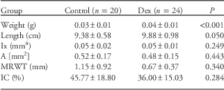
The mean body weight of the newborns from the Dex group was 4.7±0.36 g, which was significantly lower compared with the control group 5.6±0.79 g (P<0.001). Data on the femoral geometry are presented in Table 1. Treatment with Dex reduced femoral length and increased its weight. The spiny mice from the Dex-treated group had lower MRWT than the control group. Moreover, the IC in the experimental mice was also lower, compared with that in the control. However, the difference was not statistically significant.
The effect of prenatal Dexamethasone (Dex) treatment of geometry properties of femur in newborns spiny mice after birth

Ix, moment of inertia; A, cross-section area; MRWT, mean relative wall thickness; IC, cortical index.
Data are presented as mean±s.d. Control group: mice under prenatal treatment with physiological saline; Dex group: mice being under prenatal influence of Dex between the 20th day of gestation until birth.
Growth plate thickness
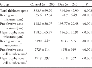
The growth plate thickness zone is presented in Table 2. Spiny mice receiving Dex had a significantly thinner femoral growth plate, compared with the control group. The total thickness was lower by 9%. Both the hypertrophy and resting zones were significantly reduced in thickness by 36 and 19%, respectively. However, Dex administration increased the proliferative zone by 23%.
The effect of prenatal dexamethasone (Dex) treatment on the growth plate

Data are presented as mean±s.d. Control group: mice under prenatal treatment with physiological saline; Dex group: mice being under prenatal influence of Dex between the 20th day of gestation until birth.
Quantitative and qualitative evaluation of growth plate and articular cartilage chondrocytes
The effect of Dex administration on the number and morphological properties of the growth plate and articular cartilage chondrocytes of the femur are presented respectively in Tables 2 and 3. Maternal Dex treatment decreased the number of cells in the articular cartilage by 27% and reduced their diameter by 5%. Moreover, the mean and max diameter of chondrocytes was significantly different between the control and the Dex-treated group. There were also significant differences in the mean intensity of chondrocyte cells of articular cartilage after Dex administration, in comparison with in the control. On the other hand, the prenatal dexamethasone treatment resulted in an almost double increase in the number of chondrocyte in all growth plate zones. The largest increase was observed in the proliferative zone by 64% in comparison to the control. After Dex treatment the number of cells in the hypertrophy and resting zone was also significantly different from the respective values in the control group.
The effect of prenatal dexamethasone (Dex) treatment on the chondrocyte number and other parameter of articular cartilage

Data are presented as mean±s.d. Control group: mice under prenatal treatment with physiological saline; Dex group: mice being under prenatal influence of Dex between the 20th day of gestation until birth.
The distribution of thick and thin collagen fibers of articular cartilage and matrix autofluorescence
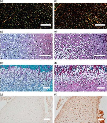
The effect of dexamethasone on the distribution of thick and thin collagen fibers of femoral articular cartilage of newborns is presented in Figs 1a and 1b. The distribution of thin and thick collagen fibers differed in both examined groups. In the control, both thin and thick fibers were almost equally distributed in all zones of articular cartilage. Dex exerted its effect mostly in the primordial articular cartilage, whereas the thin fibers were almost absent in comparison with the control group. In addition, Dex treatment significantly increased the density of thick fibers in the primordial epiphyseal and articular cartilage, unlike in the control group, where thin collagen dominated.
The effect of prenatal administration of dexamethasone on the femoral cartilage structure in newborn mice. (a) The primordial articular cartilage of the control group. (b) The primordial articular cartilage of the experimental group. Different color indicate differences between the distribution of thick (orange to red color) and thin (green color) collagen fibers of articular cartilage. (c–f) The effect of prenatal administration of dexamethasone on the femoral cartilage structure in newborn mice. (c) The primordial articular cartilage of the control group. (d) The primordial articular cartilage of the experimental group. (e) The primordial epiphyseal cartilage of the control group. (f) The primordial epiphyseal cartilage of the experimental group. Differences observed in the distribution and staining intensity of the proteoglycans in the femoral epiphyseal and articular cartilage (darker SO staining means more proteoglycans). (g and h) The effect of prenatal administration of dexamethasone on the femoral cartilage structure in newborn mice. (g) The primordial articular cartilage of the control group. (h) The primordial articular cartilage of the experimental group. The detection of the GR was showed as high or low intensity of immunostaining reaction in experimental and control group, respectively.

The content and distribution of proteoglycans in growth plate and articular cartilage
The distribution and staining intensity of proteoglycans in the femoral growth plate and articular cartilage is shown in Figs 1c–1f. Compared with the control group, the spiny mice from the experimental group had a significantly higher amount of proteoglycans (darker SO staining means more proteoglycans) in the growth plate. The greatest amounts of proteoglycans were present in the resting and hypertrophy zone. In addition, microscopic observations of articular cartilage showed a uniform distribution of proteoglycans in both the primordial epiphyseal and articular cartilage in the spiny mice after Dex administration.
Glucocorticoid receptor staining intensity in growth plate and articular cartilage
The staining of the GR in the growth plate and articular cartilage are presented in Figs 1g and 1h. Compared with the control group, prenatal Dex treatment induced a darker staining of GR in the femoral growth plate and articular cartilage than in control group.
Discussion
Directly before and after birth, adrenal hormones play a crucial role in the adaptation to a new environment in newborns. Synthetic GCs, for example dexamethasone, administered to pregnant women, or increased endogenous GCs induced by stressors, cause a number of changes different from normal physiological processes. When the GCs concentration is increased constantly, detrimental effects can be observed in the offspring.Reference Śliwa, Tatara, Nowakowski, Pierzynowski and Studziński 22 The results obtained showed that newborn spiny mice that were under the influence of prenatal dexamethasone administration were characterized by significantly lower body weight (a 17% decrease). The negative impact of glucocorticoids on body weight could be explained be the fact that DEX caused proteolysis in muscle, reduction in bone mineral mass, and increased metabolic catabolism which led to reduced growth.Reference Śliwa, Tatara, Nowakowski, Pierzynowski and Studziński 22 – Reference Tomaszewska, Dobrowolski and Puzio 24 The exposure to the excess of GCs during prenatal life leads additionally to intrauterine growth retardation (IUGR) related with low birth weight and lowered size of brain of newborns. IUGR is a common problem in human and animals and increases the risk of mortality of newborns during perinatal period.Reference Śliwa and Dobrowolski 2 Moreover, foetus have geometrically immature skeleton caused by reduced marrow cavity and in consequence reduced biomechanical parameters. Moreover, Dex reduced concentration of bone marker like osteocalcin.Reference Śliwa, Tatara and Kowalik 7 , Reference Śliwa, Kowalik and Tatara 13 At the same time, dexamethasone used in the last weeks of fetal life exerts anabolic effect, as already described in previous studies, where the weight of the femur were heavier (but still immature), compared with the bone mass in control group.Reference Śliwa, Tatara and Kowalik 7 Although there were no statistically significant differences in the length and weight of the examined bone, the administration of Dex showed a tendency to lower the length of femur. This may be considered as a catabolic (inhibitory) effect on the development of the whole body.Reference Śliwa, Kowalik and Tatara 13 The steroid used, lowered the geometric parameters as well. It retards maturation of the skeletal system, considering the values of the cross sectional area (A), IC, and the MRWT of the assessed femora.Reference Ferretti, Capozza and Mondelo 14 Earlier studies confirm that fetuses exposed to the synthetic GCs during intensive growth per last weeks of prenatal life were characterized by geometrically immature skeletal system and impaired mineralization.Reference Śliwa, Studziński and Tatara 8 Dexamethasone altered these parameters and resulted in lower thickness of the bone, compared with that in the control group. Still little is known about the effect of dexamethasone on the bone development during the prenatal period in both animals and humans.Reference Holemans, Aertis and Van Assche 9 , Reference Smith and Ozanne 25 Fetal life is a crucial period for the growth and development of systems involved in the pathology of bone metabolism.Reference Tomaszewska, Dobrowolski and Wydrych 26 Animal studies have shown that prenatal exposure to synthetic glucocorticoids, for example dexamethasone, can have also detrimental effects on the development of organs such as the kidney, heart, brain, small intestine which may, in the long term, lead to adult-onset disease.Reference Singh, Cuffe and Moritz 27 , Reference Gruver-Yates and Cidlowski 28 Moreover, the authors observed impairment in the morphological parameters of bone and articular and growth plate cartilages caused by abnormalities in thickness, number of chondrocytes, and collagens and proteoglycans of the extracellular matrix in the structure of both cartilages.Reference Śliwa and Dobrowolski 2 , Reference Tomaszewska, Dobrowolski and Puzio 24 , Reference Tomaszewska, Dobrowolski and Wydrych 26 , Reference Canalis and Delany 29 GCs can exert their action on the skeleton and related tissues in many ways. Responses to GCs can occur by genes or by non-genomic mechanisms associated with their receptors.Reference Athanasiou, Darling, Duraine, Hu and Reddi 30 GCs exert their effect directly through GR present on chondrocyte cells and indirectly through the hypothalamus–pituitary–axis.Reference Moutsatsou, Kassi and Papavassiliou 31 In the present study, more intense immunostaining of the GR was observed in femoral growth and articular cartilage after Dex administration. Dexamethasone not only inhibits the body weight gain but also acts directly on bone cells.Reference Tomaszewska, Dobrowolski and Wydrych 26 , Reference Canalis and Delany 29 Several previous studies showed that the effects of glucocorticoids were dependent on the stage of osteoblast growth and differentiation. Higher doses of GCs than the physiological range inhibit the osteoblastic function.Reference Buckingham 32 , Reference Bronner, Farach-Carson and Rodan 33 In the present study, maternal Dex treatment evoked a 27% decrease in the number of chondrocytes in the articular cartilage, although there were no statistically significant differences and their diameter was reduced by 5%. Possibly, dexamethasone reduces the number of bone-forming cells by decreasing their formation. Finally, it can damage fetal skeletal growth and development.Reference Canalis and Delany 29 , Reference Giustina, Angeli, Canalis and Manelli 34 , Reference Harrison, Woite and Kream 35 On the other hand, a physiological concentration of GCs is needed to induce cell differentiation of the osteoblastic lineage cells into mature cells.Reference Katta, Jin and Ingham 6 , Reference Tomaszewska, Dobrowolski and Wydrych 26 In addition, the results obtained have shown that prenatal treatment with dexamethasone affects many aspects of cartilage structure from the chondrocytes to the collagens and proteoglycans of the extracellular matrix.Reference Rich and Whittaker 17 , Reference Dobrowolski, Tomaszewska, Bieńko, Radzki and Pierzynowski 18 Dex as well as other GCs inhibits the synthesis of collagen and proteins, decreasing the production of osteoid in the bone.Reference Feng and McDonald 36 Collagen fibers, which give cartilage its form and tensile strength,Reference Buckwalter, Mankin and Grodzinsky 37 were affected by Dex, whereas in the control group thin and thick collagen fibers were almost equally distributed. Elevated glucocorticoid levels during fetal development may also have a negative impact on the microscopic image of articular and growth cartilage showing an almost uniform distribution of collagen after Dex administration (as evidenced by darker SO staining). Which may indicate the precocious puberty of hyaline cartilage. Glucocorticoids can affect skeletal function by modulating bone formation and resorption.Reference Ferretti, Gaffuri and Capozza 38 – Reference Wong, Rao and Ly 41 Morphological analysis of the growth plate performed in this study revealed that the thickness of the hypertrophy and resting zones were reduced and the total thickness was decreased after prenatal Dex administration. Conversely, the proliferative zone was significantly thicker in the Dex group and the number of chondrocytes was greater. Moreover, the presented results also showed an increase in the number of chondrocytes in the hypertrophy and resting zones. These findings allowed a speculation that dexamethasone applied in the period of intensive fetal growth may increase chondrocytes proliferation. Furthermore, the reduced thickness of the hypertrophy and resting zones may indicate that this process was disturbed and GCs exerted apoptotic effects on cartilage cells, as reported in earlier studies.Reference Tomaszewska, Dobrowolski and Puzio 24 , Reference Moutsatsou, Kassi and Papavassiliou 31 , Reference Smink, Gresnigt and Hamers 42 , Reference Zaman, Chrysis, Huntjens, Fadeel and Sävendahl 43
In summary, the present study has demonstrated that dexamethasone administered from day 20 to the end of the gestation period (day 40) exerts a negative effect on long bone development, especially on the cartilage of the femur. First, it reduces the birth weight and decreases the length of femur in newborns, which may be considered as a catabolic effect on the development of the whole body. Second, dexamethasone decreases the total thickness of growth plate and the number of chondrocytes in articular cartilage. Finally, Dex affects the structure and spatial distribution of thick and thin collagen fibers, lowering the proportion of thin fibers and increases the amount of proteoglycans in both articular and growth cartilages. These findings have given significant insight into the critical role of these steroid hormones in the maintenance of connective tissue homeostasis playing an important role in the pathogenesis of diseases in adult life. Finally, these investigations have a major implication, given the common clinical use of synthetic glucocorticoids during pregnancy. The research on the effects of GCs on bone metabolism in fetuses are still insufficient, and their results are questionable. Therefore, elucidation of the processes of maturation and mineralization of the fetal skeleton in Acomys cahirinus, that is a new research model characterized by rapid development and a relatively long pregnancy compared with other rodents, generates new opportunities for innovative research in the field of obstetrics and gynecology.
Acknowledgment
All authors are grateful to Alicja Gożelak for professional assistance in animal maintenance.
Financial Support
P.I., P.D., M.H.S., A.G. – Institutional Grant No. BS-P-11-010-15-1-18; E.T., A.T. – Institutional Grant No. WKF/DS1.
Contributions
Author contributions are as follows: Paulina Iwaniak (paulina-kurlakkk@o2.pl) – Conception and design, Provision of study materials, Collection and assembly of data, Analysis and interpretation of the data, Drafting of the article. Piotr Dobrowolski (piotr.dobrowolski@umcs.lulbin.pl) – Conception and design, Analysis and interpretation of the data, Statistical expertise, Critical revision of the article for important intellectual content, Administrative, technical, or logistic support, Final approval of the article. Ewa Tomaszewska (ewaRST@interial.pl) – Conception and design, Analysis and interpretation of the data, Critical revision of the article for important intellectual content, Administrative, technical, or logistic support, Final approval of the article. Monika Hułas-Stasiak (monhul@o2.pl) – Conception and design, Provision of study materials, Analysis and interpretation of the data, Critical revision of the article for important intellectual content, Administrative, technical, or logistic support. Agnieszka Tomczyk (agnieszka.up.tomczyk@o2.pl) – Analysis and interpretation of the data, Administrative, technical, or logistic support. Antoni Gawron (antoni.gawron@poczta.umcs.lublin.pl) – Critical revision of the article for important intellectual content, Final approval of the article, Administrative, technical, or logistic support.
Conflicts of Interest
None.
Ethical Standards
The experiments were performed in accordance with the Polish legal requirements under the license of the Local Ethical Committee (No. 55/2013).