Milk fat is a globular component of milk and varies with genetics, nutrition, season and health status (Bernard et al., Reference Bernard, Bonnet, Delavaud, Delosière, Ferlay, Fougère and Graulet2018, Mccarthy et al., Reference Mccarthy, Overton, Mechor, Bauman, Jenkins and Nydam2018, Ventto et al., Reference Ventto, Leskinen, Kairenius, Stefański, Bayat, Vilkki and Shingfield2017). Milk from Holstein cows averages around 3.6% milk fat. Milk fat depression is indicated by a decrease in milk fat by 0.2% or more (Mccarthy et al., Reference Mccarthy, Overton, Mechor, Bauman, Jenkins and Nydam2018). When there is a milk fat depression, the production of milk fat decreases, with possible economic consequences. We are concerned with effects of lipopolysaccharides (LPS) on milk fat content. LPS are the major component of the outer membrane of Gram-negative bacteria, contributing greatly to the structural integrity of the bacteria, and protecting the membrane from certain kinds of chemical attack. The rumen concentration of LPS increases when rumen pH decreases. Previous study showed markedly greater concentrations of rumen LPS with increasing dietary grain level and found a strong negative relationship between rumen LPS and milk fat content (Zebeli and Ametaj, Reference Zebeli and Ametaj2009). It is also known that feeding high-concentrate diets can increase the release of LPS in the rumen and pro-inflammatory cytokines in the mammary gland of dairy cows (Zhou et al., Reference Zhou, Dong, Ao, Zhang, Qiu, Wang, Wu, Erdene, Jin, Lei and Zhang2014). In addition, LPS-induced activation of the acute phase response and the inflammatory response may contribute to nutrient partitioning and re-distribution of energy, and ultimately lead to milk fat depression (Dong et al., Reference Dong, Wang, Jia, Ni, Zhang, Zhuang, Shen and Zhao2013, Stefanska et al., Reference Stefanska, Człapa, Pruszynska-Oszmałek, Szczepankiewicz, Fievez, Komisarek, Stajek and Nowak2018). However, it is not completely clear how LPS regulate triglyceride (TG) synthesis in dairy cow mammary epithelial cells (DCMECs).
Elevated interleukin 1 beta (IL-1β) level in mammary gland was observed in LPS-induced mastitis (Miao et al., Reference Miao, Fa, Gu, Zhu and Zou2012). A new study showed that IL-1β directly inhibits milk fat production in DCMECs concurrently with enlargement of cytoplasmic lipid droplets (Matsunaga et al., Reference Matsunaga, Tsugami, Kumai, Suzuki, Nishimura and Kobayashi2018). This study suggested that LPS-induced IL-1β is a key inhibitor of milk fat production in DCMECs. In addition, the regulation of milk fat synthesis involves multiple transcription factors to control the milk fat enzymatic machinery (Oppi-Williams et al., Reference Oppi-Williams, Suagee and Corl2013). Liver X receptor α (LXRα) is a nuclear receptor and transcription factor that acts as a key sensor of cholesterol and lipid homeostasis (Oppi-Williams et al., Reference Oppi-Williams, Suagee and Corl2013, Yao et al., Reference Yao, Luo, He, Xu, Li, Shi, Wang, Chen and Loor2016). LXRα target genes include SREBP1 (sterol regulatory element binding protein 1), FAS (fatty acid synthetase), ACC1 (acetyl-CoA carboxylase-1), and CD36 (cluster of differentiation 36) (Oppi-Williams et al., Reference Oppi-Williams, Suagee and Corl2013, Yao et al., Reference Yao, Luo, He, Xu, Li, Shi, Wang, Chen and Loor2016). These genes play vital roles in regulating milk fat synthesis in bovine mammary gland (Bionaz and Loor, Reference Bionaz and Loor2008). Moreover, LXRα agonist T0901317 and Platycodin D can suppress LPS-induced mastitis in mice and DCMECs (Fu et al., Reference Fu, Tian, Wei, Liu, Song, Liu, Zhang, Wang, Cao and Zhang2014, Wang et al., Reference Wang, Zhang, Wei, Wang, Zhang, Shi, Yang and Fu2017). Therefore, we hypothesize that LXRα may be a key factor in LPS-induced inhibition of triglyceride synthesis in DCMECs. In the present study, we investigate the change of LXRα pathway after LPS challenge, so as to elucidate the mechanisms of LPS regulation of triglyceride synthesis in DCMECs.
Material and methods
Establishment and characterization of the lactating DCMECs culture model
All experimental protocols involving animals were approved by the Animal Care and Use Committee of Heilongjiang Bayi Agricultural University. Primary cell cultures were established from lactating Holstein mammary parenchymal tissue obtained post mortem (cows were slaughtered according to the Humane Methods of Livestock Slaughter Act). Tissue pieces were digested with collagenase I (Invitrogen, Carlsbad, CA, USA). Then, fibroblast-free cultures were obtained by differential trypsinization according to our previous study (Xu et al., Reference Xu, Wang, He, Wang, Yang, Sun, Sun, He, Zhang and Wu2017). Pure DCMECs were used for the following experiments. Immunofluorescence was used to detect expression of cytokeratin-18 (CK-18) to identify purified DCMECs according to our previous studies (Xu et al., Reference Xu, Wang, He, Wang, Yang, Sun, Sun, He, Zhang and Wu2017, Zhao et al., Reference Zhao, Li, Wang, Xing, Wang, Wang and Wang2015). The lactating DCMECs culture model with high lipid production ability was prepared by addition of oleic acid to the culture medium. Then, the expression of the β-casein (CSN2) was analyzed by Western blotting. Primary and secondary antibodies against CK-18, CSN2, and β-actin are listed in online Supplementary Table S1. BODIPY (493/503, Invitrogen, Carlsbad, CA, USA) was used to visualize intramuscular lipid droplets in DCMECs according to Spangenburg et al. (Reference Spangenburg, Pratt, Wohlers and Lovering2011).
Measurement of TG
LPS (Sigma-Aldrich, St. Louis, MO, USA) was added to the medium in 6-well culture plates at a final concentration of 10 μg/ml during 0–48 h. Total TG (the intracellular TG and TG secreted into the medium) was measured using the triglyceride assay kit (Applygen Technologies, Beijing, China) according to previous studies (Wang et al., Reference Wang, Zhang, He, Yang, Wang, Shan, Li, Sun and Wu2018, He et al., Reference He, Lian, Zhang, Hao, Shan, Wang, Sun, Wu and Wang2019).
BODIPY493/503 staining of lipid droplets
Staining for lipid droplets in DCMECs was performed as described previously (Spangenburg et al., Reference Spangenburg, Pratt, Wohlers and Lovering2011). Briefly, DCMECs grown on coverslips at a density of 1 × 10⁵ cell/ml, were treated as indicated above. Then, the coverslips were washed with PBS, fixed with 4% paraformaldehyde for 30 min at 4°C, and incubated with BODIPY493/503 for 30 min at room temperature (RT). After further washing, the cell nuclei were counterstained with 4′,6-diamidino-2-phenylindole (DAPI, Sigma-Aldrich) for 5 min at RT. The coverslips were observed and photographed using a confocal laser-scanning microscope (Leica, Mannheim, Germany). The ratio of area integrated optical density (AIOD) of lipid droplets (yellow) to nuclei (blue) was calculated.
Immunofluorescence staining
Nuclear translocation for LXRα in DCMECs was performed as described previously (Sticozzi et al., Reference Sticozzi, Pecorelli, Belmonte and Valacchi2010). Briefly, coverslips were incubated for 1 h with primary antibody, followed by 1 h with secondary antibodies. Nuclei were stained with DAPI for 1 min after removal of secondary antibodies. Finally, the coverslips were observed and photographed using a confocal laser-scanning microscope (Leica).
Western blotting
The translation level of LXRα in DCMECs was performed as Western blotting protocol. DCMECs were harvested and protein concentration was determined by the method of Bradford (Biyotime, Shanghai, China). The protein samples were electrophoresed using a 12.5% SDS-polyacrylamide gel and transferred onto polyvinylidene difluoride membranes (Bio-Rad Laboratories, Hercules, CA). Membranes were incubated with the appropriate primary antibodies and secondary antibodies (online Supplementary Table S1). The immunoreactive bands were detected using ECL Western Blotting Substrate (Solarbio, Beijing, China). The images of the protein bands were obtained with a Bio-Rad ChemiDocTM XRS+ (Bio-Rad, Hercules, CA, USA).
Quantitative real-time reverse transcription polymerase chain reaction (qRT-PCR)
Total RNA from DCMECs was extracted with TRIzol reagent (Sigma-Aldrich). High quality RNA (OD260/280 = 1.8–2.0) was reverse-transcribed to cDNA using PrimeScript RT Reagent Kit with gDNA Eraser (TaKaRa, Dalian, China). The mRNA level of LXRα, SREBP1, FAS, ACC1, and CD36 genes were evaluated by qRT-PCR using a SYBR Green QuantiTect reverse RT-PCR Kit (Roche, Penzberg, Germany). Primer sequences used in the study are listed in online Supplementary Table S2. Quantitative RT-PCR was conducted on the CFX96 Real-Time PCR Detection System (Bio-Rad). The relative expression level of genes was calculated by normalizing to GAPDH (glyceraldehyde 3-phosphate dehydrogenase) using the comparative cycle threshold method (Taylor et al., Reference Taylor, Wakem, Dijkman, Alsarraj and Nguyen2010, Liu et al., Reference Liu, Jin, Wang, Wang, Wang and Wang2015).
Statistical analysis
Each sample was assessed in triplicate and the experimental data were expressed as the mean ± sd. Significance values were calculated by one-way analysis of variance (ANOVA) using SPSS version 19.0 software (SPSS, IBM, Armonk, NY, USA). The differences were considered significant at P-values of <0.05. All experiments were performed a minimum of three times using different samples.
Results
Characterization of the lactating DCMECs culture model
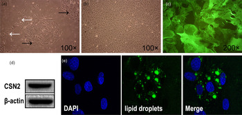
After 4 d of primary cell culture, epithelial-like cells (Fig. 1a; black arrow) were observed, which had clear boundaries with areas of fibroblast-like cells (Fig. 1a; white arrow). Mammary epithelial-like cells and fibroblasts were separated according to their differing sensitivity to trypsin. Purified DCMECs with characteristic cobblestone morphology were typically observed (Fig. 1b). Immunofluorescence staining exhibited intensely positive staining for CK-18 in the purified DCMECs' cytoplasm (Fig. 1c). We found that oleic acid could increase the lipid production in DCMECs. CSN2 was detected by western blotting in the purified DCMECs (Fig. 1d). In addition, fluorescence staining result showed that the milk lipid droplets DCMECs stained with BODIPY493/503 (yellow; Figure 1e). These results suggest that the lactating DCMECs culture model is suitable for investigating the milk fat synthesis function.
Characterization of the lactating DCMECs culture model. (a) Morphology of the primary cells cultured on the 4th day. Black arrows indicate epithelial-like cells and white arrows indicate fibroblast-like cells. (b) Morphology of the purified DCMECs. (c) Immunofluorescence staining of CK-18 in the purified DCMECs. (d) The expression level of the CSN2 in the purified DCMECs were detected by western blot. (e) The result of BODIPY493/503 staining of lipid droplets (yellow) in the purified DCMECs (400 × ).

LPS decreases lipid droplets synthesis and secretion in the lactating DCMECs
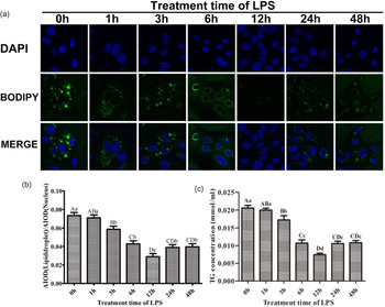
As shown in Fig. 2, both the number and the size of lipid droplets were decreased after LPS treatment (Fig. 2a). Lipid droplets in DCMECs treated with LPS for 12 h appeared to be lower than at other times (Fig. 2a and b). In addition, the number and the size of lipid droplets in DCMECs treated with LPS for 24 h and 48 h appeared to be larger than those at 12 h after LPS treatment (Fig. 2a). The result of total TG content (Fig. 2c) was consistent with the ratio of AIOD of lipid droplets to nuclei (Fig. 2b). Total TG content at 1, 3, 6, 12, 24, and 48 h after LPS treatment was 0.89-, 0.71-, 0.56-, 0.38-, 0.48-, and 0.51-fold lower than the control, respectively. Based on these data, we found that LPS could decrease lipid droplet synthesis in lactating DCMECs culture model.
LPS decreases lipid droplets synthesis and secretion in the lactating DCMECs. (a) The typical images of BODIPY493/503 staining for lipid droplets (yellow) in DCMECs (400 × ). (b) The ratio of AIOD of lipid droplets to nuclei. (c) The result of TG content in culture medium. The data with different capital letters between two groups showed very significant differences (P < 0.01); data with different lower-case letters between two groups showed significant differences (P < 0.05); data with the same letters between two groups showed no significant differences (P > 0.05).

LPS inhibits the expression of LXRα in the lactating DCMECs
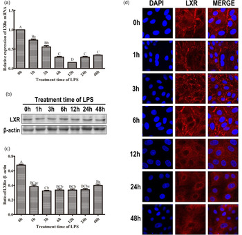
As shown in Fig. 3, the transcription level (Fig. 3a) and translation level (Fig. 3b and c) of LXRα were down-regulated in DCMECs treated with LPS, and then began to increase gradually at 12 h, and 24 h after LPS treatment respectively. The transcription level of LXRα at 12 h after LPS treatment was 6.4-fold lower than the control (P < 0.01). Consistent with the above results, LPS could inhibit the translocation of LXRα from the cytoplasm to the nucleus (Fig. 3d). These results suggest that LPS could inhibit the expression level and activity of LXRα in lactating DCMECs culture model.
LPS inhibits the transcription, translation and translocation of LXR in the lactating DCMECs. (a) The relative transcription level of LXRα gene. (b) The western blotting result of LXRα. (c) The relative expression level of LXRα. (d) The nuclear translocation of LXRα (red) in DCMECs (400 × ). The data with different capital letters between two groups showed very significant differences (P < 0.01); data with different lower-case letters between two groups showed significant differences (P < 0.05); data with the same letters between two groups showed no significant differences (P > 0.05).

LPS regulates the transcription of LXRα target genes in the lactating DCMECs
As shown in Fig. 4, LPS could inhibit the transcription of SREBP1, FAS, and ACC1 genes in a time-dependent manner. These results suggest that LPS could inhibit the de novo synthesis of fatty acids in DCMECs. However, LPS treatment could increase the transcription level of CD36 gene (Fig. 4d). The transcription level of CD36 at 6 h after LPS treatment was 3.4-fold higher than the control (P < 0.01). We hypothesize that CD36 gene may be involved in LPS-induced innate immune responses in DCMECs.
LPS regulates the transcription of LXRα target genes in the lactating DCMECs. (a) The transcription level of SREBP1 gene. (b) The transcription level of FAS gene. (c) The transcription level of ACC1 gene. (d) The transcription level of CD36 gene. The data with different capital letters between two groups showed very significant differences (P < 0.01); data with different lower-case letters between two groups showed significant differences (P < 0.05); data with the same letters between two groups showed no significant differences (P > 0.05).

Discussion
LXRs are nuclear receptors that regulate the synthesis of lipid and control cholesterol homeostasis upon activation by oxysterols or synthetic agonists. Farke et al. (Reference Farke, Meyer, Bruckmaier and Albrecht2008) identified the presence of LXRα in bovine mammary tissue, but did not elucidate its role in lipid metabolism. Mani et al. (Reference Mani, Sorensen, Sejrsen, Bruckmaier and Albrecht2009) reported that LXRα should be considered a potentially important regulator of milk fat synthesis, as expression of LXRα is increased during the transition from pregnancy to lactation. McFadden and Corl (Reference Mcfadden and Corl2010) found that activation of LXRα could enhance de novo fatty acid synthesis in bovine mammary epithelial cells. In addition, LXR activation increases SREBP1 and FAS mRNA and protein abundance. On the other hand, Oppi-Williams et al. (Reference Oppi-Williams, Suagee and Corl2013) reported that reducing LXRα mRNA abundance in mammary alveolar cells (Mac-T) did not alter mRNA abundance of genes involved in de novo lipogenesis or the rate of de novo lipogenesis. Li et al. (Reference Li, Luo, Zhu, Sun, Yao, Shi and Wang2015) found that LXRα regulated fatty acid synthase promoter activity by directly interacting with LXRα response elements by increasing SREBP1 abundance in goat mammary epithelial cells (Li et al., Reference Li, Luo, Zhu, Sun, Yao, Shi and Wang2015). Fu et al. (Reference Fu, Tian, Wei, Liu, Song, Liu, Zhang, Wang, Cao and Zhang2014) and Wang et al. (Reference Wang, Zhang, Wei, Wang, Zhang, Shi, Yang and Fu2017) reported that LXRα agonist T0901317 and Platycodin D could suppress LPS-induced mastitis in mice and DCMECs. Overall, the evidence suggests that LXRα may serve as a new target for mastitis therapy and regulation of milk fat synthesis (Hu et al., Reference Hu, Zhang and Fu2019). In the current research, our objective was to evaluate the role of LXRα in LPS-induced reduction of triglyceride synthesis in lactating DCMECs.
Cell culture is a convenient tool for studying the mechanisms of milk fat synthesis. Several immortalized bovine mammary epithelial cell lines have been established, such as BMEC + H, HH2A, ET-C, BME-UV, and Mac-T (Hosseini et al., Reference Hosseini, Sharma, Bionaz and Loor2013). However, these cell lines are not suited for studying the mechanisms of milk fat synthesis. In the present study, lactating DCMECs culture model with high lipid production ability was prepared by addition of oleic acid to the culture medium. Fluorescent dyes offer an indirect measurement for intracellular lipids in different types of cell. The most common dye for milk lipids is Nile red (Matsunaga et al., Reference Matsunaga, Tsugami, Kumai, Suzuki, Nishimura and Kobayashi2018). BODIPY staining has been used more recently as a potential alternative to Nile red (Govender et al., Reference Govender, Ramanna, Rawat and Bux2012). We found that BODIPY staining is an alternative to the Nile Red fluorescence method for the evaluation of intracellular lipids in DCMECs. The results of BODIPY staining and TG detection showed that oleic acid could induce high lipid production ability of DCMECs. The lactating DCMECs culture model is suitable for investigating the milk fat synthesis function.
The intracellular lipids and total TG were detected by BODIPY staining and enzymatic colorimetric assay in the DCMECs layer and culture medium, respectively. We found that LPS could decrease milk lipid droplets synthesis. LPS could decrease the cell viability and induce inflammatory response in DCMECs (Liu et al., Reference Liu, Zhang, Lin, Bian, Gao, Qu and Li2016). Besides cell viability and inflammatory response, the regulation of milk fat synthesis in DCMECs with LPS treatment also involves multiple transcription factors to control the milk fat enzymatic machinery. LXRα is a nuclear receptor and transcription factor that plays important role in milk fat synthesis and mastitis therapy in dairy cows (Hu et al., Reference Hu, Zhang and Fu2019). Therefore, the transcription, translation, and translocation of LXR were detected in this study. We found that LPS could inhibit the transcription, translation and nuclear translocation of LXR. The results suggest that LXRα participate in LPS-induced reduction of triglyceride synthesis in DCMECs.
SREBP1 is a membrane-bound transcription factor that directly regulates the synthesis and uptake of cholesterol and fatty acids. The promoter of SREBP1c contains a liver X response element (LXRE) (Mcfadden and Corl, Reference Mcfadden and Corl2010). ACC1 and FAS are key enzymes regulating de novo synthesis in dairy cow mammary gland. The promoters of the lipogenic enzymes ACC1, and FAS all contain a sterol response element (SRE) capable of binding SREBP1 (McFadden and Corl, Reference Mcfadden and Corl2010, Oppi-Williams et al., Reference Oppi-Williams, Suagee and Corl2013). LXR indirectly promotes the transcription of ACC1 and FAS by increasing the expression of SREBP1c. Furthermore, LXRα can increase the transcription of ACC1 and FAS by binding to LXRE found within the promoter region of these lipogenic genes (Talukdar and Hillgartner, Reference Talukdar and Hillgartner2006). CD36 is a scavenger receptor that functions in high-affinity tissue uptake of long-chain fatty acids and contributes to milk lipid accumulation (Pepino et al., Reference Pepino, Kuda, Samovski and Abumrad2014). Zhou et al. (Reference Zhou, Febbraio, Wada, Zhai, Kuruba, He, Lee, Khadem, Ren, Li, Silverstein and Xie2008) found that CD36 is a shared target of LXRα, pregnane X receptor (PXR), and peroxisome proliferator-activated receptor (PPAR) gamma. Therefore, SREBP1, FAS, ACC1, and CD36 are LXRα target genes. Consistent with the LXRα and previous report (Liu et al., Reference Liu, Zhang, Lin, Bian, Gao, Qu and Li2016), we found that LPS could inhibit the transcription of SREBP1, FAS, and ACC1 genes in a time-dependent manner. The results suggest that LPS could inhibit the de novo synthesis of fatty acids in DCMECs via inhibiting the expression and activity of the LXRα. Contrary to the LXRα and previous report (Liu et al., Reference Liu, Zhang, Lin, Bian, Gao, Qu and Li2016), we found that LPS treatment could increase the transcription level of CD36 gene. Liu et al. (Reference Liu, Zhang, Lin, Bian, Gao, Qu and Li2016) reported that LPS significantly decreased the mRNA expression levels of CD36 in DCMECs compared with the control group. Consistent with our results, Cao et al. (Reference Cao, Luo, Chen, Xu, Shi, Jing and Zang2016) reported that CD36 mRNA levels in mammary gland were significantly increased in E. coli-induced dairy goat mastitis compared with healthy goats. They also found that at concentrations of 1–10 μg/ml, LPS did not induce cell apoptosis or necrosis but up-regulated the expression of CD36 mRNA after LPS treatment for 12 h (Cao et al., Reference Cao, Luo, Chen, Xu, Shi, Jing and Zang2016). They proved that CD36 regulates LPS-induced signaling pathways and mediates the internalization of E. coli in cooperation with toll-like receptor 4 in goat mammary gland epithelial cells (Cao et al., Reference Cao, Luo, Chen, Xu, Shi, Jing and Zang2016). We hypothesize that CD36 gene may be involved in LPS-induced innate immune responses in DCMECs.
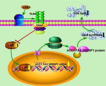
In conclusion, LPS can decrease the level of triglyceride synthesis in lactating DCMECs via down-regulating the expression and activity of the LXRα (Fig. 5). Our data may provide new insight into the mechanisms of milk fat depression caused by LPS.
A schematic drawing illustrating the proposed mechanism of LPS-induced reduction of milk lipid.

Supplementary material
The supplementary material for this article can be found at https://doi.org/10.1017/S0022029920000990
Acknowledgements
This work was financially supported by the National Natural Science Foundation of China (31472249, 31402157, 31772698, 31972747), the Natural Science Foundation of Heilongjiang province (C2018043).