





Introduction
Parasites form a critical component of resilient ecosystems, providing important ecosystem services that underscore the need for their conservation (Carlson et al., Reference Carlson, Hopkins, Bell, Dona, Godfrey, Kwak, Lafferty, Moir, Speer, Strona, Torchin and Wood2020). However, some parasites pose significant threats to human health, livestock production and wildlife conservation (Johansen et al., Reference Johansen, Lier and Sithithaworn2015; Mehmood et al., Reference Mehmood, Zhang, Sabir, Abbas, Ijaz, Durrani, Saleem, Rehman, Iqbal, Wang, Ahmad, Abbas, Hussain, Ghori, Ali, Khan and Li2017; De Oliveira et al., Reference De Oliveira, Felix-nascimento, Berenguer, Da Silva E Souza, Bernal-valle, De Souza, Acosta-jamett and Chaves2024). Furthermore, anthropogenic pressures can drastically alter parasite transmission, compromising both human and animal health (Sow et al., Reference Sow, De Vlas, Engels and Gryseels2002; Jones et al., Reference Jones, Grace, Kock, Alonso, Rushton, Said, McKeever, Mutua, Young, McDermott and Pfeiffer2013; Thompson, Reference Thompson2013; Gottdenker et al., Reference Gottdenker, Streicker, Faust and Carroll2014) and shifting the balance in parasite-mediated competition (Price et al., Reference Price, Westoby, Rice, Atsatt, Fritz, Thompson and Mobley1986; Schall, Reference Schall1992; Tompkins et al., Reference Tompkins, White and Boots2003), whereby both effects often shape community structure, biodiversity and therefore conservation efforts. Together, these contrasting roles highlight parasitism as a complex and context-dependent process with important implications for wildlife conservation and management.
The ultimate effect of parasites on host health is often dependent on co-infections with other pathogens, environmental stressors, and anthropogenic pressures. For instance, parasite-mediated immune modulation or tissue damage can facilitate concomitant or secondary infections, which ultimately drive disease severity and host mortality (Allen and Wynn, Reference Allen and Wynn2011; Demann and Wegner, Reference Demann and Wegner2019). Environmental pressures, such as temperature fluctuations and resource limitation, can additionally influence disease outcomes (Vicente-Santos et al., Reference Vicente-Santos, Willink, Nowak, Civitello and Gillespie2023). Such multifactorial dynamics have become increasingly relevant in the Anthropocene, where anthropogenic stressors, including habitat alteration, global warming, and pollution, are reshaping aquatic ecosystems and modulating host–parasite relationships, which can result in elevated host morbidity or mortality (Marcogliese and Pietrock, Reference Marcogliese and Pietrock2011; Schols et al., Reference Schols, Carolus, Hammoud, Muzarabani, Barson and Huyse2021). Trematodes exemplify this complexity: as parasites with intricate, multi-host life cycles, they are highly sensitive to environmental change, with their transmission dynamics often responding non-linearly to temperature shifts and pollution (Sures et al., Reference Sures, Nachev, Schwelm, Grabner and Selbach2023).
Artificial water bodies, such as man-made dams, are particularly prone to environmental and anthropogenic modifications, including altered physicochemical conditions and the introduction of invasive species. These can consequently intensify trematode transmission and disease risk (Morley, Reference Morley2007; Telfer and Bown, Reference Telfer and Bown2012; Carolus et al., Reference Carolus, Muzarabani, Hammoud, Schols, Volckaert, Barson and Huyse2019). For instance, the creation of the artificial Lake Kariba in Zimbabwe facilitated both the spillback and potential spillover of trematode infections in large herbivores such as the hippopotamus through a cascade of biological invasions (Carolus et al., Reference Carolus, Muzarabani, Hammoud, Schols, Volckaert, Barson and Huyse2019; Schols et al., Reference Schols, Carolus, Hammoud, Muzarabani, Barson and Huyse2021). Although comparable empirical data for terrestrial megafauna remain limited, anthropogenic modifications to the environment are increasingly linked to enhanced parasite transmission and disease burden in wildlife (Brearley et al., Reference Brearley, Rhodes, Bradley, Baxter, Seabrook, Lunney, Liu and McAlpine2013), with examples from elephants and primates (Vítovec et al., Reference Vítovec, Kotrlá, Haji and Hayles1984; Gillespie and Chapman, Reference Gillespie and Chapman2008). Therefore, understanding environmental drivers of disease burden and transmission dynamics is critical for predicting disease outcomes and developing effective conservation and management strategies in a rapidly changing world.
Fasciolosis, also known as liver rot, is a trematode-induced disease that is particularly sensitive to environmental modifications and changes in freshwater habitats (Alba et al., Reference Alba, Vazquez and Hurtrez-boussès2021). It is caused by Fasciola species, of which 3 are linked to animal disease in Africa: Fasciola hepatica (Linnaeus, 1758), Fasciola gigantica (Cobbold, 1856), and Fasciola nyanzae (Leiper, 1910) (McCully et al., Reference McCully, Van Niekerk and Kruger1967; Malatji et al., Reference Malatji, Pfukenyi and Mukaratirwa2020). They infect the liver of livestock and wild ruminants, causing anaemia, weight loss, lesions and fibrosis in the liver, haemorrhage and ultimately death (Behm and Sangster, Reference Behm, Sangster and Dalton1999). Most studies focus on livestock due to the severe economic burden (Mehmood et al., Reference Mehmood, Zhang, Sabir, Abbas, Ijaz, Durrani, Saleem, Rehman, Iqbal, Wang, Ahmad, Abbas, Hussain, Ghori, Ali, Khan and Li2017), whereas infections in wildlife remain severely neglected (Malatji et al., Reference Malatji, Pfukenyi and Mukaratirwa2020; Namirembe et al., Reference Namirembe, Huyse, Wangalwa, Tumusiime and Tolo2024). In Southern Africa, transmission occurs via freshwater snails belonging to Lymnaeidae, mostly by Radix natalensis (Krauss, 1848), Galba spp. (Schrank, 1803) (Mahulu et al., Reference Mahulu, Clewing, Stelbrink, Chibwana, Tumwebaze, Stothard and Albrecht2019) and the invasive snail Pseudosuccinea columella (Say, 1817). Upon release from the freshwater snail host, cercariae encyst on aquatic vegetation, after which they are ingested by the final host (Malatji et al., Reference Malatji, Pfukenyi and Mukaratirwa2020; Nukeri et al., Reference Nukeri, Malatji, Sengupta, Vennervald, Stensgaard, Chaisi and Mukaratirwa2022).
Amphistomes, or stomach flukes, represent another important group of trematodes infecting ruminants. Members of Paramphistomoidea (Fischoeder, 1901) cause paramphistomosis or amphistomiasis when infecting the rumen and reticulum of ruminants. The adults generally are of low pathogenicity, but immature stages cause severe clinical disease when they migrate from the small intestine to the rumen, leading to weight loss, anaemia, decreased milk production, and even death (Elelu and Eisler, Reference Elelu and Eisler2018; Pfukenyi and Mukaratirwa, Reference Pfukenyi and Mukaratirwa2018). Similar to fasciolosis, the burden of amphistomiasis on wildlife remains severely understudied, despite the disease’s wide geographic distribution and potentially great impact on animal health (Sibula et al., Reference Sibula, Nyagura, Malatji and Mukaratirwa2024b). The most widespread species, possibly a species complex (Laidemitt et al., Reference Laidemitt, Anderson, Wearing, Mutuku, Mkoji and Loker2019), causing amphistomiasis in Africa is Calicophoron microbothrium (Fischoeder, 1901), whose reported host snails are mostly Bulinus species (Pfukenyi and Mukaratirwa, Reference Pfukenyi and Mukaratirwa2018; Sibula et al., Reference Sibula, Nyagura, Malatji and Mukaratirwa2024b), although recently a natural infection in the non-native Melanoides tuberculata (O. F. Müller, 1774) has been reported (Sibula et al., Reference Sibula, Malatji, Nyahunda and Mukaratirwa2024a). Similar to liver flukes, amphistomes encyst on vegetation to reach their final host through ingestion (Ghatani and Tandon, Reference Ghatani, Tandon, Toledo and Fried2024).
Both liver and stomach flukes infect African buffalo (Syncerus caffer Sparrman, 1779) (Bengis et al., Reference Bengis, Gakuya, Munyeme, Shiferaw, Kock, Chardonnet, Caron, Cornélis, Chardonnet and Prins2023; Sibula et al., Reference Sibula, Nyagura, Malatji and Mukaratirwa2024b). This species is the largest African bovid and occurs in diverse ecosystems across the continent, from rainforests to savannas. It is water-dependent and lives in complex social herds (Taylor et al., Reference Taylor, Bennitt, Fynn, Korte, Naidoo, Roug, Cornelis, Caron, Cornélis, Chardonnet and Prins2023). As a result of habitat loss, poaching, increasing droughts and diseases, the species’ conservation status has been updated from ‘least concern’ to ‘nearly threatened’ (IUCN SSC Antelope Specialist Group 2019), despite regional disparities (Cornelis et al., Reference Cornelis, Renaud, Melletti, Fonteyn, Bonhotal, Hauptfleisch, Asefa, Breuer, Korte, Scholte, Elkan, Kohi, Mwiu, Ngene, Omondi, Tadjo, Prin, Caron, Prins, Chardonnet, Caron, Cornélis, Chardonnet and Prins2023). Besides conservation, the African buffalo is also a species of interest from a One Health perspective. Buffaloes and cattle both belong to the tribe Bovini and therefore have many common pathogens that cause severe disease in cattle, such as bovine tuberculosis, brucellosis and several trematodiases (Le Roex et al., Reference Le Roex, Cooper, Van Helden, Hoal and Jolles2016; Pfukenyi and Mukaratirwa, Reference Pfukenyi and Mukaratirwa2018; Bengis et al., Reference Bengis, Gakuya, Munyeme, Shiferaw, Kock, Chardonnet, Caron, Cornélis, Chardonnet and Prins2023). The latter can cause mortality in S. caffer either directly or indirectly through interactions with other pathogens (Ezenwa and Jolles, Reference Ezenwa and Jolles2015; Woodburn et al., Reference Woodburn, Steyl, Du Plessis, Last, Reininghaus and Mitchell2021).
In the early cold season (June–July 2023), 26 wild ruminants died at the Imire Rhino & Wildlife Conservancy in Zimbabwe, a sharp contrast to the limited animal losses in 2022. Especially the loss of 4 young African buffaloes caused concern to the management team. Earlier post-mortem examinations of 2 greater Kudus (Tragelaphus strepsiceros Pallas, 1766) by veterinarians listed snail-borne parasites as one of the potential causes of these unexplained deaths. Therefore, a snail survey combined with a post-mortem examination of a culled buffalo was conducted to identify and monitor trematode infections that potentially contributed to the observed increase in buffalo mortality.
Materials and methods
Study area
A snail and trematode survey was conducted in Imire Rhino and Wildlife Conservancy (referred to as Imire) during the cold dry season (winter) in August 2023. Imire is located in the northern part of Hwedza District, Zimbabwe, bordering Marondera District. The region experiences a temperate highland tropical climate, characterised by distinct dry and rainy seasons, with a mean annual precipitation of approximately 675 mm and an annual average temperature of 21.5 °C. Imire consists of 3 fenced conservation areas and an adjacent zone dedicated to irrigated tobacco farming, a land-use practice that is widespread in this region of Zimbabwe. To secure water supplies for irrigation and wildlife, particularly under increasing climatic variability, several rivers and creeks within Imire have been dammed, resulting in multiple artificial water bodies. One artificial reservoir (Site W) was only created in 2018 and is permanently topped up with borehole water. Sampling was conducted in the Lodge section, the northernmost conservation section, which directly borders the agricultural area (Figure 1). This section encompasses the full range of the resident buffalo herd, as well as other large mammals, including elephant, wildebeest, antelope and baboons. Accordingly, all water bodies within this section (1 natural creek and 3 man-made dams) were surveyed for freshwater snails.
Study area and sampling locations at Imire Rhino and Wildlife Conservancy, Hwedza District, Zimbabwe. (A) Location of the study area within Zimbabwe, Southern Africa. (B) Position of Imire within Hwedza District. (C) Spatial partitioning of Imire into 4 management sections from south to north: Chiwawe, Middle section, Farm section and Lodge section. (D) Detailed map of the Lodge section showing freshwater bodies sampled for snail intermediate hosts, indicated as black dots with their respective site letter codes.

Figure 1 Long description
A) Map showing the location of Imire Rhino and Wildlife Conservancy within Zimbabwe, Southern Africa. B) Map indicating the position of Imire within Hwedza District, bordered by Marondera District. C) Map illustrating the spatial partitioning of Imire into four management sections from south to north: Chiwawe, Middle Section, Farm Section and Lodge Section. D) Detailed map of the Lodge Section, highlighting freshwater bodies sampled for snail intermediate hosts, marked by black dots with site letter codes. The map includes symbols for wildlife sanctuaries and irrigation areas, with labeled sections such as Wildlife Sanctuary and Irrigated Farm Section. The map also shows the proximity to Marondera and Hwedza, with directional arrows indicating north. Scale bars are present for distance reference.
Trematode recovery from buffalo and snail hosts
A post-mortem examination was conducted on 18 August 2023 at 10:30 on a female African buffalo, estimated 8–10 years old, approximately 1 h after being culled in close proximity to Site G (Mermaid’s dam). The buffalo herd was scheduled for their annual fluke treatment the following week, typically administered immediately before or after the winter or rainy season. Treatment consists of Flukazole C (Virbac, Triclabendazole 12% mass/volume and Oxfendazole 4.53% mass/volume) drenched cubes for oral intake. The herd had been treated with an undetermined insecticide against ticks. Conservation managers reported that the culled buffalo had exhibited markedly poor health during the week preceding culling, including difficulty remaining upright. On the day of culling, the animal was unable to stand and was found recumbent near the water’s edge at Site G. It allowed close human approach and was euthanized with a shot to the temple. During the necropsy, general health indicators, including body mass, coat condition, body fat reserves, organ condition and the presence of cysts or lesions, were assessed. The liver, stomach compartments and mesenteric veins were inspected for the presence of trematode parasites. The liver was screened for liver flukes by opening and inspecting the bile ducts and by sectioning the liver tissue using a scalpel. All stomach compartments were opened, and the mucosal surfaces were visually inspected for the presence of stomach flukes. Finally, the mesenteric veins were screened for blood flukes by transillumination, i.e. holding a bright light behind the tissue to detect (moving) adult schistosomes. During collection in the field, live stomach flukes were first relaxed in 50 mL tubes containing warm saline solution (approximately 70 °C; 9 g NaCl per litre of borehole water) and fixed in 70% ethanol for morphological and subsequent molecular identification.
Freshwater snails were collected in a standardized way by 2 persons sampling for 30 min per site using scooping nets with a mesh size of 1 mm. Snails were transported alive to the field lab in ambient water, where they were identified to species or genus level using keys by Mandahl-Barth (Reference Mandahl-Barth1962), Brown (Reference Brown1994), and Appleton and Miranda (Reference Appleton and Miranda2015). Identification was additionally supported by previous morphological and molecular studies of snails at the same study sites (Mudavanhu et al., Reference Mudavanhu, Goossens, Schols, Manyangadze, Nhiwatiwa, Lemmens, Huyse and Brendonck2024a, Reference Mudavanhu, Schols, Goossens, Nhiwatiwa, Manyangadze, Brendonck and Huyse2024b). Snails were grouped per morphotype and stored in the dark overnight in multiwell plates filled with borehole water. Before sunrise, all wells were inspected using a stereomicroscope for the emergence of cercariae, which are the larval trematode stages, from the snails. At sunrise, shedding experiments started, whereby the release of cercariae was stimulated by exposing the snails to indirect sunlight at the windowsill combined with artificial lighting. All wells were checked for the emergence of cercariae using a stereomicroscope at 3 time points: at sunrise (06:30), before noon (08:00–10:00) and at the end of the shedding experiment in the late afternoon (15:00–17:00). As a consequence of these time frames, however, C. aff. microbothrium cercariae had already encysted on the surface of the plastic multiwell plates after shedding. Amphistome cercariae encyst on vegetation following release from their snail intermediate host in natural settings, and have been shown to encyst on various other biotic and abiotic surfaces, including plastic multiwell plates (LeSage and Fried, Reference LeSage and Fried2011). Therefore, subsequent analysis and reporting were based on the metacercarial form. Snails, cercariae and metacercariae were subsequently stored in 80% ethanol and transported to Belgium for molecular analysis.
Imaging
Imaging was performed for digital archiving and to document and assess diagnostic morphological traits of snails and trematode parasites. Adult parasites and snails were photographed using the focus-stacking technique described in Brecko et al. (Reference Brecko, Mathys, Dekoninck, Leponce, VandenSpiegel and Semal2014) and Schols et al. (Reference Schols, Carolus, Hammoud, Muzarabani, Barson and Huyse2021). Histological sections of the stomach flukes were digitized with a Leica DMLB using Objective Imaging through the Surveyor software (version 8.0.0.2) at 5× magnification and saved as bitmap files with ×2 scaling to reduce file size. Key diagnostic features, including the genital pore, pharynx, and acetabulum, were imaged at 10× magnification. Multiple images of each specimen were captured at successive focal depths and subsequently merged into a single high-depth-of-field image using Zerene Stacker and the Pmax option (version 1.04 Build T2022-04-21-0715). Cercariae and metacercariae were washed in water, isolated on microscopic slides, and photographed using a BMS camera (model number: XFCAM1080PHD) and associated BMS Pix3 software (version 4.12.24621.20240204). Subsequently, cercarial types were morphologically classified according to Frandsen and Christensen (Reference Frandsen and Christensen1984). Final image processing was performed in Adobe Photoshop (version 26.9.0) to remove background artefacts, add scale bars and assemble multiple orientations of individual specimens into a single figure. Morphometric measurements were obtained from images of 80% ethanol-fixed cercariae and metacercariae using ImageJ version 1.54f.
Morphological analysis of stomach flukes
Stomach flukes were morphologically identified based on median sagittal sections and scanning electron microscopy (SEM) following keys by Sey (Reference Sey1991) and Eduardo (Reference Eduardo1983). Median sagittal sections were made to examine the internal morphological traits of the collected stomach flukes. First, samples were submerged in 100% ethanol for 2 times 1 h. Next, they were incubated in 1:1 ethanol/butanol (1 h), 100% butanol (1 h), 100% butanol/paraffin (overnight) and 100% paraffin (4 h). After being embedded in paraffin, sections of 10 µm were cut with a Leica SM 2000R sliding microtome and collected on glass slides. Then the sections were dried on a warm plate and deparaffinized gradually to dH2O by first washing them 3 times with xylene and subsequently in a descending series of ethanol: 100% absolute ethanol (2 times), followed by 96%, 90%, 75% and 50% ethanol each for 15 min. Finally, the sections were submerged in dH2O for 10 min. The deparaffinized sections were then submerged in haematoxylin Ehrlich for 20 min and rinsed with tap water for 15 min. Next, they were stained with erythrosin 0.1% in dH2O for 10 min. The sections were dehydrated in an ascending series of ethanol: 50%, 75%, 80% and 90% (a few seconds each), followed by 96% (10 min) and 100% absolute ethanol (2 times 15 min). Lastly, the sections were submerged in xylene for 2 times 20 min and mounted in Depex.
Two amphistome specimens were examined using SEM following Schols et al. (Reference Schols, Henrard, Brecko, Mudavanhu, Goossens, Steffanie, Clegg, Vanhove and Huyse2025) to visualize the tegumental papillae and the genital pore region. The specimens were first dehydrated overnight in 100% ethanol, then submerged in 100% ether in a watch glass and incubated on a heating block (low heat) until dry. The resulting specimens were mounted on aluminium stubs using copper tape, coated with gold and examined using a JEOL JSM-6480LV SEM. The contrast of the resulting images was subsequently optimized in Adobe Photoshop.
Molecular analysis
Adult parasite DNA extraction
All 18 preserved liver flukes and a subset of 15 out of 35 stomach flukes (due to the more invasive tissue sampling in these smaller specimens) were selected for DNA extraction. A sterile scalpel was used to excise one-quarter to one-half of the acetabulum for 5 stomach flukes. However, since the acetabulum serves as a diagnostic morphological feature, a section from the dorsal tegument was taken from the remaining 10 stomach flukes. A tissue section of approximately 5 mm2 was isolated from the lateral margin of liver flukes. Tissue samples were homogenized using a sterile scalpel and air-dried on sterile absorbent paper. The work surface, gloves and equipment were sterilized between specimens using DNA AWAYTM (Thermo FisherTM). Subsequently, genomic DNA extraction proceeded according to the manufacturer’s guidelines using the proteinase K-based E.Z.N.A. Tissue DNA Kit (OMEGA Bio-tek). DNA was eluted in 2 elution steps using 75 µL of elution buffer. The eluate was subsequently diluted by adding 10 µL of the extract to 90 µL of sterile Milli-Q water (Rockland). From the remaining extract, 100 µL was dried on GenTegraTM DNA-Tubes for curation in the Royal Museum for Central Africa (RMCA) Biobank, while the remaining 40 µL was stored at −20 °C.
Snail DNA extraction
The snail tissue was carefully separated from the shell using thin tweezers and placed on absorbent paper to remove excess ethanol. The entire soft body was used for DNA extraction, while the shells were retained for curation in the RMCA collection. To prevent cross-contamination, all instruments were sterilized between specimens using ethanol and a flame. All collected snails were extracted, except for Physella acuta, for which a maximum of 50 individuals per site were extracted, as this invasive snail occurred in high numbers and is usually not infected with the targeted trematodes, while still allowing a sufficient sampling effort to detect trematode infections. DNA extraction was performed using the E.Z.N.A. Mollusk DNA Kit (OMEGA Bio-tek), following the manufacturer’s protocol and using 2 elution steps, each with 75 μL elution buffer. Of the 150 μL eluate, 10 μL was added to 100 μL water to obtain a 1:10 dilution for downstream analysis. From the remaining extract, 100 µL was dried onto GenTegraTM DNA-Tubes for curation in the RMCA Biobank, and the remaining 40 µL was stored at −20 °C.
Cercariae DNA extraction
Four cercariae per shedding snail were transferred from their respective tube into a watch glass and washed in distilled water. Individual cercariae were then pipetted into wells of a PCR plate, each containing 5 µL of Ultrapure sterile water (Rockland). Next, a lysis buffer was prepared as in Zietara et al. (Reference Zietara, Arndt, Geets, Hellemans and Volckaert2000), and 10 µL of lysis buffer containing 1 µL of proteinase K was added to each well. The plate was gently swirled, briefly centrifuged and incubated at 65 °C for 25 min, followed by 96 °C for 10 min. Wells were then visually inspected to confirm complete lysis of the cercariae. For metacercariae, however, the above-mentioned protocol proved ineffective. Therefore, a freeze-thaw cycle was used to weaken the metacercarial cyst wall, followed by DNA extraction using the E.Z.N.A. Tissue Kit. The samples were frozen at −70 °C for 5 min and subsequently incubated at 90 °C for 5 min at 1500 rpm. This cycle was repeated 5 times consecutively. Following thermal treatment, the weakened cyst walls were mechanically disrupted by gently compressing the cyst between a glass slide and a cover slip prior to DNA extraction. For each shedding snail, DNA concentration was quantified using a Qubit™ fluorometer for 2 of the 4 extracted cercariae. All DNA extracts were stored undiluted at −20 °C.
Rapid diagnostic PCR (RD-PCR)
All snail DNA extracts were first screened using a rapid diagnostic PCR (RD-PCR) protocol as described in Carolus et al. (Reference Carolus, Muzarabani, Hammoud, Schols, Volckaert, Barson and Huyse2019). This multiplex assay enables the simultaneous detection of any trematode infection and specific infections by Fasciola spp., using a general trematode marker (18S rDNA), an internal control marker targeting snails (18S rDNA) and a Fasciola-specific, high-copy non-coding nuclear repeat (a 124 bp repetitive element) that facilitates sensitive detection of Fasciola spp. (Kaplan et al., Reference Kaplan, Dame, Reddy and Courtney1995; Krämer and Schnieder, Reference Krämer and Schnieder1998). PCR reactions were performed in a volume of 15 μL containing 1 μL DNA, 5 μL dH2O, 1.5 μL primer mix and 7.5 μL Qiagen Multiplex Mastermix. The final mix primer concentrations were 0.2 μM for 18S_Digenea_F and 18S_Digenea_R, 0.1 μM for FW_SNAIL2 and REV_SNAIL2 and 0.6 μM for Kaplan_F and Kaplan_R. Each PCR plate included positive controls consisting of DNA from an adult F. gigantica specimen and a mixed DNA sample containing both P. columella and F. gigantica DNA, as well as a negative control containing nuclease-free water. Amplification products were separated by gel electrophoresis on a 3% agarose gel run at 120 V for 105 min. Bands were visualized using Midori Green Direct staining and UV illumination.
Species identification and DNA barcoding
All adult trematodes and cercariae for which DNA had been extracted and all trematode infections detected through the RD-PCR assay were selected for molecular identification. This was done by sequencing segments of the mitochondrial cytochrome c oxidase subunit I (COI) gene and the nuclear ribosomal DNA (rDNA) region, including the complete internal transcribed spacer 1 and 2 (ITS1 and 2) and the 5.8S rDNA. For snail species identification, a fragment of the mitochondrial COI gene was amplified. A maximum of 2 snails per morphotype per site were selected for sequencing. All primer combinations and PCR protocols followed those described in Schols et al. (Reference Schols, Carolus, Hammoud, Muzarabani, Barson and Huyse2021). Amplifications were carried out using the Qiagen™ Taq DNA Polymerase kit in 20 µL reaction volumes, composed of 2 µL DNA extract, 2 µL 10× PCR buffer, 0.6 µL deoxyribonucleoside triphosphate (dNTP) mix (5 mM), 1.2 µL MgCl2 (25 mM), 0.8 µL of each primer (20 µM), 12.68 µL Milli-Q water and 0.12 µL of Taq Polymerase. Thermal cycling was conducted on a Biometra™ TProfessional thermocycler and consisted of an initial denaturation at 94 °C for 5 min, followed by 39 cycles of denaturation at 94 °C for 30 s, primer-specific annealing and elongation steps (Schols et al., Reference Schols, Carolus, Hammoud, Muzarabani, Barson and Huyse2021), and a final elongation step at 72 °C for 10 min. For the parasites, primers used for the COI barcoding region were COI1_DIG_F, COI1_DIG_R, COI2_DIG_F, COI2_DIG_R and NASMIT_R (Hammoud et al., Reference Hammoud, Mulero, Van Bocxlaer, Boissier, Verschuren, Albrecht and Huyse2022; 0.8 μM forward primer and 0.8 μM reverse primer). For amplification of the ITS region, we used ITS2_DIG_F and ITS2_DIG_R (Hammoud et al., Reference Hammoud, Mulero, Van Bocxlaer, Boissier, Verschuren, Albrecht and Huyse2022; 0.8 μM forward primer and 0.8 μM reverse primer), as well as ITS4 and ITS5 (White et al., Reference White, Bruns, Lee and Taylor1990; 0.8 μM forward primer and 0.8 μM reverse primer). For snails, universal HCO and LCO primers were used (Folmer et al., Reference Folmer, Black, Hoeh, Lutz and Vrijenhoek1994). All primer sequences and annealing temperatures are listed in Schols et al. (Reference Schols, Carolus, Hammoud, Muzarabani, Barson and Huyse2021). PCR products were visualized on a 1.5% agarose gel stained with Midori Green Direct® and examined under UV light. Samples that showed a clear band were purified using the ExoSAP-IT™ PCR Product Cleanup protocol and sent to Macrogen™ Europe for bidirectional Sanger sequencing using the same primers as those used for amplification.
Phylogenetic analysis
Sequences were processed in Geneious Prime® (v.2025.1.2); chromatograms were visually inspected and ambiguous base calls were resolved or coded using the International Union of Pure and Applied Chemistry (IUPAC) ambiguity codes prior to and following consensus sequence generation. Sequences of the coding marker COI were inspected for the absence of stop codons and the correct start codon. All sequences are available on GenBank (see Data Availability Statement). Alignments were made using MAFFT v.7.526 (Katoh et al., Reference Katoh, Misawa, Kuma and Miyata2002). Uncorrected pairwise distances were calculated using MEGA11 (Tamura et al., Reference Tamura, Stecher and Kumar2021). Thresholds for species differentiation were p-distance values >5% for COI and >1% for ITS sequences (Vilas et al., Reference Vilas, Criscione and Blouin2005; Lawton et al., Reference Lawton, Lim, Dukes, Kett, Cook, Walker and Kirk2015). For multiple sequences belonging to the same species, the longest sequence was selected for species identification using the Basic Local Alignment Search Tool (BLAST) on GenBank (Altschul et al., Reference Altschul, Gish, Miller, Myers and Lipman1990). Since BLAST results did not unambiguously confirm Calicophoron species identity, published sequences of all members of Calicophoron represented by overlapping COI or ITS2 fragments were downloaded for further phylogenetic analysis along with a representative subset of our samples. The related species Gigantocotyle gigantocotyle (Brandes in Otto, 1896) Näsmark, 1937, a member of the same family (Paramphistomidae), was selected as the outgroup for phylogenetic inference because previous analyses place it basally relative to Calicophoron (Schols et al., Reference Schols, Henrard, Brecko, Mudavanhu, Goossens, Steffanie, Clegg, Vanhove and Huyse2025). Sequences of the resulting 16 taxa were first aligned per marker in Aliview v.1.26 (Larsson, Reference Larsson2014) using MAFFT and visually inspected.
Substitution saturation was inspected for each codon position of the rapidly mutating COI marker by inspecting the Iss and Iss.c values obtained through Xia’s method, which is an information entropy-based index of substitution saturation. In addition, the suitability of the outgroup was assessed through Steel’s method, which is based on the parsimony method, in DAMBE v.7.3.37, https://dambe.bio.uottawa.ca/DAMBE/dambe.aspx (Xia, Reference Xia2018). The outgroup was suitable, and no substitution saturation was detected for codon positions 1 and 2. However, codon position 3 did reveal nonsignificant (P = 0.43) substitution saturation (Iss = 0.6151 > Iss.c = 0.5890) for a symmetrical tree topology and a significant (P = 0) substitution saturation (Iss = 0.6151 > Iss.c = 0.3588) for an asymmetrical tree topology. This was taken into consideration during the subsequent analyses by providing a separate substitution model for codon position 3 of the COI marker (see below).
The ITS2 and COI alignments were then curated using the least stringent settings in the online web interface of Gblocks (https://ngphylogeny.fr/; Lemoine et al., Reference Lemoine, Correia, Lefort, Doppelt-azeroual, Mareuil, Cohen-boulakia and Gascuel2019). Next, a concatenated alignment of both markers (COI: 519 sites, ITS2: 270 sites) was generated using Mesquite v.3.5 (Maddison and Maddison, Reference Maddison and Maddison2023). Using the greedy search algorithm and the Bayesian Information Criterion (BIC), the best partition scheme and best-fit substitution models were estimated in PartitionFinder 2 (Guindon et al., Reference Guindon, Dufayard, Lefort, Anisimova, Hordijk and Gascuel2010; Lanfear et al., Reference Lanfear, Calcott, Ho and Guindon2012, Reference Lanfear, Frandsen, Wright, Senfeld and Calcott2017). Partition 1 contained the first and second COI codon positions and the ITS2 sites (688 sites) and was analysed using the Hasegawa–Kishino–Yano substitution model (Hasegawa et al., Reference Hasegawa, Kishino and Yano1985) with an invariant site correction. Partition 2 contained the third codon position of COI (173 sites) and was analysed using the transition (TIM) substitution model with a gamma site correction (Posada, Reference Posada2003).
Phylogenetic reconstructions were performed using maximum likelihood (ML) and Bayesian inference through the CIPRES Science Gateway v.3.3; https://www.phylo.org/por-tal2 (Miller et al., Reference Miller, Pfeiffer and Schwartz2010). ML analyses were conducted in GARLI v.2.10 (Zwickl, Reference Zwickl2006). Multiple GARLI runs to determine the best tree converged on the same likelihood (−2,816.12), indicating a stable ML solution. Next, node supports were assessed from 1,000 bootstrap replicates obtained from 20 independent runs of 50 replicates each. Bootstrap values were then summarized on the best ML tree, and nodes with a nodal support value below 50% were collapsed using SumTree v.4.6.1, run in DendroPy v.4.6.1 (Moreno et al., Reference Moreno, Holder and Sukumaran2024). Bayesian inference was performed using MrBayes v.3.2.7a (Ronquist et al., Reference Ronquist, Teslenko, Van der Mark, Ayres, Darling, Hhna, Larget, Liu, Suchard and Huelsenbeck2012), with 2 parallel runs containing 4 chains each for 10 million generations. Parameters were estimated independently for each partition [unlink statefreq = (all) revmat = (all) shape = (all) pinvar = (all) tratio = (all)]. Trees were sampled every 100th generation and were used to reconstruct a 50% majority rule consensus tree after discarding the first 25% as burn-in. The MrBayes log files were inspected in Tracer v.1.7.1 (Rambaut et al., Reference Rambaut, Drummond, Xie, Baele and Suchard2018) to ensure an effective sample size above 200 for all parameters and a caterpillar-like pattern in the trace plot. The resulting trees were rooted using G. gigantocotyle as the outgroup in FigTree v.1.4.4. Final adjustments were made in Inkscape v.1.3, including repositioning misplaced support values, adding the bootstrap support values to the Bayesian inference and italicizing species names.
The phylogenetic signal for each separate marker was assessed to confirm that the concatenated analysis is justified. The Gblocks-curated COI and ITS2 alignments were analysed using the ML method in MEGA X (Kumar et al., Reference Kumar, Stecher, Li, Knyaz and Tamura2018) with the same best-fit substitution models as the concatenated analysis and with 100 bootstrap replicates. For COI, the dataset was analysed with all codons, only codons 1 and 2, and finally with codon 3 only. COI codon 3 did not follow the substitution model of the concatenated analysis, as MEGA X does not offer TIM. The General Time-Reversible model (GTR) (Rodríguez et al., Reference Rodríguez, Oliver, Marín and Medina1990) with a gamma correction showed only marginally different BIC and Akaike Information Criterion (AIC) values and was therefore used to analyse this dataset. Sequences spanning less than 75% of the alignment were removed. For COI, all 5 sequences from our specimens were retained in the alignment given the substantial genetic variation between them. In contrast, the longest ITS2 sequence was chosen to represent all our specimens, as they were 100% identical. This resulted in 12 taxa being included for COI, spanning 519 bp, and 10 taxa being included for ITS2, spanning 315 bp.
Similarly, trematode infections detected in snails with inconclusive BLAST output were integrated with Aliview (MAFFT) into the curated partial COI dataset of Schols et al. (Reference Schols, Mudavanhu, Carolus, Hammoud, Muzarabani, Barson and Huyse2020) to obtain higher-level taxonomic information. The resulting dataset was analysed using the ML method in MEGA X with 1,000 bootstrap replicates and the GTR model with a gamma correction. This resulted in sequences of 27 taxa in an alignment of 306 bp.
Results
Buffalo post-mortem and infecting trematodes
The animal was severely emaciated, with an estimated body mass of 250–300 kg compared to a typical healthy weight of 450–680 kg (Grobler, Reference Grobler and Penzhorn1996), prominently protruding hip bones and an almost complete absence of subcutaneous/body fat. The coat was almost completely absent. No ticks were found on the animal. Post-mortem examination revealed extensive accumulation of extracellular fluid within the body cavity, consistent with effusion. The lung showed clear signs of trauma, likely associated with ribcage damage; however, it remains unclear whether this injury occurred ante- or post-mortem, as the carcass had been moved using a vehicle. Urine was clear and odourless. Multiple light-yellow-coloured abscesses or cyst-like structures were observed on the intestinal wall.
No blood flukes were detected in the mesenteric veins, although only a limited section of the vasculature was examined due to time constraints. In the stomach, trematodes were present exclusively in the rumen, where a total of 35 stomach flukes were recovered, rinsed in saline solution and preserved in 70% ethanol. The liver exhibited marked calcification, and dense aggregations of liver flukes were observed within the bile ducts, in some cases appearing to obstruct bile flow. Approximately 50 liver flukes were present in and collected from the liver bile ducts. The initial batch of liver flukes (est. n = 30) disintegrated during relaxation in the warm saline solution; therefore, a second batch (n = 18) was preserved directly in 70% ethanol without prior relaxation.
Trematode identification
Identification of stomach flukes
Class Trematoda Rudolphi, 1808
Order Plagiorchiida La Rue, 1957
Superfamily Paramphistomoidea Fischoeder, 1901
Family Paramphistomidae Fischoeder, 1901
Genus Calicophoron Näsmark, 1937
Calicophoron aff. microbothrium (Fischoeder, 1901)
Synonyms and literature of C. microbothrium
Paramphistomum microbothrium Fischoeder, 1901; Näsmark (Reference Näsmark1937), pp 450–452; Eduardo (Reference Eduardo1983), pp 36–41; Sey (Reference Sey1991), pp 374–377; Chingwena et al. (Reference Chingwena, Mukaratirwa, Kristensen and Chimberi2002); Pfukenyi and Mukaratirwa (Reference Pfukenyi and Mukaratirwa2018); and Sibula et al. (Reference Sibula, Nyagura, Malatji and Mukaratirwa2024b).
Diagnosis of C. microbothrium
Diagnosis: length: 3.1–13.0 mm, breadth: 1.4–4.3 mm. Acetabulum subterminal, of the Paramphistomum type (sensu Näsmark, Reference Näsmark1937; Sey, Reference Sey1991, Fig. 173, p. 86); diameter: 1.28–2.42 mm; number of muscle units, dorsal exterior circular muscle units 1, 10–17; dorsal exterior circular muscle units 2, 23–39; dorsal interior circular muscle units, 29–53; ventral exterior circular muscle units, 10–19; ventral interior circular muscle units, 39–64; and middle exterior circular muscle units, 9–15. Pharynx of the Calicophoron type (sensu Dinnik, 1964; Sey, Reference Sey1991, Fig. 58, p. 35); length: 0.38–1.42 mm; and breadth: 0.66–0.93 mm. Terminal genitalium of the Microbothrium type (sensu Näsmark, Reference Näsmark1937; Sey, Reference Sey1991, Fig. 122, p. 63), located posterior to the oesophageal bifurcation. Pars prostatica almost as long as wide, 0.13–3.67 mm × 0.13–3.46 mm. Large papillae around oral opening extending ventrally to mid-body; small papillae around acetabulum. Excretory pore opening on the dorsal surface at the posterior level of the posterior testis and anterior to the opening of Laurer’s canal. Testes tandem, posterior ½ of body: Ant. 0.14–1.79 mm × 0.25–2.76 mm; and Post. 0.62–2.00 mm × 1.14–2.87 mm. Vitellaria lateral; from pharynx to acetabulum; not confluent dorsomedially. Caeca with dorso-ventral bends reach the acetabulum, with blind ends facing dorsally and meeting medially. Mature cercariae are heavily pigmented with 2 eyespots; metacercariae are dome-shaped, measuring 0.14–0.26 mm in diameter (Sey, Reference Sey1991).
Definitive hosts: S. caffer (Sparrman, 1779); Bos taurus Linnaeus, 1758; Bubalus bubalis Linnaeus, 1758; Capra nubiana Cuvier, 1825; Capra hircus Linnaeus, 1758; Damaliscus pygargus (Pallas, 1767); Damaliscus korrigum (Ogilby, 1837); Gazella gazella (Pallas, 1766); Eudorcas thomsonii (Günther, 1884); Eudorcas rufifrons (Gray, 1846); Hippotragus equinus (É. Geoffroy Saint-Hilaire, 1803); Kobus defassa (Rüppell, 1835); Kobus kob (Erxleben, 1777); Redunca redunca (Pallas, 1767); Ourebia ourebi (Zimmermann, 1783); Kobus leche Gray, 1850; Kobus vardonii (Livingstone, 1857); Kobus ellipsiprymnus (Ogilby, 1833); Ovis aries Linnaeus, 1758; Oryx gazella (Linnaeus, 1758); Redunca arundinum (Boddaert, 1785); Taurotragus oryx (Pallas, 1766); Tragelaphus angasii Angas, 1849; Capra ibex Linnaeus, 1758; and Camelus dromedarius Linnaeus, 1758.
Infection site: rumen and reticulum.
Intermediate host: Bulinus tropicus (Krauss, 1848); Bulinus truncatus (Audouin, 1827); Bulinus liratus (Tristram, 1863); Bulinus forskalii (Ehrenberg, 1831); Biomphalaria pfeifferi (Krauss, 1848); Bulinus globosus (Morelet, 1866); and M. tuberculata (Müller, 1774).
Type locality: Africa/Sudan. Other localities: Africa: Egypt, Kenya, DRC, Chad, Ethiopia, Angola, Zambia, South Africa, Tanzania, Cameroon, Central Africa, Zimbabwe, Uganda, Senegal, Botswana, Mozambique and Lesotho; Europe: Italy, Portugal and France; and Asia: Iraq, Israel and Iran.
Material examined
ZIMBABWE • 2 adult specimens, median sagittal sectioning; Africa: Zimbabwe: Imire Rhino and Wildlife Conservancy; 2023; Schols & Mudavanhu leg.; host: S. caffer; host organ: rumen; GenBank: PZ494265 and PZ494149; and RMCA accession codes: RMCA_VERMES_44928 and RMCA_VERMES_44929.
ZIMBABWE • 10 adult specimens, mounted; Africa: Zimbabwe: Imire Rhino and Wildlife Conservancy; 2023; Schols & Mudavanhu leg.; host: S. caffer; host organ: rumen; GenBank: PZ494266–PZ494274, PZ494095–PZ494099 and PZ494152–PZ494154; and RMCA accession codes: RMCA_VERMES_44932–RMCA_VERMES_44941.
ZIMBABWE • 2 adult specimens, SEM; Africa: Zimbabwe: Imire Rhino and Wildlife Conservancy; 2023; Schols & Mudavanhu leg.; host: S. caffer; host organ: rumen; GenBank: PZ494275, PZ494276, PZ494100 and PZ494101; and RMCA accession codes: RMCA_VERMES_44942–RMCA_VERMES_44943.
ZIMBABWE • 1 metacercaria; Africa: Zimbabwe: Imire Rhino and Wildlife Conservancy; 2023; Schols & Mudavanhu leg.; intermediate host: P. columella; GenBank: PZ494094 and PZ494286; and RMCA accession code: RMCA_VERMES_44931.
Morphological identification
The crossing of the excretory duct and Laurer’s canal is in line with members of Paramphistominae (Sey, Reference Sey1991; key to the subfamily of Paramphistomidae p. 308). The conical body with a medium-sized acetabulum, absent genital sucker, developed pars musculosa and a pharynx of the Calicophoron type (sensu Dinnik, 1964; i.e. anterior, posterior and lip sphincters absent, inner surface of pharynx not covered in papillae, and exterior and middle circular unit weakly developed) confirm that our specimens belong to Calicophoron (Sey, Reference Sey1991; key to the Paramphistominae p. 342). The vertical position of the testes, absence of a true ventral atrium, a terminal genitalium of the Clavula/Microbothrium type (sensu Näsmark, Reference Näsmark1937; Figure 2), located posterior to oesophageal bifurcation, absent esophageal bulb, tegumental papillae large around the oral opening and extending ventrally to mid-body, and the blind cecal ends directed dorsally and meeting medially are all in line with the identification of our specimens as C. microbothrium as examined by SEM, whole mounts and median sagittal sectioning (Figure 2, Supplementary Figure 1; Sey (Reference Sey1991); key to the species of Calicophoron p. 387). Furthermore, the diameter of the metacercarial cyst (0.25 mm) falls within the size range for C. microbothrium reported by Sey (Reference Sey1991), and is heavily pigmented with eyespots visible (Figure 4A).
Imaging (A), SEM (B–D) and median sagittal sectioning (E) to visualize different diagnostic traits of C. aff. microbothrium. (A) Overview of the whole worm, fixed in 70% ethanol. The anterior oral opening is indicated by the left arrow, the terminal genitalium by the middle arrow and the acetabulum by the right arrow. (B) The location and extent of the tegumental papillae (left arrow) in RMCA_VERMES_44942: large around the oral opening (right arrow) and extending ventrally to mid-body. (C) The fine ridges visible near the terminal genitalium of RMCA_VERMES_44942, indicated by an arrow. (D) The frontal view of the terminal genitalium of RMCA_VERMES_44943. (E) Overview of the general morphology of RMCA_VERMES_44929, with the terminal genitalium of RMCA_VERMES_44928 presented as an inset and both terminal genitalia indicated by the left arrows. The right arrow indicates the acetabulum.

Notably, nearly all morphological traits overlap to some extent between C. microbothrium and the closely related species Calicophoron clavula (Näsmark, Reference Näsmark1937), with the sole exception that the caeca meet medially in C. microbothrium. Several characters traditionally used to differentiate these species appear ambiguous. The differentiation of the genital atria of the Clavula and Microbothrium types is considered to be unclear, as the genital sphincter is described as ‘enormously developed’ for the Clavula type in Sey (Reference Sey1991), Fig. 104, p. 57, while drawings do not appear to represent a significantly different size. Furthermore, Eduardo (Reference Eduardo1983) and Sey (Reference Sey1991) do not describe tegumental papillae on the ventral side of the body for C. clavula (Näsmark, Reference Näsmark1937), while Soliman et al. (Reference Soliman, Montaser, Ashour, Soliman and Nigm2019) do (with matching ITS sequences to C. microbothrium from Laidemitt et al. (Reference Laidemitt, Zawadzki, Brant, Mutuku, Mkoji and Loker2016), identical to ours; see below under Genetic results).
However, the dorsal exterior circular muscle unit counts (n = 10 in RMCA_VERMES_44929 and 18 in RMCA_VERMES_44928) fall outside the known range of C. microbothrium (23–39), the mostly obliquely tandem position of the testes, the ridges around the terminal genitalium as examined by SEM (Figure 2C) and the level of the excretory pore posterior to the posterior testes in specimen RMCA_VERMES_44928 do not match the known description of C. microbothrium (although this could be due to the noticeably small testes size in this specimen). These differences, yet for the rest, the closest resemblance to C. microbothrium, warrant the tentative identification as C. aff. microbothrium.
Genetic insights on C. aff. microbothrium
The partial COI region was successfully amplified for 5 C. aff. microbothrium specimens with both primers (GenBank: PZ494149 and PZ494151–54), and for the remaining 10 specimens with a single primer (NASMIT_R, GenBank: PZ494094–PZ494103). Overall, the quality of the sequences generated by the forward primer COI_DIG_F was poor. However, the quality of all non-consensus sequences except one was sufficiently high to be included in the analysis. The resulting sequence lengths varied between 695 and 850 bp, with 0–39 differences between each of the specimens, corresponding to pairwise distances of 0.0–5.2%. The ITS1-5.8S-ITS2 region of 14 out of 15 specimens was successfully sequenced using the ITS4 primer (GenBank: PZ494265–PZ494278). The strands generated by the ITS5 primer were of poorer quality and were excluded from the analysis. The resulting 14 sequences were 100% identical across the overlapping 891 bp.
For COI sequences, all specimens showed the most similar BLAST hits with 2 Calicophoron sp. from Zimbabwe (GenBank: PP556552 and MT994280, query coverage 88–90%, host: B. truncatus and B. taurus) with identity scores ranging from 94.8% (RMCA_VERMES_44942 and GenBank: MT994280) to 99.2% (RMCA_VERMES_44945 and GenBank: MT994280). All other GenBank records showed a low overlap (query coverage <50%) or low similarity (identity scores < 90.6%), with the closest match being C. clavula (GenBank: KX670139, query coverage 68–72%, country: Kenya) with identity scores ranging from 90.2% to 90.5%.
For the ITS1-5.8S-ITS2 region, the most similar BLAST hits were with Paramphistomum sp., Nilocotyle praesphinctris, Calicophoron raja and Paramphistomum leydeni (resp., GenBank: OQ459353, LC660653, LC633276 and PP754894; query coverage 49–52%; all members of Paramphistomidae; hosts: Canis lupus familiaris, Hippopotamus amphibius, Connochaetes gnou and B. taurus) with identity scores of 99.3%. Notably, only C. raja, Calicophoron microbothrioides and Calicophoron daubneyi were included as members of Calicophoron in the 100 best BLAST results. Pairwise alignment with C. clavula and C. microbothrium (resp., GenBank: MK416145 and KP639632) revealed 1–4 and 4 mismatches, respectively. However, 1 mismatch in C. clavula and 4 in C. microbothrium occur at the first nucleotide positions of the GenBank sequences and also mismatch with all other paramphistomid sequences available on GenBank, suggesting they could be erroneous. Excluding these nucleotides reveals a 100% similarity between C. microbothrium and all our specimens, and 0–3 mismatches between our specimens and C. clavula.
Phylogenetic inference using the ML method did not reveal drastically different tree topologies between the separate marker datasets, justifying the concatenated analysis (Supplementary Figures 2–5). Furthermore, most nodes were characterized by low nodal support values. This remained in the concatenated analysis (Figure 3), whereby most internal relationships within Calicophoron remained weakly supported and collapsed in the majority-rule consensus tree (posterior probability < 50%). Nevertheless, the sequences from C. aff. microbothrium generated in this study formed a clade separate from available sequences on GenBank.
Bayesian phylogenetic inference based on the concatenated COI (519 bp) and ITS2 (341 bp) sequences of Calicophoron (860 bp). Gigantocotyle gigantocotyle from Paramphistomidae was used as an outgroup to root the tree. Nodal support is indicated as posterior probabilities and bootstrap percentages, respectively, before and after the ‘/’ separator. Nodes with nodal support values below 50 were collapsed. The scale bar indicates 0.03 substitutions per site.

Figure 3 Long description
The phylogenetic tree is rooted with Gigantocotyle gigantocotyle at the base. The tree branches out with Calicophoron sukari as the first divergence. Following this, Calicophoron microbothrioides and Calicophoron phillerouxi branch off. Calicophoron raja branches off next, followed by Calicophoron shillongensis. Calicophoron daubneyi and Calicophoron calicophorum are next. Calicophoron clavula branches off next, followed by Calicophoron sp. together with Calicophoron microbothrium ,shown with a nodal support value of 66. The top of the tree includes a clade of the samples labeled 2023IM13AUGStomach1 through 2023IM13AUGStomach5, with nodal support values of 100/76 and 83/-. The scale bar indicates 0.03 substitutions per site.
Identification of liver flukes
The liver flukes were morphologically identified as F. gigantica (Figure 4E). COI sequencing was successful for 17 out of 18 liver fluke samples (GenBank: PZ494155–PZ494170 and PZ494104), with pairwise distances between the obtained sequences ranging between 0% and 3% for COI (>683 bp), while ITS1-5.8S-ITS2 sequencing was successful for all samples (GenBank: PZ494287–PZ494304), and the resulting sequences were 100% identical (911 bp), confirming that all liver fluke samples belong to the same species (Ai et al., Reference Ai, Chen, Alasaad, Elsheikha, Li, Li, Lin, Zou, Zhu and Chen2011). The selected representative COI sequence was 100% identical to 2 F. gigantica sequences from cattle in Zimbabwe (GenBank: MT994269 and MK330627). The representative ITS1-5.8S-ITS2 sequence was 100% identical to several F. gigantica sequences from across Africa, including Chad, Kenya and Côte d’Ivoire. BLAST results for both COI and ITS1-5.8S-ITS2 support the identification as F. gigantica and do not indicate hybridization.
Overview image showing the different freshwater snail species linked to the larval trematode stages, final hosts and adult trematode stages found within the Buffalo-containing section of Imire. (A) P. columella was found shedding C. aff. microbothrium, of which adults were collected from the buffalo. The picture shows a metacercaria that encysted after shedding. (B) R. natalensis was found shedding P. maculosus (top) and Plagiorchis sp. (bottom) cercariae. (C) A Lentorbis-like planorbid was found to shed cercariae of a member of the order Plagiorchiida. (D) P. acuta did not release any cercariae. (E) F. gigantica was only found in the adult stage within the buffalo and could not be linked to active infections in any of the collected snails. (F) Scale bar. Molecular screening additionally revealed that both P. columella and R. natalensis were also infected with an Echinostoma sp. (non-shedding infections).

Figure 4 Long description
The table has four columns: Intermediate host, Larval trematode stage, Final host, Adult trematode stage and is showing a scale bar. Row A: Intermediate host shows three snail shells; larval trematode stage shows a circular structure; final host is a buffalo; adult trematode stage shows two elongated structures. Row B: Intermediate host shows three snail shells; larval trematode stage shows two elongated structures; final host and adult trematode stage are noted as ′Not recorded in this study′. Row C: Intermediate host shows three small snail shells; larval trematode stage shows an elongated structure; final host and adult trematode stage are noted as ′Not recorded in this study′. Row D: Intermediate host shows three snail shells; larval trematode stage, final host and adult trematode stage are noted as ′Not recorded in this study′. Row E: Intermediate host, larval trematode stage and final host are noted as ′Not recorded in this study′; adult trematode stage shows two elongated structures. Row F: Scale bar shows measurements of 5 millimeters, 100 micrometers, NA and 5 millimeters.
Snail identification
A total of 728 snails were collected and morphologically identified as the following species: R. natalensis, P. acuta, P. columella and a Lentorbis-like planorbid (Figure 4). COI sequencing was successful for all snails, and morphological identification of P. acuta, R. natalensis and P. columella was well supported by the BLAST results, with BLAST identities >99% and query coverage >98%. One planorbid remains with an uncertain identification. Morphologically, the Lentorbis-like planorbid closely resembles the description of Lentorbis junodi (Connolly, 1922): shell height <2 mm, lentiform shell, the absence of internal septa and glossy reddish-brown shell colour (DBL-WHO, 1977, 1986; Brown and Kristensen, Reference Brown and Kristensen1989). Genetically, all 10 specimens have 100% identical COI sequences across 562 bp (GenBank: PZ494122–PZ494131). The closest BLAST match was with Planorbidae sp. (GenBank: LC491289, country: Kenya) with 96.9% identity and 100% query coverage. The second-closest BLAST match was with Polypylis sp. (GenBank: LC429542, country: Japan, Planorbidae) with 96% identity and 100% query coverage. It should be noted that no COI sequences belonging to representatives of Lentorbis and the morphologically similar Segmentorbis are currently available on GenBank. However, we received access to unpublished sequences of a Segmentorbis sp. (pers. comm. Christian Albrecht) and a tentatively identified Lentorbis sp. (pers. comm. Fiona Allan), allowing direct comparison of COI sequences across 554 bp. Pairwise distances with Segmentorbis sp. were 3.3–4.1% (29683–29684, Christian Albrecht, country: Uganda), and 15.4% with the tentatively identified Lentorbis sp. (CQJ265_17772656 and CQJ268_17772687, Fiona Allan, country: Angola).
Trematode diversity in snails and linking life cycles
Six out of the 728 (0.8%) collected snails shed cercariae, while RD-PCR revealed that 76 out of 338 (22.5%) contained trematode DNA. Table 1 shows snail abundance and infection prevalence per snail species per site. Out of the 76 detected infections through RD-PCR, only 9 yielded good-quality sequences for the COI marker, of which 6 also yielded good-quality ITS1-5.8S-ITS2 sequences. None of the collected snails was found infected with F. gigantica. In contrast, 1 P. columella was infected with C. aff. microbothrium. This was confirmed by the COI sequence of the metacercaria obtained from this snail (GenBank: PZ494094), which was between 94.9% and 98.9% identical to the sequences of the adult stomach flukes. These distances are in line with the 1.6–5.2% pairwise distance seen in the 5 adult C. aff. microbothrium. The ITS1-5.8S-ITS2 sequence (GenBank: PZ494286) was 100% identical to the sequences of the adults infecting the buffalo. Furthermore, the top COI BLAST hit was the same Calicophoron sp. sequence as the adults (99.1% identical, GenBank: MT994280, larval specimen from Zimbabwe), and the top BLAST hits for ITS2 were Calicophoron sp. sequences from Uganda (100% identical, 98% query coverage, GenBank: OQ548160– OQ548168) and C. microbothrium from Egypt and Southern Africa (100% identical, 77–80% query coverage, GenBank: OK216192 and KP639635, adults).
Overview of the number of snails per species per site. Columns show the total number of snails that were sampled and how many of those were tested, the number of snails shedding cercariae, and the number of snails that were infected as detected by RD-PCR

Table 1 Long description
Counts are given by site and snail species for sampled and tested totals, numbers shedding cercariae, and numbers infected as detected by RD PCR. For Physella acuta, site G lists 59 sampled with 50 tested, 0 shedding, and 9 infected; site H lists 10 sampled with 10 tested, 0 shedding, and 0 infected; site W lists 431 sampled with 50 tested, 0 shedding, and 2 infected; site Q lists 4 sampled with 4 tested, 0 shedding, and 1 infected. For Pseudosuccinea columella, site G lists 13 sampled with 13 tested, 0 shedding, and 11 infected with marker d, plus an additional line for site G with 39 sampled with 39 tested marked with an asterisk, 2 shedding marked with an asterisk and marker a, and 4 infected marked with an asterisk; site H lists 7 sampled with 7 tested, 0 shedding, and 2 infected; sites W and Q list 0 for sampled, shedding, and infected. For Radix natalensis, site H lists 1 sampled with 1 tested, 0 shedding, and 0 infected; site W lists 149 sampled with 149 tested, 2 shedding with markers b and c, and 42 infected with markers b, c, and e; site Q lists 5 sampled with 5 tested, 0 shedding, and 2 infected; site G lists 0 for sampled, shedding, and infected. For Lentorbis-like planorbid, only site Q has nonzero values, with 10 sampled with 10 tested, 2 shedding with marker f, and 3 infected; sites G, H, and W list 0 for sampled, shedding, and infected.
a C. aff. microbothrium (1 patent infection genetically confirmed, 1 patent infection not genetically confirmed).
b P. maculosus (1 patent and 1 prepatent infection genetically confirmed).
c Plagiorchis sp. (1 patent and 2 prepatent infections genetically confirmed).
d Echinostoma sp. (1 prepatent infection genetically confirmed).
e Echinostoma sp. (2 prepatent infections genetically confirmed).
f A member of Plagiorchiida (2 patent infections genetically confirmed).
* Were sampled outside of the standardized sampling time frame.
One R. natalensis individual was shedding xiphidiocercariae that were genetically similar to a prepatent infection in a second R. natalensis individual (COI: 99.4% identical, GenBank: PZ494148 and PZ494115; ITS1-5.8S-ITS2: 100% identical, GenBank: PZ494284, PZ494279 and PZ494280). Both infections were identified with caution as Plagiorchis maculosus based on BLAST results (COI: >99.2% identical and 100% query coverage, GenBank: NC_042482, adult; and ITS1-5.8S-ITS2: >99.6% identical and 100% query coverage, GenBank: PV650291, life stage unknown). Another R. natalensis individual was also shedding xiphidiocercariae that were genetically identical to infections in 2 other non-shedding individuals for both the COI and ITS1-5.8S-ITS2 sequences (GenBank: PZ494150, PZ494116, PZ494117, PZ494281 and PZ494285). This trematode was identified as Plagiorchis sp. given that the longest cercarial ITS sequence (1097 bp) was 100% identical to an adult Plagiorchis sp. sequence from mice in Senegal (Catalano et al., Reference Catalano, Nadler, Fall, Marsh, Léger, Sène, Priestnall, Wood, Diouf, Bâ and Webster2019), and the second-best hits all belonged to Plagiorchis sp. as well. The BLAST results for COI also pointed towards Plagiorchis, with top hits all being Plagiorchis sp. and between 87.3% and 97.9% identical to our sequence. Cercariae of both Plagiorchis spp. were xiphidiocercariae and shared the following characteristics: a clear stylet and a simple tail without finfolds, shorter than the body (Figure 4B). A third trematode infecting 2 R. natalensis and 1 P. columella snail was only revealed through RD-PCR, as the 3 infected snails did not shed any cercariae (GenBank: PZ494118, PZ494092, PZ494093 and PZ494282). The resulting COI sequences revealed that the trematodes causing these 3 infections belong to the same species (176 bp, COI: 99.4–100%, ITS1-5.8S-ITS2 sequencing was only successful for 1 out of 3 infections). The longest COI fragment (413 bp from P. columella) showed the closest affinity to the Echinostoma revolutum complex (91–98.6% identical, adults), and the ITS1-5.8S-ITS2 sequence from the same P. columella snail showed close affinity to E. revolutum as well (97.6–99.1% identical, adults). However, this is a species complex comprising more than 57 species, and only E. caproni and E. deserticum have been reported from the African continent (Chai et al., Reference Chai, Cho, Chang, Jung and Sohn2020). Therefore, this trematode is identified as Echinostoma sp.
The cercariae and infections isolated from the Lentorbis-like planorbid were successfully sequenced and are curated in the RMCA collection (GenBank: PZ494147 and PZ494283; RMCA codes: 3 cercariae as RMCA_VERMES_44925 parasites of BE_RMCA_MOL.Gas.923936 and 1 cercaria as RMCA_VERMES_44926 parasites of BE_RMCA_MOL.Gas.923939). All sequenced cercariae and infections were 100% identical across specimens and markers (COI and ITS1-5.8S-ITS2). However, the sequences yielded no significant BLAST matches, with the closest match for COI being Paragonimus mexicanus (82.1% identical). The ITS1-5.8S-ITS2 sequences were 98.7% identical to Lepocreadium trulla (GenBank: KU527433, host: marine ray-finned fish Lutjanus campechanus, country: USA, order: Plagiorchiida), although query coverage was only 41%. The closest match with 100% coverage was Ribeiroia sp. (88.9% identical, GenBank: OQ549868, host: freshwater snail B. tropicus, country: Uganda, order: Plagiorchiida). Phylogenetic inference further supports the classification of this parasite as a member of the order Plagiorchiida (Supplementary Figure 6). The cercariae were xiphidiocercariae (Figure 4C) with a clear stylet, a tail without finfolds and the ventral sucker smaller than the oral sucker (28.8 and 45.7 µm, respectively). The total body length was 287 µm, almost equal to the tail length (290 µm).
Discussion
This work integrates wildlife parasitology, molecular approaches and classical snail surveys to elucidate trematode epidemiology across life stages within a managed wildlife conservancy. Moreover, the co-occurrence of multiple trematode taxa within shared snail and vertebrate hosts is increasingly recognized as a key modulator of infection intensity, transmission success and disease outcome (Sousa, Reference Sousa2015; Laidemitt et al., Reference Laidemitt, Anderson, Wearing, Mutuku, Mkoji and Loker2019). Therefore, a comprehensive understanding of the entire snail and trematode community within a system is imperative for identifying the drivers behind infection outcome.
Two trematode species were found infecting the weakened buffalo: F. gigantica and C. aff. microbothrium. The integrative taxonomic approach in this study resulted in the designation of the stomach flukes as C. aff. microbothrium, but also highlighted inconsistencies in certain diagnostic morphological traits within Calicophoron, such as the genital atria and tegumental papillae. As expected, the applied molecular markers showed contrasting patterns from a species identification perspective. The ITS region displayed very limited variation both within species (all sequences identical) and between species (pairwise distance 0.3–2.5%), supporting its usefulness as a conservative marker for confirming species identity (Laidemitt et al., Reference Laidemitt, Zawadzki, Brant, Mutuku, Mkoji and Loker2016). In contrast, COI exhibited substantially higher divergence between species (8.3–15%) and notable intraspecific variation (up to 5.2%). This higher variability is consistent with the rapid accumulation of genetic variation reported for COI in amphistomes (Laidemitt et al., Reference Laidemitt, Zawadzki, Brant, Mutuku, Mkoji and Loker2016) and other animal groups (Mueller, Reference Mueller2006). While such variability can complicate the establishment of clear genetic thresholds, it also provides discriminatory power at the species and population level. The concatenated analysis recovered C. aff. microbothrium as a single clade, clearly separated from previously generated C. clavula and C. microbothrium sequences. Although clustering of specimens from the same locality is not unexpected, the combined COI and ITS dataset supports their consistent assignment to a single taxonomic entity. Given the persistent uncertainty surrounding species boundaries in Calicophoron and other amphistomes (Laidemitt et al., Reference Laidemitt, Zawadzki, Brant, Mutuku, Mkoji and Loker2016; Schols et al., Reference Schols, Henrard, Brecko, Mudavanhu, Goossens, Steffanie, Clegg, Vanhove and Huyse2025), the continued use of COI and ITS in a barcoding framework remains valuable, provided that thresholds are applied cautiously and within an integrative taxonomic context including morphological and ecological data. When the same dataset is considered from a phylogenetic perspective, however, its limitations become apparent. Most internal relationships within Calicophoron remained weakly supported and collapsed in the concatenated majority-rule consensus tree (posterior probability < 50%), indicating a lack of phylogenetic signal for resolving deeper relationships among species. Completely abandoning standard DNA barcoding regions is not necessarily warranted, but expanding marker choice, including, for example, 28S rDNA, would likely improve phylogenetic resolution and contribute to more robust reference databases (Blasco-Costa et al., Reference Blasco-Costa, Cutmore, Miller and Nolan2016).
Calicophoron microbothrium, possibly a species complex (Laidemitt et al., Reference Laidemitt, Anderson, Wearing, Mutuku, Mkoji and Loker2019), is the most widespread amphistome species in Africa and has been reported to infect 24 other African wild ruminant species besides African buffalo, as well as cattle, goats and sheep (Sey, Reference Sey1991; Sibula et al., Reference Sibula, Nyagura, Malatji and Mukaratirwa2024b). Despite their widespread and increasing prevalence in domestic ruminants, the economic impact of amphistome infections remains poorly understood and is likely underestimated in Eastern and Southern Africa, underscoring the need for further research in the region (Pfukenyi and Mukaratirwa, Reference Pfukenyi and Mukaratirwa2018). To our knowledge, we provide the first report of a natural and patent infection of a member of Calicophoron, C. aff. microbothrium, in the invasive exotic North American snail P. columella and with extension to any member of Lymnaeidae (Dinnik, Reference Dinnik1962; Chingwena et al., Reference Chingwena, Mukaratirwa, Kristensen and Chimberi2002). So far, only C. daubneyi (Dinnik, Reference Dinnik1962) has been shown to experimentally infect P. columella, although infection was only sustained in immature snails in a laboratory environment (Dar et al., Reference Dar, Rondelaud, Vignoles and Dreyfuss2015). This finding could have great implications for the transmission of C. aff. microbothrium and consequently impact the health of domestic and wild ruminant populations across the invasive range of P. columella (countries and territories with confirmed records of P. columella: Egypt, Eswatini, Kenya, La Réunion, Malawi, Mayotte, Namibia, South Africa, Zambia and Zimbabwe; Jones et al., Reference Jones, Juhász, Makaula, Cunningham, Archer, Nkolokosa, Namacha, Kambewa, Lally, Kapira, Chammudzi, Kayuni, Musaya and Stothard2024; Schols and Huyse, Reference Schols and Huyse2024). It highlights the potential for a similar scenario to that documented for P. columella and Fasciola spp. in Africa, where the invasive snail acts as a highly susceptible intermediate host, driving parasite spillback and boosting transmission to local final hosts, with implications for both cattle and wildlife health (Grabner et al., Reference Grabner, Mohamed, Nachev, Méabed, Sabry and Sures2014; Malatji and Mukaratirwa, Reference Malatji and Mukaratirwa2020; Schols et al., Reference Schols, Carolus, Hammoud, Muzarabani, Barson and Huyse2021; Outa et al., Reference Outa, Bhika and Avenant-Oldewage2024). The absence of prior Calicophoron records in P. columella likely reflects historical detection bias, as molecular xenomonitoring for amphistomes in freshwater snails across Africa has only recently gained traction (Sibula et al., Reference Sibula, Malatji, Nyahunda and Mukaratirwa2024a). Furthermore, established paradigms prioritizing intermediate host species of Bulinus, Biomphalaria and Galba (Laidemitt et al., Reference Laidemitt, Zawadzki, Brant, Mutuku, Mkoji and Loker2016; Sibula et al., Reference Sibula, Nyagura, Malatji and Mukaratirwa2024b), reliance on conventional visual shedding assays and the rapid encystment of Calicophoron cercariae have likely caused this association to be systematically overlooked.
In Imire, no Fasciola spp. infections have been reported in P. columella so far, despite its reported role in Fasciola transmission and the numerous fasciolosis cases within the conservancy (this study; Mudavanhu et al., Reference Mudavanhu, Goossens, Schols, Manyangadze, Nhiwatiwa, Lemmens, Huyse and Brendonck2024a; pers. comm. Karl van Laeren and autopsy report Jacqueline De Grant). Notably, Fasciola infections were also absent in snails sampled in August 2021 (Mudavanhu et al., Reference Mudavanhu, Schols, Goossens, Nhiwatiwa, Manyangadze, Brendonck and Huyse2024b), while Mudavanhu et al. (Reference Mudavanhu, Goossens, Schols, Manyangadze, Nhiwatiwa, Lemmens, Huyse and Brendonck2024a) identified a single F. gigantica infection in R. natalensis in the middle section at Imire in the warm, wet season of 2022. Fasciola prevalence in snails is generally low compared to the high infection prevalence in the final hosts, although this is the case for all trematode species (Pfukenyi et al., Reference Pfukenyi, Mukaratirwa, Willingham and Monrad2006; Hadebe et al., Reference Hadebe, Manyangadze, Kalinda, Mindu and Chimbari2023). Moreover, seasonal variation is a typical phenomenon, with Fasciola egg hatching and development being optimal between 20 °C and 30 °C (stopping below 10 °C), while miracidia hatching and cercarial development times increase significantly with decreasing temperature (Modabbernia et al., Reference Modabbernia, Meshgi and Kinsley2024). As such, the cold, dry season in which sampling took place could be suboptimal for F. gigantica development, which possibly explains the absence of infected snails. However, in samples collected during the same period from the central section of the conservancy, multiple specimens of both R. natalensis and P. columella were found infected with Fasciola spp. using the same RD-PCR protocol (unpublished results). This suggests that seasonality alone is unlikely to account for the absence of infected snails in the present study. Furthermore, host–parasite incompatibility is unlikely, as invasive African P. columella populations belong to a globally distributed, highly susceptible clone (Lounnas et al., Reference Lounnas, Correa, Vázquez, Dia, Escobar, Nicot, Arenas, Ayaqui, Dubois, Gimenez, Gutiérrez, González-ramírez, Noya, Prepelitchi, Uribe, Wisnivesky-colli, Yong, David, Loker, Jarne, Pointier and Hurtrez-boussès2017). Instead, this discrepancy underscores the focal nature of Fasciola transmission and low prevalence in snail intermediate hosts. Field screenings frequently fail to detect infected snails even in active foci, a pattern well documented in endemic settings where screening hundreds of lymnaeid snails by PCR returned zero infections despite >50% prevalence in local livestock (Hoang Quang et al., Reference Hoang Quang, Levecke, Dung, Binh, Hien, Ha, Thanh, Devleesschauwer, De Jong, Paredis, De Wilde, Casaert, Polman, Callens, Dorny and Dermauw2026).
Another consideration is the yearly treatment of the conservancy’s ruminants with the anthelmintic Flukazole C, a highly effective drug against liver flukes if dosed correctly (Hutchinson et al., Reference Hutchinson, Dawson, Fitzgibbon and Martin2009), which could affect the number of eggs that are released and subsequently infect snails (Kouadio et al., Reference Kouadio, Giovanoli Evack, Achi, Balmer, Utzinger, N’goran, Bonfoh, Hattendorf and Zinsstag2021). In this case, however, administration is done annually through supplemental feeding, which makes accurate dosing nearly impossible for free-roaming animals. Furthermore, no literature describing the efficacy of this drug against amphistomes was found, and other flukicides tend to show only limited efficacy against amphistomes, especially against immature stages (Huson et al., Reference Huson, Oliver and Robinson2017). Therefore, F. gigantica and C. aff. microbothrium will likely be able to persist and co-infect local ruminants in the coming years unless treatment methods change.
Shedding experiments and RD-PCR confirmed the presence of additional trematodes. As expected, RD-PCR revealed more trematode infections than shedding (Born-Torrijos et al., Reference Born-Torrijos, Poulin, Raga and Holzer2014). However, many of the amplification and sequencing attempts for the molecularly detected infections failed. This is probably due to the limited set of available COI primers for African trematodes, combined with low trematode DNA template within snail tissue. Therefore, isolating trematode parthenitae from dissected snails might be beneficial in future studies. Radix natalensis was infected with P. maculosus and an unidentified Plagiorchis species. Trematodes of this genus are widely distributed and infect freshwater snails as first intermediate hosts, insect larvae as second intermediate hosts and a diverse range of final hosts, including birds and mammals, and some species are zoonotic (Chai, Reference Chai2019). Knowledge of Plagiorchis spp. in Africa is scarce; however, P. maculosus has been reported once from Hirundo rustica in Togo (Chai, Reference Chai2019). Given that only a single top BLAST match corresponds to a morphologically verified adult specimen, this identification should be interpreted with caution. Our Plagiorchis sp. sequences were identical to those of a previously undescribed Plagiorchis species in R. natalensis and mice from Senegal, reported by Catalano et al. (Reference Catalano, Nadler, Fall, Marsh, Léger, Sène, Priestnall, Wood, Diouf, Bâ and Webster2019).
Echinostoma sp. was found infecting R. natalensis and P. columella. Echinostomatids typically infect freshwater snails as a first intermediate host; a broad range of aquatic organisms as second intermediate hosts, including but not limited to snails, frogs, clams and fish; and birds and mammals as definitive hosts (Toledo and Fried, Reference Toledo and Fried2019). The host snails reported here are consistent with those reported in the literature (Grabner et al., Reference Grabner, Mohamed, Nachev, Méabed, Sabry and Sures2014; Chibwana and Nkwengulila, Reference Chibwana and Nkwengulila2019).
Finally, the Lentorbis-like planorbid was found infected with a species of the order Plagiorchiida, which could not be further identified. Morphologically, the Lentorbis-like planorbid closely resembles the description of L. junodi, which is known to occur, albeit rarely, from Ethiopia to South Africa, matching its occurrence in Zimbabwe (DBL-WHO, 1977, 1986; Brown and Kristensen, Reference Brown and Kristensen1989). The pairwise distance of 3.3–4.1% with Segmentorbis sp. is close to the intergeneric threshold of 4–6% frequently seen for Planorbidae (pers. comm. Christian Albrecht). Combined with the absence of internal septa, the genetic distance indicates that the Lentorbis-like planorbid belongs to a genus closely related to Segmentorbis, which could potentially be Lentorbis. Combined, this suggests that our specimen could be identified as Lentorbis cf. junodi. However, the pairwise distance of 15.4% with the tentatively identified Lentorbis sp. (Angola) suggests that one of the specimens is wrongly identified as Lentorbis. Soft tissue morphology, together with comparison to type material, should provide a definite species identification (Brown and Kristensen, Reference Brown and Kristensen1989). Therefore, the classification of our specimens remains as Lentorbis-like planorbid. Clearly, African small planorbids and their parasites remain largely unexplored, and this underrepresentation has also been reported in Europe (Schwelm et al., Reference Schwelm, Soldánová, Vyhlídalová, Sures and Selbach2018). A taxonomic effort is required to fully map their diversity across Africa. In turn, this will facilitate future barcoding efforts and provide the basis for a more profound understanding of snail host–parasite interactions across Africa (Brooks, Reference Brooks1988; Kamiya et al., Reference Kamiya, O’dwyer, Nakagawa and Poulin2014; Engelstädter and Fortuna, Reference Engelstädter and Fortuna2019).
Although buffalo mortality is typically higher during winter months due to limited resources and low temperatures (Prins et al., Reference Prins, Ottenburghs, Hooft, Caron, Cornélis, Chardonnet and Prins2023; Taylor et al., Reference Taylor, Bennitt, Fynn, Korte, Naidoo, Roug, Cornelis, Caron, Cornélis, Chardonnet and Prins2023), the number of fatalities observed in the winter of 2023 (26 wild ruminants, including 4 juvenile buffaloes) was substantially higher than in previous years. This increase occurred despite reportedly milder conditions, with frost starting approximately 1 month later than usual and the provision of supplemental feeding. The presented data do not directly confirm trematode infections as the main cause of this individual buffalo’s condition, either. These observations suggest that additional factors may have contributed to the observed increase in mortality. Malnutrition and stress reduce buffaloes’ resilience to disease, and increased parasite and pathogen transmission is more likely to occur during droughts when animals aggregate around water sources (Kock et al., Reference Kock, Bengis, Shiferaw, Gakuya, Mdetele, Prins, Caron, Kock, Caron, Cornélis, Chardonnet and Prins2023). In addition, trematode infections have been linked to an increased susceptibility to pathogens and more severe disease in buffaloes and cattle (Abruzzi and Fried, Reference Abruzzi, Fried, Rollinson and Hay2011; Ezenwa and Jolles, Reference Ezenwa and Jolles2011; Howell et al., Reference Howell, Tongue, Currie, Evans, Williams and McNeilly2018). For instance, buffaloes with bovine tuberculosis that were also infected with helminths suffered higher mortality compared to those treated with anthelmintics (Ezenwa and Jolles, Reference Ezenwa and Jolles2015). Environmental stress is known to affect parasite burden in other vertebrates as well (Oppliger et al., Reference Oppliger, Clobert, Lecomte, Lorenzon, Boudjemadi and John-alder1998; Lafferty and Kuris, Reference Lafferty and Kuris1999; Vicente-Santos et al., Reference Vicente-Santos, Willink, Nowak, Civitello and Gillespie2023). For instance, pesticide exposure suppresses immune responses in frogs, leading to increased trematode infections (Kiesecker, Reference Kiesecker2002). Therefore, we propose that trematode infections in large African herbivores may act as an additional stressor, most pronounced under conditions of environmental stress and exacerbated by anthropogenic pressures and co-infections with other pathogens. A multiple-stressor approach is required to test this hypothesis, in which individual-level samples are collected to measure physiological and immunological phenotypes in order to determine patterns of wildlife health across stressful time periods and landscapes (Pirotta et al., Reference Pirotta, Thomas, Costa, Hall, Harris, Harwood, Kraus, Miller, Moore, Photopoulou, Rolland, Schwacke, Simmons, Southall and Tyack2022; Simonis et al., Reference Simonis, Ciarrachi, Dyer, Allira, Demory, Zubayr, Van Parys, Whitmore, Fitzgerald, Castle, Dewey, O’keefe, Bernard, Chumchal, Haase, Foster and Becker2025). Nevertheless, it will be a challenging effort; solutions such as non-invasive (stool or blood) sampling are needed to facilitate ethical parasite sampling in living mammals, complex interactions need to be considered and confounding snail and parasite identification needs to be overcome.
In conclusion, this study highlights the complex interactions between trematodes, invasive snail species and wildlife health in an intensively managed wildlife conservancy. We reported F. gigantica and C. aff. microbothrium in a deceased African buffalo and provide novel evidence that the invasive snail P. columella can naturally sustain and transmit C. aff. microbothrium, a major cause of amphistomiasis of African ruminants. This finding further highlights the potential role of this invasive snail in parasite spillback and elevated trematode transmission. Based on these results, we suggest that the role of trematodes as additional stressors should not be underestimated when environmental stress is high or potential pathogen co-infections occur. Finally, this study encountered persistent taxonomic and knowledge gaps in African trematodes and freshwater snails, emphasizing the need for frequent monitoring using an integrated molecular, morphological and ecological approach to better understand parasite diversity, transmission dynamics and their implications for wildlife health in rapidly changing ecosystems.
Supplementary material
The supplementary material for this article can be found at https://doi.org/10.1017/S0031182026102728.
Data availability statement
DNA and specimen vouchers of all studied parasites and snails are stored in the RMCA’s collection (RMCA_VERMES_44925–RMCA_VERMES_44964, BE_RMCA_MOL.Gas.923936–BE_RMCA_MOL.Gas.923939 and BE_RMCA_MOL.Gas.923949–BE_RMCA_MOL.Gas.924205) and are available with a completed Materials Transfer Agreement. All sequences generated in this study are available on the public repository NCBI GenBank under the accession numbers PZ494092–PZ494104, PZ494115–PZ494170, PZ494279–PZ494304 and PZ494265–PZ494278.
Acknowledgements
The authors want to thank Reilly Travers, the manager of Imire, for facilitating the sampling effort within the reserve. They also acknowledge Kalle Lambaerts’ assistance with the molecular work, Rudy Jocqué for his help with SEM imaging, Natascha Steffanie for preparing the stomach fluke sections and Arnaud Henrard for curating the samples in the RMCA collection. Noortje Verbruggen is thanked for her help with sampling, and Karl Van Laeren and Jacqueline De Grant are thanked for their veterinary perspectives and input. Fiona Allen, Christian Albrecht and Maxim Vinarski are thanked for their time and assistance in identifying the Lentorbis-like planorbid specimens, and Martina Laidemitt for her perspective on the C. aff. microbothrium identification and additional imaging efforts.
Author contributions
Conceptualization: E.G., R.S., A.M. and T.H.; data collection: R.S., A.M. and T.M.; formal analysis: E.G. and R.S.; investigation: E.G. and R.S.; resources: T.M. and T.H.; funding acquisition: E.G., R.S., A.M., T.M., J.M., M.V. and T.H.; data curation: E.G. and R.S.; writing – original draft: E.G. and R.S.; writing – review and editing: E.G., R.S., A.M., T.M., J.M., M.V. and T.H.; and supervision: M.V. and T.H. All authors read and approved the final manuscript. E.G. and R.S. contributed equally.
Financial support
Fieldwork was supported by the Research Foundation-Flanders (FWO-Vlaanderen) as a grant for a short study visit abroad (K225223N). R.S. was supported by BRAIN-be 2.0 under the MicroResist project (B2/191/P1/ MicroResist) and through the KU Leuven (Grant No. PDMT2/24/045). E.G. was partly supported through the MicroResist Project and partly by the FWO PhD fellowship in fundamental research (11A0D25N) and the Special Research Fund (BOF) of Hasselt University BOF24INCENT23. Part of the infrastructure used was supported by EMBRC Belgium, FWO project GOH3817N. A.M. was supported by VLIRUOS through its Global Minds PhD fellowship 3E210232.
Competing interests
The authors declare that there are no conflicts of interest.
Ethical standards
Ethical permission to conduct the study in Zimbabwe was obtained through the National Animal Research Ethics Committee (NAREC, Contract No: 006/2020). This approval covered the collection of freshwater snails and their associated trematode larval stages, as well as the recovery of adult flukes from abattoirs and from vertebrates that were culled or found dead within wildlife conservancies. The Biosafety Export permit was obtained through the National Biotechnology Authority (permit code: BEP0009234). The import permit to Belgium was authorized by the Federal Agency for the Safety of the Food Chain (Federaal Agentschap voor de Veiligheid van de Voedselketen, FAVV; permit code: 2023DBP258).


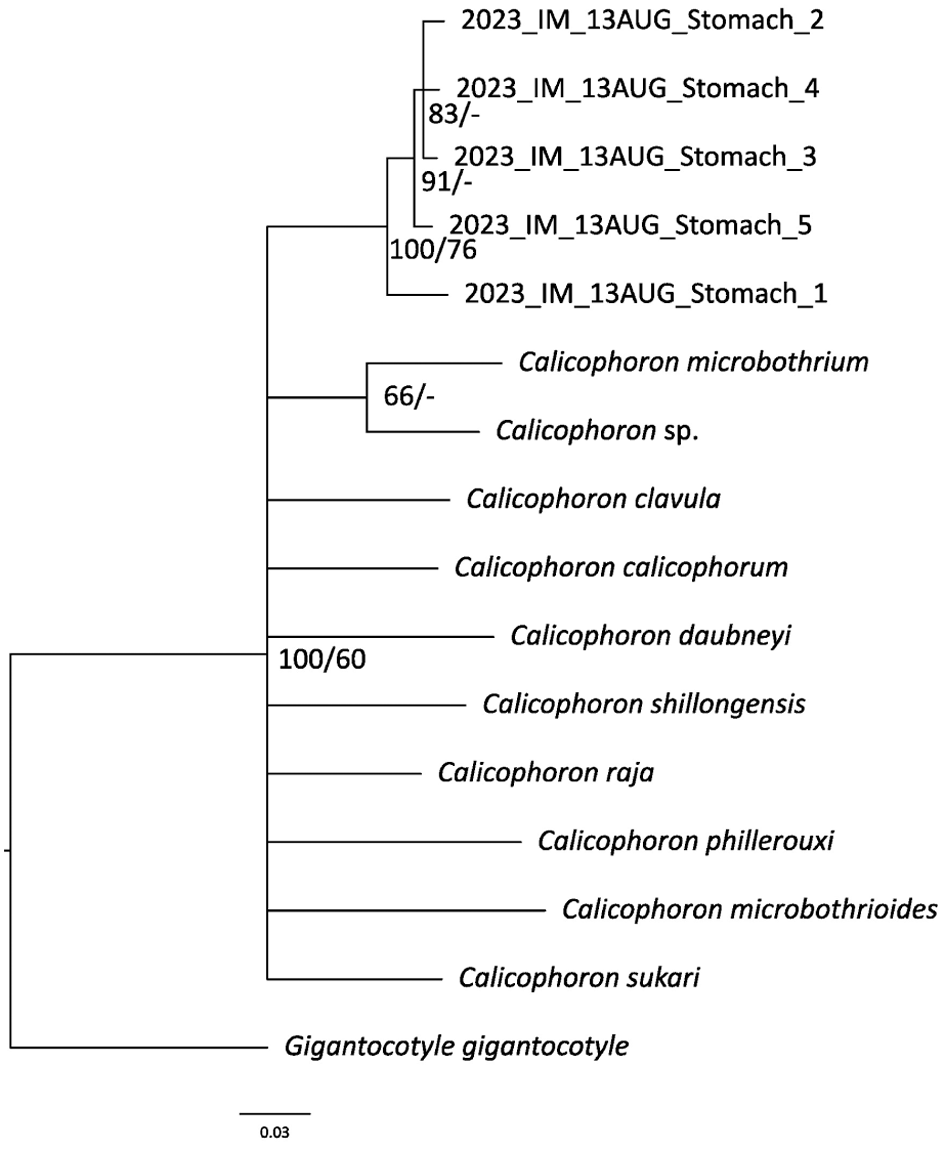


 Open access

