Introduction
The human branchial apparatus consists of five paired mesodermal arches separated by four pairs of endodermal and ectodermal pouches/clefts. Branchial fistulae are formed from the remnants of pouches and clefts following rupture of the interposing branchial plate.Reference Chaudhary, Gupta, Motwani and Kumar1 More than 90 per cent of branchial anomalies arise from the second arch, and 8 per cent from the first arch. Anomalies of the third and fourth branchial apparatus, although rare, usually present as sinuses or incomplete fistulae of the pyriform sinus, or as recurrent, suppurative thyroiditis.Reference Jaka and Singh2 Complete fistulae of the third and fourth branchial arches are extremely rare.Reference Jaka and Singh2, Reference James, Stewart, Warrick, Tzifa and Forte3
We hereby report a case of complete congenital third branchial arch fistula with an extremely unusual site of cervical opening.
Case report
A nine-year-old boy presented with a history of an opening on the left side of the anterior neck with intermittent mucoid discharge since birth. Recently, the discharge had increased on taking food, associated with painful swelling around the opening, of one week's duration. A similar episode had occurred one year previously. There was no history of trauma, spontaneous rupture or surgery, or of dysphagia, hoarseness, halitosis, recurrent cough or respiratory distress. The patient had no other openings or swellings in his neck. There was no similar history within the family.
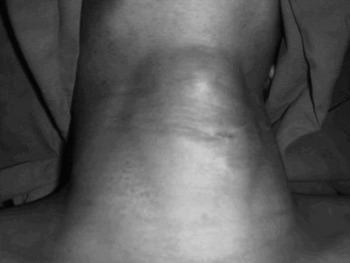
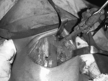
On examination, a 2 mm diameter opening was seen on the middle third of the anterior neck, 1.5 cm left of the midline (Figure 1). The skin around the opening was hyperpigmented, with mild erythema and oedema. The opening of the tract moved on swallowing but not on tongue protrusion. The rest of the physical examination was unremarkable.
Clinical photograph of the third branchial fistula; the external skin opening is seen on the middle third of the anterior neck, 1.5 cm left of the midline.

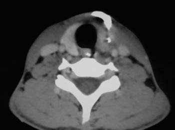
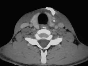
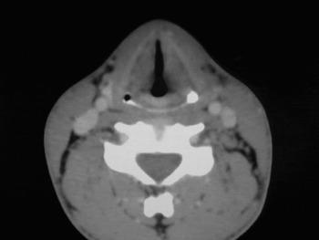
Computed tomography (CT) and fistulography revealed a patent, fistulous tract extending from the neck skin (Figure 2) through the left lobe of the thyroid gland (Figure 3) to the left pyriform sinus, where spillage of contrast was noted (Figure 4). No other neck abnormality was detected. Ultrasonography revealed a heterogeneous, hypoechoic area involving the soft tissues of the anterior neck at the level of the thyroid on the left side. Direct hypopharyngoscopy showed an opening at the lateral wall of the pyriform sinus and not at the apex. Thyroid function tests were normal. An iodine-131 scan revealed decreased uptake in the left thyroid lobe.
Axial computed tomography scan following external contrast injection, revealing a collection of contrast material just beneath the skin opening.

Axial computed tomography scan showing the passage of contrast along the tract, which passes through the left lobe of the thyroid from the external injection site.

Axial computed tomography scan revealing spillage of contrast into the pyriform sinus following injection through the external neck opening.

Under general anaesthesia, a transverse, elliptical incision was made around the external opening and the dissection continued carefully on either side of the tract. The tract passed through the left lobe of the thyroid gland up to the superior pole. A left hemithyroidectomy was performed in continuity with the tract.
At this stage, under rigid laryngoscopic guidance, 1 per cent methylene blue was injected into the aperture of the pyriform sinus. This made localisation of the tract easier during dissection (Figure 5). The tract passed behind the common and internal carotid arteries to enter the thyrohyoid membrane above the hypoglossal nerve and open into the base of the pyriform sinus.
Intra-operative photograph showing the appearance of 1 per cent methylene blue in the tract following injection into the aperture of the pyriform sinus under rigid endoscopic guidance, and after left hemithyroidectomy in continuity with the tract.

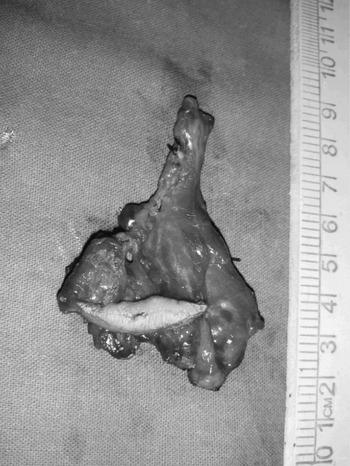
The entire tract was excised up to the pyriform sinus (Figure 6). Deeper pharyngeal mucosal defects were closed efficiently using purse string sutures, and the muscle layers were closed with strengthened sutures to avoid pharyngeal leakage. A suction drainage tube was inserted and kept in place for 48 hours.
Surgical specimen following third branchial fistula excision up to the level of the pyriform sinus, coupled with left hemithyroidectomy.

The post-operative course was uneventful.
Histopathological examination of excised tissue showed a hyperplastic epidermis with acute and chronic inflammation, and with ulceration and extensive granulation tissue replacing the dermis.
The patient was followed up regularly for 22 months showed no recurrence.
Discussion
In 1832, Ascherson published the first description of a branchial cleft lesion.Reference Nicoucar, Giger, Pope, Jaecklin and Dulguerov4 In 1933, Raven described an incomplete fistula of the third branchial arch in a neonate with left cervical swelling who had died of asphyxia. On autopsy, Raven found a congenital tract extending from the pyriform sinus to the cervical cyst.Reference Nicoucar, Giger, Pope, Jaecklin and Dulguerov4–Reference Kubota, Suita, Kamimura and Zaizen6 Kubota et al. were the first to make the clinical observation that pyriform sinus fistulae may cause suppurative thyroiditis in infants and children.Reference Kubota, Suita, Kamimura and Zaizen6
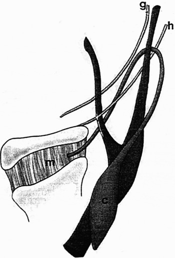
Figure 7 shows third branchial cleft anomaly. The cyst (C) is posterior to the sternocleidomastoid muscle, and the tract ascends posterior to the internal carotid artery. It then courses medially to pass between the hypoglossal nerve (H) and the glossopharyngeal nerve (G). It pierces the thyroid membrane (M) to enter the pyriform sinus.

Third and fourth branchial arch cysts and fistulae are very rare.Reference Chaudhary, Gupta, Motwani and Kumar1, Reference Nicoucar, Giger, Pope, Jaecklin and Dulguerov4 They are formed during the fifth week of gestation when the second arch enlarges and grows over the second, third and fourth clefts, forming an ectodermal depression named the cervical sinus. Failure of complete obliteration of the sinus results in branchial cyst formation. During embryological development, the third and fourth branchial pouches are connected to the pharynx by the pharyngobranchial duct, which degenerates by the seventh week. Persistence of this duct results in formation of a sinus tract that communicates with the pyriform sinus.Reference Chaudhary, Gupta, Motwani and Kumar1, Reference Liberman, Kay, Emil, Flageole, Nguyen and Tewfik7
The course of a third branchial arch fistula presumably originates from the cephalic region of the pyriform fossa, anterior to the fold made by the internal laryngeal nerve, as in our case.Reference Nicoucar, Giger, Pope, Jaecklin and Dulguerov4, Reference Mali and Prabhakaran5 The fistula then pierces the thyrohyoid membrane cranial to the superior laryngeal nerve and passes over the hypoglossal nerve. It courses behind the internal carotid artery and runs superficial to the superior laryngeal nerve, then passes through the platysma to terminate, commonly along the anterior border of the sternocleidomastoid muscle at the junction of the upper two-thirds and lower one-third of the neck.Reference Jaka and Singh2, Reference Mali and Prabhakaran5 In our patient, the tract mostly followed the classical course described above; however, it passed through the left lobe of the thyroid gland to end on the middle third of the neck just left of the midline, which is extremely rare.
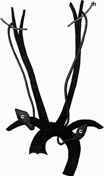
Figure 8 shows fourth branchial cleft anomaly. The cysts (C) are located anterior to the aortic arch on either side. The tract hooks either the subclavian or the aortic arch, depending on the side, and ascends to loop over the hypoglossal nerve (H).

Fourth branchial fistulae follow a ‘two-loop course’, originating from the caudal end of the pyriform fossa, posterior to the fold made by the internal laryngeal nerve, and coursing inferiorly along the tracheoesophageal groove, posterior to the thyroid gland, into the mediastinum, to loop around the aorta (if on the left side) or the subclavian artery (if on the right). The fistula then ascends cephalad to pass over the hypoglossal nerve before piercing the platysma to end on the cervical neck.Reference Chaudhary, Gupta, Motwani and Kumar1, Reference Nicoucar, Giger, Pope, Jaecklin and Dulguerov4, Reference Mali and Prabhakaran5
First branchial arch anomalies are rare, occurring in only 8 per cent of cases. Second arch anomalies account for more than 90 per cent of branchial arch lesions. Third and fourth branchial arch anomalies have been described in all age groups, even in utero, with an incidence of 2–8 and 1–2 per cent, respectively.
The clinical presentation of branchial arch anomalies depends in neonates on the presence of a mass effect and respiratory compromise, and in older patients on the presence of recurrent infection secondary to a persistent sinus tract.Reference Mali and Prabhakaran5, Reference Liberman, Kay, Emil, Flageole, Nguyen and Tewfik7
Branchial arch anomalies can be fatal in neonates due to the risk of tract enlargement during swallowing, with consequent tracheal compression and airway compromise.Reference Jaka and Singh2, Reference Liberman, Kay, Emil, Flageole, Nguyen and Tewfik7
The left-sided predominance of these lesions could be due either to asymmetrical derivatives of the branchial arch, or to suppressed embryogenesis of the ultimobranchial body on the right side.Reference Chaudhary, Gupta, Motwani and Kumar1, Reference Mali and Prabhakaran5
An external sinus opening, or pseudofistula, may develop at the site of previous surgical drainage, rupture or incomplete excision.Reference James, Stewart, Warrick, Tzifa and Forte3 In our case, no such history was present, confirming the congenital nature of the unusually sited cervical neck opening. Since our patient's external opening site was at the level of the left thyroid lobe, intrauterine rupture of a cervical cyst of branchial origin was also a possibility.
A barium swallow investigation is diagnostic in patients with a branchial fistula, and should be performed in any case of suppurative neck inflammation of unknown origin. The tract may not be delineated clearly in the presence of active infection, in which case CT scanning is preferable. A repeated barium swallow when the infection is quiescent may be useful in such circumstances.Reference Mali and Prabhakaran5, Reference Garrel, Jouzdani, Gardiner, Makeieff, Mondain and Hagen8
A CT scan performed after barium swallow or contrast injection through the cannulated external opening will be more useful in delineating the entire tract than a plain CT scan.Reference Mali and Prabhakaran5 The presence of air near the lesser horn of the thyroid cartilage on CT scanning is considered pathognomonic even during inflammation; however, this sign is inconstant.Reference Garrel, Jouzdani, Gardiner, Makeieff, Mondain and Hagen8
In our patient, cannulation of the fistulous opening followed by contrast injection and CT scanning provided excellent delineation of the entire tract, with spillage of contrast into the left pyriform sinus.
The use of modified Valsalva and trumpet manoeuvres during CT scanning and ultrasonography enables air to be used as a contrast agent, tracing the course of the tract.Reference Nicoucar, Giger, Pope, Jaecklin and Dulguerov4, Reference Mali and Prabhakaran5 Air–fluid levels within the thyroid gland or surrounding soft tissues suggest abscess formation.Reference Mali and Prabhakaran5 Direct laryngoscopic examination and visualisation of the internal pyriform sinus opening with cannulation may also be useful for diagnosis.Reference Mali and Prabhakaran5, Reference Garrel, Jouzdani, Gardiner, Makeieff, Mondain and Hagen8
Complete excision of the fistulous tract with partial or hemithyroidectomy, during a quiescent period, is the treatment of choice.Reference Chaudhary, Gupta, Motwani and Kumar1–Reference Nicoucar, Giger, Pope, Jaecklin and Dulguerov4 Adequate exposure may be provided by the Woodman approach, or by retracting or excising a vertical strip of the posterior border of the thyroid ala. Some investigators believe that complete disconnection of the tract from the pyriform sinus at the level of the thyrohyoid membrane is sufficient.Reference Mali and Prabhakaran5 Intra-operative cannulation, with or without injection of 1 per cent methylene blue into the pyriform sinus opening under endoscopic or laryngoscopic guidance, is effective in localising the course of the fistulous tract.Reference James, Stewart, Warrick, Tzifa and Forte3, Reference Nicoucar, Giger, Pope, Jaecklin and Dulguerov4
• This paper describes a rare case of a complete congenital third branchial arch fistula in a nine-year-old boy
• Anomalies of the third and fourth branchial apparatus, although rare, usually present as sinuses or incomplete fistulae of the pyriform sinus, or as recurrent, suppurative thyroiditis
• In the presented patient, computed tomography fistulography and injection of dye into the pyriform sinus were used to delineate the tract intra-operatively
• Recurrence is possible many years after surgery, usually following inadequate excision or excision during an acute episode of suppurative thyroiditis
Post-operative complications are more common in children younger than eight years of age, and include temporary vocal fold paralysis, salivary fistula and wound infection.Reference Nicoucar, Giger, Pope, Jaecklin and Dulguerov4 Recurrence is possible many years after surgery, usually following inadequate excision or excision during an acute episode of suppurative thyroiditis. Hence, long term follow up is necessary.Reference Mali and Prabhakaran5 Some authors suggest the use of endoscopic electrocauterisation and endoscopic fibrin glue after simple cervical drainage. Recently, chemocauterisation with trichloroacetic acid has been reported.Reference Jaka and Singh2, Reference Liberman, Kay, Emil, Flageole, Nguyen and Tewfik7, Reference Garrel, Jouzdani, Gardiner, Makeieff, Mondain and Hagen8