Introduction
Motor neuron disease refers to a spectrum of disorders resulting from degeneration of the upper or lower motor neurons or both. There are numerous motor neuron diseases with overlapping clinical features, sometimes making it difficult to properly identify and classify subpopulations of the condition. Amyotrophic lateral sclerosis is the most common form of motor neuron disease. This condition is a devastating, untreatable neurodegenerative disorder of the neurons of the motor cortex and anterior horn cells of the spinal cord and of the brainstem motor nuclei, which leads to progressive muscle weakness, atrophy and pyramidal signs.Reference Leigh, Abrahams, Al-Chalabi, Ampong, Goldstein and Johnson1
Amyotrophic lateral sclerosis occurs both sporadically (i.e. sporadic amyotrophic lateral sclerosis) and, in about 5–10 per cent of cases, as a familial disease (i.e. familial amyotrophic lateral sclerosis).Reference Leigh, Abrahams, Al-Chalabi, Ampong, Goldstein and Johnson1 It has been well known since 1993.Reference Rosen, Siddique, Patterson, Figlewicz, Sapp and Hentati2 Mutations of Cu-Zn superoxide dismutase type 1 are responsible for familial amyotrophic lateral sclerosis. The degeneration of motor neurons seen in sporadic amyotrophic lateral sclerosis is also related to oxidative stress, deficient neurotrophic factors, glutaminergic excitotoxicity, and immunological or viral agents, as well as to excess heavy metals, environmental toxins and smoking. The mechanism underlying the characteristic selective degeneration and death of motor neurons in amyotrophic lateral sclerosis remains unknown. However, the aetiology of sporadic amyotrophic lateral sclerosis is likely to be multifactorial.Reference Strong3
The typical onset of amyotrophic lateral sclerosis is between the fifth and seventh decade.Reference Leigh, Abrahams, Al-Chalabi, Ampong, Goldstein and Johnson1 The diagnosis of amyotrophic lateral sclerosis is usually based on the clinical neurological examination. This examination confirms the presence of symptoms characteristic for the combination of upper and lower motor neuron degeneration in different anatomical regions of the body (i.e. bulbar, cervical, thoracic and lumbar regions), as well as in the progressive course of the disease, according to the criteria recommended by the World Federation of Neurology.Reference Brooks4
The clinical diagnosis of amyotrophic lateral sclerosis is always confirmed by electromyography (EMG).Reference Brooks4 Neuroimaging has been used as a measure of motor neuron degeneration and corticospinal tract degeneration, with variable success, as a diagnostic aid in amyotrophic lateral sclerosis.Reference Kalra and Arnold5 Routine magnetic resonance imaging has not been useful in identifying new amyotrophic lateral sclerosis cases, although it can assist in ruling out diseases that mimic the condition.Reference Brooks4
At present, the clinical symptoms of the disorders, such as muscle atrophy, weakness or fasciculations, spasticity, and speech and swallowing disturbances, are used for diagnostic procedures.
There are two different types of onset of amyotrophic lateral sclerosis: limb and bulbar. Limb onset is more common; a combination of upper and lower motor neuron degeneration signs (i.e. spasticity, brisk reflexes and Babinski sign, together with muscular atrophy, weakness and fasciculation) are observed in the extremities, enabling neurological diagnosis.
However, in about 25 per cent of amyotrophic lateral sclerosis cases, the disease presents initially with bulbar symptoms and signs (i.e. bulbar onset). This presentation is associated with a worse prognosis – the average survival is around two years.Reference Hillel and Miller6, Reference Mitsumoto7 The bulbar presentation of amyotrophic lateral sclerosis includes symptoms that originate from the disabled oropharyngeal muscles, such as difficulty with speech, swallowing, salivation and aspiration. Hypernasality, dysphonia, breathiness, articulation defects and voice harshness, as well as difficulty in swallowing, should all be seen as possible early signs of bulbar onset amyotrophic lateral sclerosis. It may fall to the otolaryngologist to recognise the significance of these signs.Reference Strand, Miller, Yorkston and Hillel8–Reference Watts and Vanryckeghem10
The laryngological presentation of amyotrophic lateral sclerosis has been seldom described in the literature,Reference Hillel, Dray, Miller, Yorkston, Konikow and Strande9–Reference Isozaki, Hayashi, Hayashida, Oda and Hirai13 particularly with regards to the comparison of laryngological abnormalities in the bulbar versus limb onset conditions. Some recent studies have emphasised the importance of laryngological examination in the diagnosis of amyotrophic lateral sclerosis.Reference Hillel, Dray, Miller, Yorkston, Konikow and Strande9, Reference Chen and Garrett14
In the present study, we investigated the profile of laryngological abnormalities in bulbar and limb onset amyotrophic lateral sclerosis patients.
Material and methods
Thirty-five consecutive amyotrophic lateral sclerosis patients were diagnosed according to the El Escorial criteriaReference Brooks4 at the Krakow Motor Neuron Disease Centre in the department of neurology, Jagiellonian University, Krakow, between January 2004 and January 2005. Based on the initial clinical presence or absence of bulbar signs, the patients were divided into two groups: bulbar onset and limb onset.
Bulbar and/or pseudobulbar signs occurred in all of the bulbar onset patients, e.g. dysphonia, dysarthria, swallowing difficulties and emotional lability. On neurological examination, there was an increase in brainstem reflexes and a loss of lingual motor function, or, alternately, fasciculation and atrophy of tongue muscles and a decrease in brainstem reflexes.
Limb onset patients presented with signs of upper and lower motor neuron involvement in the limbs only, such as limb muscle weakness and atrophy, fasciculations, loss of dexterity, brisk tendon reflexes, and pathological reflexes. None of these patients presented with bulbar symptoms or signs at the point of laryngological examination.
Laryngological examinations were conducted in all these patients by the same otolaryngologist in the otolaryngology department of Jagiellonian University, Krakow. Examinations of the ears, nose, throat and larynx were performed. During the inspection of the oral cavity and the throat, attention was paid to the mobility of the tongue and the soft palate and to palatal symmetry, as well as to palatal and pharyngeal reflexes. The laryngological evaluation was carried out by means of indirect laryngoscopy (using a mirror) and fibroscopy (using a flexible fibroscope; Olympus America, Melville, NY). Once a good view of the vocal folds was obtained, the patient was asked to vocalise, cough and forcibly exhale. A stroboscopic examination (using a videostroboscope instrument; Karl Storz, Tuttlingen, Germany) was performed in several individuals (five out of the 35) to assess the character of vocal fold vibrations, their mobility and phonatory closure. In each case, the examinations were supplemented with perceptual phoniatric assessment.
A computer database was used to record epidemiological data (i.e. the age of onset, time and type of initial amyotrophic lateral sclerosis symptoms, and duration of the disease) as well as details of the neurological and laryngological examinations. The results were separately analysed for both groups of amyotrophic lateral sclerosis patients.
Results
Of the amyotrophic lateral sclerosis patients, 24 were bulbar onset (13 women and 11 men; age range 49–73 years; mean age, 54.5±10.4 years) and 11 were limb onset (four women and seven men; age range 47–69 years; mean age, 51.7±9.3 years). The disease duration, calculated from the time of first symptom onset, ranged from four to 28 months in the bulbar onset patients (mean duration, 9.5±14.9 months) and from nine to 45 months in the limb onset patients (mean duration, 16.4±13.7 months).
In the bulbar onset patients, analysis of neurological findings as well as inspection of the oral cavity, face and lips allowed a subdivision into two subgroups: patients with predominantly lower motor neuron presentation, and those with predominantly upper motor neuron presentation.
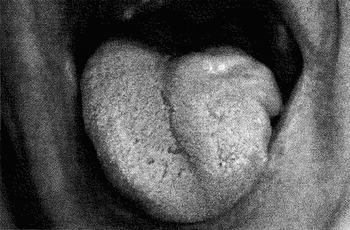
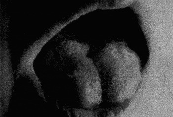
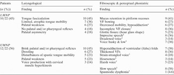
Patients with predominantly lower motor neuron presentation (10 out of the 24 bulbar onset cases) presented with an atrophic tongue with limited mobility (Figures 1 and 2), palatal asymmetry, and weak or no palatal or pharyngeal reflexes, as well as retention of mucus in the piriform recesses. However, a brisk jaw jerk was observed in some cases. Detailed fibroscopic examination revealed that the vocal folds were smooth, with decreased mobility and adduction during the respiratory phase (to the intermedial position). During phonation, some patients showed lack of complete vocal fold closure, with an hour glass shaped fissure of the glottis. In these cases, phoniatric perceptual examination revealed a diminished voice, sometimes husky and low. Table I shows details of the fibroscopic examinations of patients with a predominantly lower motor neuron presentation.
Hemiatrophy and lateral deviation of the tongue in bulbar onset amyotrophic lateral sclerosis case.

General atrophy of the tongue with loss of tongue movements in bulbar onset amyotrophic lateral sclerosis case.

Bulbar onset amyotrophic lateral sclerosis patients: examination results

* Respiratory phase; †on phonation; ‡on perceptual phoniatric examination. LMNP=predominantly lower motor neuron presentation; VF=vocal fold; UMNP=predominantly upper motor neuron presentation
The remaining 14 patients were classified as predominantly upper motor neuron presentation. In two of these, laryngological examination was not performed due to the brisk palatal and pharyngeal reflexes and mandibular clonus. In the remaining 12 patients, the predominant features were brisk palatal and pharyngeal reflexes or jaw jerk, and disturbances of spastic tongue mobility. However, we also observed slowness of tongue movements, with fasciculation and minor atrophy in some cases. During fibroscopic examination of the vocal folds, slight disturbances of their mobility were seen, with hypoadduction of the vocal folds and hyperadduction of the ventricular folds. The vocal folds were thicker, and the voice was produced by cervical muscle hyperkinesis. The perceptual phoniatric examination revealed hypernasality, strain-strangled voice and harshness. Spasmodic dysphonia was diagnosed in one of these patients (see Table I for details).
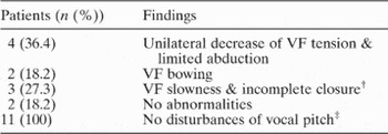
In the limb onset amyotrophic lateral sclerosis patients, a detailed fibroscopic evaluation of vocal fold mobility revealed slight deviation from normal in nine out of 11 patients. Slowness of both vocal folds and lack of complete closure during phonation were detected in three patients. There was a unilateral decrease in tension and mobility of the vocal fold in four cases and vocal fold bowing in two cases (see Table II for details). However, perceptual phoniatric assessment of limb onset patients revealed no disturbance of vocal pitch.
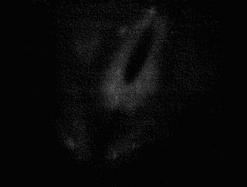
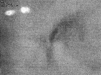
Bulbar onset amyotrophic lateral sclerosis patient with severe dysarthria. Videostroboscopy examination shows lack of complete vocal fold closure during phonation.

Limb onset amyotrophic lateral sclerosis patient. Videostroboscopy examination shows supraglottic hyperfunction and vocal fold bowing.

Limb onset amyotrophic lateral sclerosis patients: fibroscopic and perceptual phoniatric examination results

* n=11. †On phonation; ‡on perceptual phoniatric examination. VF=vocal fold
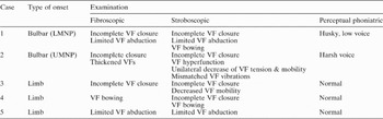
Fibroscopic examination was supplemented by stroboscopy in two bulbar onset and three limb onset patients. The details are given in Table III and Figures 3 and 4.
Selected amyotrophic lateral sclerosis patients: examination results

LMNP=predominantly lower motor neuron presentation; VF=vocal fold; UMNP=predominantly upper motor neuron presentation
Discussion
The above results of laryngological examination in amyotrophic lateral sclerosis patients confirmed the occurrence of vocal fold dysfunction (i.e. vagus nerve lesion) in bulbar onset patients. This dysfunction was accompanied by speech changes, as per perceptual phoniatric assessment, in all bulbar onset patients studied. However, we also discovered ‘silent’ vocal fold dysfunction (such as uni- or bilateral slowness of vocal folds, decrease in tension, limited abduction, vocal fold bowing and incomplete closure) in limb onset patients without any overt bulbar signs. Phoniatric examination did not detect any voice changes in these cases.
In amyotrophic lateral sclerosis patients, it is well known that bulbar dysfunction affecting the control and strength of the pharyngeal and laryngeal muscles (due to corticobulbar pathway or brainstem degeneration) may cause the most significant clinical problems, and may contribute to complications such as loss of communication, dysphagia and aspiration of secretions.Reference Strand, Miller, Yorkston and Hillel8, Reference Watts and Vanryckeghem10 Although the first clinical presentation of amyotrophic lateral sclerosis is usually either at the limb or bulbar level only,Reference Tomik, Krupinski, Glodzik-Sobanska, Bala-Slodowska, Wszolek and Kusiak15–Reference Przedborski17 some data suggest that degeneration of the motor neurons of the motor cortex, brainstem and spinal cord can occur simultaneously in amyotrophic lateral sclerosis cases. Our finding of vocal fold dysfunction in limb onset patients supports this hypothesis.
It is important to emphasise that vocal fold abnormalities were previously seldom investigated in amyotrophic lateral sclerosis patients, although epiglottic obstruction of the airway was recognised.Reference Robbins, Scanlan and Brooks18 Polkey et al.Reference Polkey, Lyall, Green, Leigh and Moxham19 described two patients with vocal fold abnormalities: one with unilateral vocal fold paralysis, and the other with unexpected bilateral fold closure during both rapid inspiratory and expiratory manoeuvres. A few reports of direct and indirect laryngeal observation described the following: aperiodic vocal fold vibration; paradoxical vocal fold adduction patterns; hyperadduction of the ventricular folds; and hypoadduction of the vocal folds, as well as phonatory disruptions.Reference Watts and Vanryckeghem10–Reference Isozaki, Hayashi, Hayashida, Oda and Hirai13, Reference Klasner, Yorkston and Strand20, Reference Aronson, Ramig, Winholtz and Silber21 Interesting results were presented by Isozaki et al.;Reference Isozaki, Hayashi, Hayashida, Oda and Hirai13 these authors investigated the mechanism of vocal fold abductor paralysis in different neurodegenerative diseases and histologically examined the intrinsic laryngeal muscles (i.e. the cricothyroid, the interarytenoid and the posterior cricoarytenoid muscles), obtained during autopsy. They found the totally paralytic form of vocal fold abductor paralysis in 10 amyotrophic lateral sclerosis patients and showed that all three muscles had developed neurogenic atrophy.
The importance of laryngological and stroboscopic examination in amyotrophic lateral sclerosis patients was stated by Hillel et al. in 1999Reference Hillel, Dray, Miller, Yorkston, Konikow and Strande9 and by Chen and Garrett in 2005.Reference Chen and Garrett14 Until the latter publication, there had been no systematic studies focusing on this area. It has been pointed out that patients with early symptoms of bulbar amyotrophic lateral sclerosis are usually referred to the otolaryngologist without a diagnosis.Reference Watts and Vanryckeghem10 Thus, a physical examination, including the vocal folds, should be undertaken, together with careful examination of speech quality.Reference Watts and Vanryckeghem10 Dysarthria, dysphagia, tongue fasciculation and incomplete vocal fold closure were common findings in Chen and Garrett's study of 19 amyotrophic lateral sclerosis patients initially presenting to an otolaryngologist.Reference Chen and Garrett14
Finally, in our study, the vocal fold examination was found to be abnormal in both bulbar and limb onset amyotrophic lateral sclerosis patients. This confirms that vocal fold dysfunction is a feature of established amyotrophic lateral sclerosis, and it emphasises the laryngological and phoniatric aspects of the diagnosis. We suggest that the standard laryngological examination, carefully conducted, may reveal signs of early dysfunction of the vagus nerves, thus demonstrating preclinical presentation of bulbar failure in limb onset amyotrophic lateral sclerosis patients.
• Laryngological evaluation of vocal fold changes in neurodegenerative disease such as amyotrophic lateral sclerosis is seldom described in the literature
• This study compared the profile of laryngological abnormalities in bulbar onset amyotrophic lateral sclerosis (with dysarthria and dysphagia) and limb onset amyotrophic lateral sclerosis (without dysarthria and dysphagia)
• In limb onset amyotrophic lateral sclerosis patients, there were signs of early, preclinical vocal fold dysfunction (i.e. vagus nerve lesion)
• The importance of laryngological evaluation in the early stages of limb onset amyotrophic lateral sclerosis is emphasised
In the light of the above, the otolaryngologist could be the primary diagnostician of amyotrophic lateral sclerosis. For example, in the Krakow Motor Neuron Disease database, out of a total of 630 amyotrophic lateral sclerosis patients, 22 presented with dysphonia, voice changes, dysarthria or swallowing problems during an initial evaluation by an otolaryngologist, before being referred for neurological examination, and amyotrophic lateral sclerosis was diagnosed in 19. Thus, we wish to make the case for the importance of laryngological evaluation in the early stages of limb onset amyotrophic lateral sclerosis.
Future directions
We have described the profile of laryngological abnormalities in amyotrophic lateral sclerosis patients with bulbar onset and limb onset of the disease. Based on our findings, we plan to perform stroboscopic examinations as well as a successive cycle of laryngological and fibroscopic assessments, in order to monitor the progression of vocal fold mobility changes as the disease advances.
Acknowledgement
The present results are the part of ongoing work supported by Jagiellonian grant 501/NKL/194/L. We would like to thank D A Figlewicz, Department of Neurology, University of Michigan, for critical reading of the manuscript.