

Introduction
Strenuous exercise is associated with an increase in oxygen consumption to provide fuel to working musculature through aerobic pathways. This increase potentially overwhelms the electron transport chain, causing electrons to leak into the mitochondrial space and react with components of the cell. These reactions give rise to reactive oxygen species (ROS) in the body, thereby causing an increase in oxidative stressReference Powers and Hamilton1. The generation of ROS during exercise is probably a cause of muscular disturbances, inflammation and pain in performance animalsReference Avellini, Chiaradia and Gaiti2. There is increased interest in providing supplemental dietary antioxidants to exercising subjects to reduce free radical levels in the body, thereby reducing the damage done to the animal by these substances. However, the benefits of these supplements are controversial. Popularly used measures of oxidative stress are often variables that are measurable in the blood. Very little work in horses has looked at muscle parameters that might be used to indicate oxidative stress. One such parameter is the protein carbonyl assay.
ROS are produced normally in skeletal muscle fibres in low amounts where they function during muscle contractilityReference Barreiro, Gea, Di Falco, Kriazhev, James and Hussain3. At elevated concentrations, ROS are neutralized by the intracellular antioxidant defence system. Accumulation of ROS disrupts muscle cellular functions including action potential conduction, excitation coupling, contractile proteins and mitochondrial respirationReference Barreiro, Gea, Di Falco, Kriazhev, James and Hussain3. Carbonyls result from oxidation of arginine, lysine, threonine or proline amino acids. Carbonylation of amino acids can occur in different ways. Hydroxyl radicals are highly reactive and thought to be generated in vivo by catalytic action of transition metals such as iron and copper, which will bind to specific sites of proteins and modify nearby amino acid residuesReference Goto, Nakamura, Radak, Nakamoto, Takahashi, Yasuda, Sakurai and Ishii4. Carbonyl groups also result from glycation/glycoxidation of lysine, cysteine or histidine amino acids with α- or β-unsaturated aldehydes formed during the peroxidation of polyunsaturated fatty acidsReference Barreiro, Gea, Di Falco, Kriazhev, James and Hussain3.
Oxidation of proteins causes them to lose their function and become targets for proteolytic degradationReference Radak, Kaneko, Tahara, Nakamoto, Ohno, Sasvari, Nyakas and Goto5. Goto et al. Reference Goto, Nakamura, Radak, Nakamoto, Takahashi, Yasuda, Sakurai and Ishii4 suggested that α-actin and myosin heavy chain are carbonylated in skeletal muscle. Proteins of the quadriceps muscles of rats trained for 4 weeks at 4000 m altitude oxygen pressure exhibited a significantly higher extent of carbonylation than untrained rats or rats trained at sea levelReference Radak, Kaneko, Tahara, Nakamoto, Ohno, Sasvari, Nyakas and Goto5. Proteins that showed a marked increase in signal intensity by Western blot were actins, as determined by their molecular weight and abundance. Actin and myosin heavy chains in the cells of human arteries and veins also are highly carbonylated, indicating that contractile proteins are susceptible to oxidationReference Goto, Nakamura, Radak, Nakamoto, Takahashi, Yasuda, Sakurai and Ishii4. However, while protein oxidation in rats trained at altitude did increase due to exercise, lipid peroxidation measured by thiobarbituric acid reactive substances and amount of lipid peroxides did not differ between animals exposed to sea level and high-altitude conditions. This suggests that protein oxidation occurs independently of lipid oxidation. Radak et al. Reference Radak, Takahashi, Kumiyama, Nakamoto, Ohno, Ookawara and Goto6 stated that oxidized proteins accumulated at a much higher rate (5–10% of total cellular proteins) than lipid or DNA ( < 0.1% at steady state level). Goto et al. Reference Goto, Nakamura, Radak, Nakamoto, Takahashi, Yasuda, Sakurai and Ishii4 also found a 40% increase in protein carbonyls in the lung following exhaustive running of rats.
The purpose of this study was first to determine whether protein carbonyls could be isolated in equine muscle tissue taken from horses pre- and post-exercise. The second objective was to determine whether exercising horses supplemented with vitamin E above 1989 NRC recommendations had decreased measures of oxidative stress in muscle tissue following a single strenuous bout of exercise, as measured by protein carbonyls.
Materials and methods
Animals
Eight untrained healthy Thoroughbred geldings between the ages of 3 and 10 years were used in a complete repeated measures (crossover) design. All horses were maintained between body condition scores of 4.5–5.0. All animal procedures were conducted within the guidelines of, and approved by, the University of Florida Institutional Animal Care and Use Committee.
Diets and adaptation
Prior to being divided into treatment groups, all horses were put on the control diet for 3 weeks and familiarized with the treadmill. A control baseline standard exercise test (SET) was performed at the end of the adaptation period. Subsequently, the animals were randomly divided into control and vitamin E-supplemented groups and acclimated to the respective diets for 7 weeks to achieve a consistent level of vitamin E in both blood and tissue. The horses on the control diet were fed a 12% crude protein textured oat-based feed at a rate of 1.25 kg feed per 100 kg body weight and ad libitum coastal Bermuda grass hay. Vitamin E content of the grain was 80 IU kg DM− 1, as recommended by the NRC7 for horses undergoing moderate to intense levels of work. The horses receiving vitamin E supplementation were given the same diet as the control but were supplemented with 3000 IU vitamin E day− 1 in the form of dl-α-tocopheryl acetate. The horses were fed the concentrate mixture twice daily. Once training began, the amount of concentrate fed was increased from 1.25% of body weight to 1.5% of body weight divided equally between the feedings. The treatment group received supplementation as a daily evening top dressing (68.2 g of a vitamin E premix containing 44 IU vitamin E g− 1). The premix was composed of dl-α-tocopheryl acetate mixed with a rice meal carrier. The control group received 68.2 g of a placebo (rice meal feed). In addition, all horses received ad libitum access to coastal Bermuda grass hay. The horses were housed at the University of Florida in 4 × 4 m stalls or in an adjacent dry lot turnout paddock. The use of the dry lot paddock eliminated grazing and helped control dietary intake. The horses were rotated daily in groups of four, with four being kept in stalls and four being kept in the turnout paddock. On days of blood sampling, all horses were kept in stalls beginning on the evening prior to collection to eliminate variations in temperature and climate.
Washout and crossover period
Prior to the crossover, all horses underwent a 15-week washout period during which they were turned out to pasture, fed the control diet and left untrained. The horses were then crossed over and the experiment repeated beginning with the adaptation period. Due to mechanical problems with the treadmill, the adaptation period for the second half of the experiment lasted 10 weeks rather than 7. During the extra 3 weeks, the horses were fed their respective diets and left untrained. Blood was collected both at the end of 7 weeks and at the end of 10 weeks of adaptation, and vitamin E analysis was run and statistically compared to ensure that there were no baseline differences between weeks 7 and 10 due to the difference in times on treatment diets. The horses followed the exercise schedule performed in the first half of the experiment.
Training period
The training and SET were conducted at the University of Florida Veterinary Medicine Teaching Hospital in a climate-controlled room equipped with a treadmill. The training period consisted of 8 weeks of gradually increasing workloads. Treadmill exercise was performed 3–4 days per week, depending on the availability of the treadmill. For the first 3 days, the horses trotted at 4 m s− 1 for 0.6 km on a flat surface, galloped at 8 m s− 1 for 1.0 km on a flat surface and cooled down by trotting at 4 m s− 1 for 0.6 km on a flat surface. The gallop distance was increased by 0.5 km every 3 days so that, by the end of 3 weeks, the horses were performing the following exercise: trotting at 4 m s− 1 for 0.6 km, galloping at 8 m s− 1 for 3.0 km and trotting at 4 m s− 1 for 0.4 km, all on a flat surface. Starting on week 5, the gallop phase was performed twice a week on a flat surface and twice a week on a 6° incline for the remainder of the study. A SET was performed at the end of each 8-week conditioning period. The protocol was as follows: 4 m s− 1 for 2 min on a flat surface, 8 m s− 1 for 1 min at a 6° incline, 9 m s− 1 for 1 min, 10 m s− 1 for 1 min, 11 m s− 1 for 1 min and 12 m s− 1 until they would not continue with moderate persuasion.
Tissue sampling
Following the eighth week of training, all horses performed a SET with muscle tissue collected before and after exercise. Muscle tissue (size of approximately 500 mg wet weight) was taken from the middle gluteal muscle (gluteus medius) by punch biopsy following local lidocaine anaesthesia. Muscle tissue was immediately divided for future analyses, placed in cryogenic tubes in liquid nitrogen and stored at − 80°C.
Vitamin E assay
Vitamin E was analysed in plasma samples at the initial date (following the adaptation period but prior to the beginning of any exercise) and on week 8 pre-exercise (Michigan State University Diagnostic Center for Population and Animal Health, East Lansing, MI, USA). Vitamin E was extracted from the plasma and analysed by high-performance liquid chromatography.
Muscle analyses
Myofibril preparation
To isolate myofibrils, frozen muscle was placed in a homogenization buffer on ice containing the following: 75 mM KCl, 10 mM Tris (pH 6.8), 2 mM EGTA, 2 mM MgCl2, 0.1 mM PMSF and 0.1% Triton X-100. After 1 h, the samples were homogenized with a polytron, centrifuged at 1000 × g, rinsed with homogenization buffer and centrifuged two more times, as describedReference Gordillo, Atalay, Roy and Sen8. The final pellet was resuspended in homogenization buffer, minus the Triton X-100 and EGTA, containing 50% glycerol. Myofibrils were stored frozen at − 20°C. Protein concentration was determined using the Bio-Rad Protein Assay (Bio-Rad Laboratories, Inc., Hercules, CA, USA).
Western blot
Protein carbonyls were measured in myofibrils by Western blot (Oxiblot™ Protein Oxidation Detection Kit, Chemicon International, Temecula, CA, USA). Briefly, 20 μg of proteins were electrophoretically separated through 10% polyacrylamide gels and transferred to nitrocellulose membrane. Membranes were blocked with 5% bovine serum albumin in PBS containing 0.1% Tween 20 (PBS-T) and incubated with antibodies according to the manufacturer's recommendations. Blots were washed with PBS-T and incubated with goat anti-mouse peroxidase (1:5000, Vector Labs, Burlingame, CA, USA). After a final wash with PBS-T, immune complexes were visualized by enhanced chemiluminescence and X-ray exposure. For the detection of ubiquitin, mouse anti-ubiquitin (1:1000, Santa Cruz Biotechnology, Sacramento, CA, USA) was used.
Statistical analysis
Analysis was performed with SAS version 6.12. The blood data for vitamin E were analysed by ANOVA with repeated measures over time according to treatments. Muscle data for carbonylation and ubiquitin were analysed by either the presence or absence of the compound on Western blots. Statistical analysis was run using SAS by Fischer's exact test using proc freq, with a chi-square value P < 0.05 being considered statistically significant.
Results
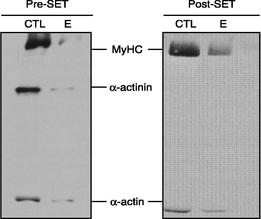
No differences in plasma vitamin E between control and vitamin E-supplemented horses were observed. However, three major carbonylated proteins were apparent prior to SET exercise, which corresponded to the approximate sizes of myosin heavy chain, α-actinin and α-actin (Fig. 1). The presence of carbonylation tended to be lower in vitamin E-supplemented horses (P < 0.07) in pre-exercise samples. Following the SET, only two major carbonylated proteins were apparent. These corresponded to the approximate sizes of myosin heavy chain and α-actin. The relative amount of carbonylated myosin heavy chain and actin remained lower for the vitamin E-supplemented group (P < 0.02). Ubiquitin was not detected in purified myofibrils at any point in time, indicating clearance of damaged muscle fibres by another mechanism. Ubiquitinated proteins were, however, present in the supernatant collected from isolate myofibrils, demonstrating that ubiquitin pathways are active.
Purified gluteus medius myofibrillar proteins are carbonylated. Myofibril proteins were purified from muscle biopsies obtained from the gluteus medius pre-SET and post-SET from control (CTL) and vitamin E (E)-treated horses. Equal amounts of protein were analysed by Western blot using mouse anti-carbonyl. Immune complexes were visualized by enhanced Chemiluminescence. Representative blots are shown. Predicted myosin heavy chain (MyHC), α-actinin and α-actin protein sizes are shown

Discussion
Strenuous exercise can lead to the formation of carbonylated proteins in muscle fibres. Carbonyl groups are formed as a consequence of ROS accumulation and may contribute to loss of contractile function. The presence and extent of carbonylation was examined in purified myofibrillar proteins isolated from the gluteus medius of control and vitamin E-supplemented horses before and after a single bout of strenuous exercise. Carbonylation was measured by Western blot. Statistical significance was based on either the presence or absence of carbonyl groups in all horses undergoing the SET. Three major carbonylated proteins were apparent prior to exercise that corresponded to the approximate sizes of myosin heavy chain, α-actinin and α-actin. The fact that carbonylation of the proteins tended to be lower pre-exercise and significantly lower post-exercise in vitamin E-supplemented horses indicates that potentially vitamin E did protect the working muscle from damage by free radicals.
It is unclear why carbonylation of pre-exercise samples was more extensive than that of post-exercise samples. Carbonylated proteins have been shown to be present in the gluteus medius of horses 4 h after exerciseReference Kinnunen, Hyyppa, Lappalainen, Oksala, Venojarvi, Nakao, Hanninen, Sen and Atalay9, Reference Kinnunen, Hyyppa, Lehmuskero, Oksala, Maenpaa, Hanninen and Atalay10. In the two studies by Kinnunen et al. Reference Kinnunen, Hyyppa, Lappalainen, Oksala, Venojarvi, Nakao, Hanninen, Sen and Atalay9, Reference Kinnunen, Hyyppa, Lehmuskero, Oksala, Maenpaa, Hanninen and Atalay10, carbonyls in plasma were shown to increase post-exercise, were the highest at 4 h post-exercise and had not returned to baseline values at 24 h post-exercise. In the muscle tissue, 4-HNE-modified proteins (marker of lipid peroxidation) did not increase between pre-exercise samples and at 4 h post-exercise. This demonstrates that measures of lipid peroxidation do not always follow the same trend as markers of protein oxidation. Protein carbonyl levels remained high at 24 h post-exercise as compared with pre-exercise samplesReference Kinnunen, Hyyppa, Lappalainen, Oksala, Venojarvi, Nakao, Hanninen, Sen and Atalay9. It is possible, therefore, that the more extensive carbonylated proteins in our pre-exercise samples actually represent carbonylation from chronic exercise training. These modified proteins may be rapidly eliminated upon commencement of exercise. The means by which muscle protein carbonyls are degraded is unknown, but probably does not involve the ubiquitin system.
Implications
The results of this preliminary study indicate that it is possible to isolate protein carbonyls from myofibrils of exercising horses. Additionally, vitamin E, a dietary antioxidant, does appear to influence the amount of carbonylation present in muscle tissue of horses undergoing intense exercise. Differences were also noticed in the extent of carbonylation between pre- and post-exercise samples in horses undergoing a bout of extreme exercise. Other studies have shown that measures of lipid peroxidation are not always correlated to measures of muscle oxidation. Future studies on oxidative stress and response to antioxidant treatment may want to consider the use of oxidative stress parameters in muscle tissue.