Introduction
Companion animals, such as dogs, frequently harbour intestinal parasites that can cause human infection. Although zoonotic parasites can cause significant morbidity in all groups of the human population, they are of particular importance in vulnerable groups, such as children, the elderly, and the immunocompromised (Robertson et al., Reference Robertson, Irwin, Lymbery and Thompson2000; Irwin, Reference Irwin2002). Among a number of zoonotic parasites that infect dogs, Toxocara canis and Ancylostoma sp. are of particular importance to humans (Macpherson, Reference Macpherson2005). Other zoonotic parasites include Echinococcus granulosus as well as emerging and re-emerging infections caused by Cryptosporidium spp. and Giardia spp. Toxocara canis does not normally establish intestinal infections in humans, but migrating larvae can cause visceral larva migrans (VLM), ocular larva migrans (OLM) or both. These can seriously compromise the health of children (Vidal et al., Reference Vidal, Sztajnbok and Seguro2003). Ancylostoma caninum had been reported as a parasite of humans causing a disease called eosinophilic enteritis (Croese et al., Reference Croese, Prociv, Maguire and Crawford1990; Prociv & Croese, Reference Prociv and Croese1990; Sanford & Prociv, Reference Sanford and Prociv1991; Croese et al., Reference Croese, Loukas, Opdebeeck, Fairley and Prociv1994).
The role of dogs as companion animals and the close relationship between humans and dogs, although offering significant benefits to many people, also represent a potential public health risk, since natural transmission of parasitic infections from dogs to humans may occur, directly or indirectly via environmental factors. All kinds of dogs (owned and stray dogs) are involved in transmission, even if the particular implication of each population is not clearly established (Eguia-Aguilar et al., Reference Eguia-Aguilar, Cruz-Reyes and Martinez-Maya2005).
Several studies of canine intestinal parasites have been reported in Nigeria and many other parts of the world (Ugochukwu & Ejimadu, Reference Ugochukwu and Ejimadu1985; Onyenwe & Ikpegbu, Reference Onyenwe and Ikpegbu2004; Ramirez-Barrios et al., Reference Ramirez-Barrios, Barboza-Mena, Munoz, Angulo-Cubillan, Henandez, Gonzalez and Escalona2004; Martinez-Moreno et al., Reference Martinez-Moreno, Hernandez, Lopez-Cobos, Becerra, Acosta and Martinez-Moreno2007; Sowemimo & Asaolu, Reference Sowemimo and Asaolu2008). However, current information on prevalence and intensity is essential for development and modification of control measures in animal and public health. The aim of this investigation was to determine the prevalence and intensity of gastrointestinal parasites of dogs in Ile-Ife, a peri-urban community in south-western Nigeria.
Materials and methods
Faecal sampling
The study was carried out in Ile-Ife, which has been described in my earlier report (Sowemimo, Reference Sowemimo2007). Faecal samples were collected randomly from 269 dogs between January and December 2004. Information was obtained on the approximate age, sex, breed, and mode of life of each dog, as well as the occupation of the dog's owner. Samples were placed in clean 30 ml bottles. About 10 g of faeces from each dog were mixed thoroughly with 10% aqueous formaldehyde for preservation. Samples were examined for parasite eggs in the laboratory by means of the modified Kato–Katz procedure (Forrester & Scott, Reference Forrester and Scott1990). Each parasite egg was identified using established structural and morphometric criteria (Bowman, Reference Bowman1999). In addition to qualitative diagnosis, an indirect measure of parasite intensity was obtained by counting eggs, expressed as eggs per gram of faeces (epg).
Statistical analysis
Statistical tests were performed using SPSS 11.0 (SPSS Inc., Chicago, Illinois, USA). Chi-squared tests were used to study the relationship between parasite prevalence and host age, sex, breed and mode of life. Mann–Whitney U tests were used to explore the relationship between egg intensity and sex and mode of life, while Kruskal–Wallis tests were used for variables with more than two levels, i.e. the relationship between the egg intensity and host age and breed.
Results
The overall prevalence of infection with intestinal parasites was 55.0%. Eggs of five species of nematodes and two species of cestodes were identified. The nematodes included Toxocara canis, Ancylostoma sp., Toxascaris leonina, Trichuris vulpis and Uncinaria stenocephala, while the cestodes were Dipylidium caninum and Taenia sp. The most prevalent intestinal parasite was the hookworm, Ancylostoma sp. (34.6%), followed by the ascarid worm, T. canis, with a prevalence of 33.8%. The prevalences of other intestinal parasites were T. leonina 3.3%, T. vulpis 3.7%, D. caninum 4.1%, U. stenocephala 0.7% and Taenia sp. 1.1%.
The overall mean intensities ( ± SEM) for the various intestinal parasites were T. canis 393.8 ± 83.4, Ancylostoma sp. 101.5 ± 32.8, T. leonina 14.3 ± 7.9, T. vulpis 3.4 ± 1.5, U. stenocephala 0.2 ± 0.2, D. caninum 2.2 ± 0.8 and Taenia sp. 5.4 ± 4.7.
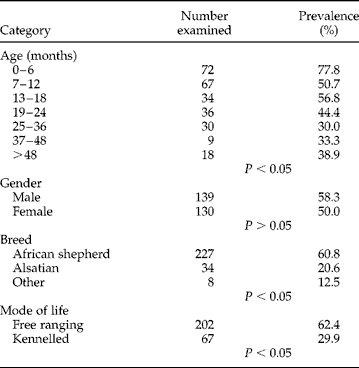
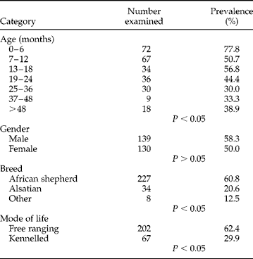
The prevalence of intestinal parasites was significantly higher (P < 0.05) in dogs less than 6 months old compared to dogs of older age groups. However, there was no significant difference in prevalence between male and female dogs (P>0.05) (table 1). The prevalence of intestinal parasites was also significantly higher (P < 0.05) in free-ranging dogs (62.4%) than in kennelled dogs (29.9%). Similarly, the prevalence was significantly higher (P < 0.05) in African shepherd dogs (60.8%) than in Alsatians (20.6%) and other exotic breeds (12.5%) (table 1).
Prevalence (%) of intestinal parasites relative to host age, gender, breed and mode of life.
Prevalence and intensity of Ancyclostoma sp
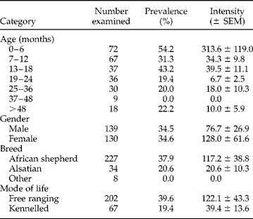
The prevalence of Ancylostoma sp. in dogs aged 0–6 months was comparable with that in dogs of age 13–18 months, but it was significantly higher (P < 0.05) than that in dogs of age 7–12 months and above 18 months. However, the intensity of infection was significantly higher (P < 0.05) among dogs of age 0–6 months than in older age groups (table 2). The prevalence of Ancylostoma sp. did not show any significant differences between male (34.5%) and female (34.6%) dogs (P>0.05). Similarly, there was no significant difference in the intensity of infection between females (128.0 ± 61.6) and males (76.7 ± 26.9) (P>0.05).
Prevalence (%) and intensity of Ancylostoma sp. relative to host age, gender, breed and mode of life.
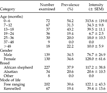
The prevalence and intensity of Ancylostoma sp. infection were significantly higher in African shepherd dogs than in Alsatian or other exotic breeds (P < 0.05) (table 2). Also, the prevalence and intensity of Ancylostoma sp. were significantly higher (P < 0.05) in free-ranging dogs than in kennelled dogs (table 2).
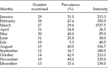
The prevalence and intensity of Ancylostoma sp. recorded in the different months for the period of study are shown in table 3. The pattern of prevalence was such that the prevalence of infection decreased from April until it reached the lowest level in July and then rose in August (this period corresponds to the rainy season, i.e. April–September). Similarly, the prevalence of infection also decreased between October and December and then rose from January to February (this period corresponds to the dry season, i.e. October–March). The overall prevalence during the rainy season (N = 138) was 33.3% while the overall prevalence during the dry season (N = 131) was 35.9%; however, there was no significant difference between the prevalences of the two seasons (P>0.05).
Monthly variation in the prevalence (%) and intensity of Ancylostoma sp. in 2004.
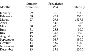
The pattern of intensity was such that lower values were recorded between April and July while higher values were recorded between August and December and from January to March. The highest intensity was recorded in March (table 3). The seasonal differences in intensity between the dry and rainy seasons were also not significantly different (P>0.05).
Prevalence and intensity of other intestinal parasites
The prevalence and intensity of T. canis relative to age, sex and season has been described earlier (Sowemimo, Reference Sowemimo2007). The prevalence and intensity of Trichuris vulpis were significantly higher (P < 0.05) in female than in male dogs, while there were no significant differences in prevalences and intensities of other intestinal parasites between male and female dogs (table 4).
Prevalence (%), intensity (I) and range (R) of helminth eggs in the faeces of dogs of different sexes.

Discussion
The overall prevalence of intestinal parasites obtained in this study was 55.0%, revealing a high level of infection which may continue to rise over time due to the lack of functional veterinary clinics for dog care in Ile-Ife. The highest prevalence of intestinal parasites so far reported in Nigeria was 86.9% (Ugochukwu & Ejimadu, Reference Ugochukwu and Ejimadu1985). Studies in Nigeria and other parts of the world have shown lower prevalences of intestinal parasites in ‘well-cared-for’ dogs. In a recent study carried out in two veterinary clinics at Ibadan, Oyo State, Nigeria, Sowemimo & Asaolu (Reference Sowemimo and Asaolu2008) reported an overall prevalence of 24.7% among 959 dogs examined, while in Venezuela, Ramirez-Barrios et al. (Reference Ramirez-Barrios, Barboza-Mena, Munoz, Angulo-Cubillan, Henandez, Gonzalez and Escalona2004) reported an overall prevalence of 35.5% among 614 dogs.
The results of this study also showed that the prevalence of intestinal parasites in free-ranging dogs was higher than in kennelled dogs. This is in agreement with findings in previous studies in which higher prevalences were recorded among homeless dogs than housed dogs (Pandey et al., Reference Pandey, Dakkak and Elmamoune1987; Illescas-Gomez et al., Reference Illescas-Gomez, Rodriguez-Osorio, Granados-Tejero, Fernandez-Valdivia and Gomez-Morales1989; Minaar et al., Reference Minaar, Krecek and Fourie2002; Martinez-Moreno et al., Reference Martinez-Moreno, Hernandez, Lopez-Cobos, Becerra, Acosta and Martinez-Moreno2007). The high prevalence recorded in free-ranging dogs may be due to lack of health control measures and also because of their scavenging habits which expose them to natural infection more than kennelled dogs.
The eggs of Ancylostoma sp. were the most prevalent out of all the eggs of intestinal parasites observed in this study. This finding is in agreement with previous studies in Nigeria and other parts of the world (Idowu et al., Reference Idowu, Okon and Dipeolu1977; Ugochukwu & Ejimadu, Reference Ugochukwu and Ejimadu1985; Blagburn, Reference Blagburn2001; Ramirez-Barrios et al., Reference Ramirez-Barrios, Barboza-Mena, Munoz, Angulo-Cubillan, Henandez, Gonzalez and Escalona2004; Sowemimo & Asaolu, Reference Sowemimo and Asaolu2008). It was also observed that dogs less than 6 months old had a higher prevalence of hookworms than dogs of older age groups. This is in agreement with findings of previous studies in Nigeria and other parts of the world (Idowu et al., Reference Idowu, Okon and Dipeolu1977; Blagburn, Reference Blagburn2001; Sowemimo & Asaolu, Reference Sowemimo and Asaolu2008). It was suggested by previous investigators that dogs less than 6 months old might have acquired the infection prenatally from bitches. The intensity of hookworm was higher among dogs less than 6 months old than in dogs of older age groups. This is similar to the findings of a recent study in Ibadan, Nigeria (Sowemimo & Asaolu, Reference Sowemimo and Asaolu2008). The eggs of the ascarid worm T. canis were the next most prevalent in the present study; however, the public health importance of this has been discussed extensively in an earlier report (Sowemimo, Reference Sowemimo2007). Uncinaria stenocephala is another hookworm observed in this study, with a very low prevalence of 0.7%. A similar finding was reported in a recent study in Ibadan, Nigeria (Sowemimo & Asaolu, Reference Sowemimo and Asaolu2008), where a prevalence of 0.4% was reported for the parasite. The prevalence of T. vulpis (3.7%) recorded in this study was far below the range (13.9–56.7%) reported in previous studies in Nigeria (Dada et al., Reference Dada, Adegboye and Mohammed1979; Arene, Reference Arene1984; Ugochukwu & Ejimadu, Reference Ugochukwu and Ejimadu1985). The seasonal variations in the prevalence and intensity of Ancylostoma sp. were not pronounced. It was observed in this study that there was no significance difference in the prevalence and intensity of Ancylostoma sp. between the rainy and dry seasons (P>0.05). This is in contrast with the reports from previous studies where seasonal distribution differences for Ancylostoma sp. were established (Lightner et al., Reference Lightner, Christensen and Beran1978; Kirkpatrick, Reference Kirkpatrick1988).
Toxocara canis and Ancylostoma sp. are the main parasites that are likely to pose public health risks in Ile-Ife, because they are responsible for causing human disease such as visceral larva migrans (VLM) and cutaneous larva migrans (CLM) or eosonophilic enteritis, respectively. Therefore the high prevalences of these zoonotic parasites in Ile-Ife constitute a threat to the people living in the area. The prevalences of these parasites may continue to rise unless urgent steps are taken by the appropriate authorities to ensure provision of functional veterinary clinics for dog care in the Ile-Ife community.
Acknowledgements
The author would like to thank dog owners from Ile-Ife for help and co-operation during faecal collections from the dogs. This research was funded partly through an Obafemi Awolowo University Postgraduate Research Grant. I thank the Obafemi Awolowo University Research Committee for providing part of the funds used in executing this project