Introduction
The genus Belondira was proposed by Thorne (Reference Thorne1939) and B. apitica was described as its type species. So far, the genus contains 43 valid species (Naz & Ahmad, Reference Naz and Ahmad2009) and is the largest genus in the subfamily Belondirinae (Dorylaimida, Belondiridae). In the literature, including the report by Andrássy (Reference Andrássy2009) before Naz & Ahmad (Reference Naz and Ahmad2009), Belondira was described as characterized by the rounded lip region, a slender odonotostyle, a moderately sclerotized cephalic framework, female gonad opisthodelphic, male with 1–2 widely spaced supplements and the shape of the tail. Naz & Ahmad (Reference Naz and Ahmad2009) proposed two new species and re-described four known species; and gave the first description of the robust odontostyle. The fusiform swelling located at the anterior part of the pharynx was also described as bearing distinct sclerotized valve plates in B. wajahati and B. porta, bearing weakly developed valve plates in B. syedi, and lacking valve plates in B. provulva, B. paraclava and B. microdora. Up to now, Belondira spp. have been reported in America, Sumatra, Brazil, Africa and India (Naz & Ahmad, Reference Naz and Ahmad2009), and, in China, Belondira has been reported in Hebei Province (Wan et al., Reference Wan, Li, Cao and Li2014), Guangdong Province (Tong et al., Reference Tong, Xie, Chen, Li, Chen, Liu, Li, Li, Wang and Deng2010), Jiangsu Province (Liu et al., Reference Liu, Ye, Li, Chen, Ran, Shen and Hu2015) and Zhejiang Province (Sun & Chen, Reference Sun and Chen1991), but the descriptions of the corresponding populations were lacking.
During research into the nematode populations of Bagongshan Forest Park, Anhui Province, China, a species was identified as belonging to the genus Belondira, with detailed examination based on light microscopy, described below, suggesting it to be a new species, Belondira bagongshanensis n. sp. Scanning electron microscopy (SEM) observations and molecular studies on 18S rDNA and the D2–D3 region of 28S rDNA of the new species are presented here.
Materials and methods
Collection and examination of nematodes
Soil samples were collected from the rhizosphere of unidentified grasses in Bagongshan Forest Park, Anhui Province, China (GPS coordinate 116°80.915E, 32°63.4563N; collected by X. Huang and H. Xie in August, 2014). Nematode populations were extracted from the soil samples using the modified Baermann funnel method. Specimens were killed by gentle heat, fixed in 4% FG fixative (40% formaldehyde:glycerol:distilled water = 10:1:89), dehydrated by using the glycerol–ethanol method and then mounted on permanent slides (Xie, Reference Xie2005). Observations were made and photomicrographs were taken using a ZEISS Scope.A1 microscope equipped with a digital camera (AxioCam MRm, Zeiss, Jena, Germany) and specialized software (ZEN 2012, Zeiss). Measurements were made under a Nikon Eclipse 90i microscope equipped with a digitizing tablet (NIS-Elements BR 4.10) and a digital camera (DS-RiI, Nikon, Tokyo, Japan). The scanned files of drawings were obtained using an Olympus CX40 microscope with a drawing tube (Olympus, Tokyo, Japan) and managed by Photoshop CS3. For SEM observations, fixed nematodes were prepared following the methodology described in Abolafia & Peña-Santiago (Reference Abolafia and Peña-Santiago2005) and observed with a FEI XL-30-ESEM electron microscope (Philips, Amsterdam, The Netherlands) at 10 kV.
Molecular analysis
A single nematode was picked into 10 μl of mixed solution (distilled water:2 × buffer for KOD FX (high efficiency and high fidelity polymerase chain reaction enzyme; Toyobo, Osaka, Japan) = 1:1) and cut using a sterilized needle. One microlitre of 20 μg/ml proteinase K was added and then reacted at 65°C for 1 h and 95°C for 15 min to extract the genomic DNA. Polymerase chain reactions (PCR) were performed in a 10 μl reaction mixture containing 5 μl of 2 × buffer for KOD FX, 0.3 μl of each primer (10 μm), 2 μl of deoxynucleoside triphosphates (dNTPs; 200 μm), 1 μl of DNA, 1.2 μl of distilled water and 0.2 μl of KOD FX (1 U/μl). Two overlapping fragments of the 18S rDNA were amplified using primer sets 988F (5′–CTCAAAGATTAAGCCATGC–3′) and 1912R (5′–TTTACGGTCAGAACTAGGG–3′) for the first fragment, and 1813F (5′–CTGCGTGAGAGGTGAAAT–3′) and 2646R (5′–GCTACCTTGTTACGACTTTT–3′) for the second one (Holterman et al., Reference Holterman, Wurff, Elsen, Megen, Bongers, Holovachov, Bakker and Helder2006; Nedelchev et al., Reference Nedelchev, Elshishka, Lazarova, Radoslavov, Hristov and Peneva2014). D2A (5′–ACAAGTACCGTGAGGGAAAGTTG–3′) and D3B (5′–TCGGAAGGAACCAGCTACTA–3′) (Castillo et al., Reference Castillo, Vovlas, Subbotin and Troccoli2003) were used for the D2–D3 region of the 28S rDNA amplifications. The PCR reactions were carried out as follows: one denaturation cycle at 94°C for 2 min; followed by 34 cycles at 94°C for 10 s, annealing at 59.4°C and 57.8°C, respectively, for the 18S region and D2–D3 region for 30 s and extension at 68°C for 1 min; and a final extension step at 68°C for 7 min. Electrophoresis was performed on 1% Tris–acetate–EDTA (TAE) agarose gels and observed under ultraviolet illumination (AlphaImager® EP; Alpha, Silicon Valley, California, USA). Sequencing of PCR products was carried out by Sangon Biotech (Shanghai) Co. Ltd. The sequences obtained were deposited in GenBank.
Results
Description of Belondira bagongshanensis n. sp
Female
Medium sized nematode, habitus almost straight following heat relaxation and fixation (figs 1–3). Cuticle with fine transverse striations, 0.9–2.1 μm thick in anterior region, 1.2–2.8 μm at mid-body and 4.6–7.2 μm on tail. Lateral body pores indistinct. Lip region rounded, continuous with body contour, about twice as wide as high, or about 0.2 times as wide as body diameter at posterior end of neck region. Cephalic framework moderately sclerotized. Amphidial foveae cup-shaped, opening about 0.7 times as wide as lip region width. Odontostyle robust with a distinct lumen, length about four-fifths of lip region width and its aperture about half its length. Guiding ring single, usually indistinct. Odontophore linear, about 1.2 times as long as the odontostyle. Nerve ring situated at 24.7–29.8% of the neck length. Anterior part of pharynx slender, with a distinct fusiform swelling bearing distinct sclerotized valve plates. Basal expanded part of pharynx about three-fifths of the total neck length, enclosed in a dextral muscular sheath. Cardia rounded to conoid. Genital system mono-opisthodelphic. Anterior branch degenerates into a simple uterine sac, 0.8–1.6 times the corresponding body diameter. Posterior branch well developed, ovary reflexed, 70–251 μm long. Oocytes arranged in a single row, except for the germinative zone. Oviduct consists of a long, slender part and a wider pars dilatata, 122.5–165 μm long. Distinct sphincter present at the junction of oviduct and uterus. Uterus 42–60.0 μm long, differentiated into two parts: one wider portion with an expanded lumen and one narrower distal portion. Vulva transverse. Vagina equatorial, extending inwards about two-fifths of the corresponding body diameter, with a lumen showing ‘+’ shape in ventral view. Pars proximalis vaginae with convex walls, 15–19 μm long and 15–21 μm wide, pars refringens lacking. Sperm can be observed in both anterior uterine sac and posterior uterus. Pre-rectum 3.0–4.7 times and rectum 0.6–1.0 times the body diameter at anus. Tail short, hemispheroid.
Belondira bagongshanensis n. sp. Female: (A) entire body; (C) anterior region; (D) amphidial fovea; (E) vulva in lateral view; (F) posterior region; (H) basal expanded part of pharynx; (I) genital system. Male: (B) entire body; (G) posterior region.

Belondira bagongshanensis n. sp. Female: (A) entire body; (C) amphidial aperture and fovea; (D) anterior region; (E) dextral muscular sheath enclosing the basal expanded part of pharynx; (F) cardia; (G) posterior genital branch; (H, I) vulva in lateral view; (J) vaginal lumen in ventral view; (K, L) anterior uterine sac; (M) anterior region; (N) posterior region. Male: (B) entire body; (O) spermatozoa; (P) spicules; (Q) posterior region showing position of ventromedial supplements (arrows). Scale bars: (A, B) 200 mm; (F, G, K–Q) 20 μm; (C–E, H–J) 10 μm.

Belondira bagongshanensis n. sp. Female: (A) anterior regions showing amphidial aperture (arrow); (B) vulva in ventromedial view. Male: (C) posterior region in ventromedial view showing positions of posterior ventromedial supplement pair and adanal pair (arrows). Scale bars: (A) 2 μm; (B) 5 μm; (C) 10 μm.

Male
Similar to female in general morphology, except the posterior region of the body, which is more ventrally curved (figs 1–3). Ventromedial supplements two, widely spaced, the posterior pair situated at 60–81 μm from the adanal pair. Spicules dorylaimoid, with lateral guiding pieces, 1.4–1.7 times the body diameter at anus in length. Length of pre-rectum 3.5–6.3 times, and rectum 1.1–1.7 times, the anal body width. Tail similar to that of female.
Type material
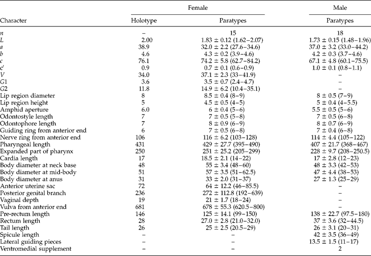
A female holotype, 15 female and 18 male paratype specimens (specimen numbers: An18-1 to An18-13) are deposited in the Laboratory of Plant Nematology/Research Center of Nematodes of Plant Quarantine, South China Agricultural University, Guangzhou, Guangdong 510642, China. See table 1 for detailed morphometrics.
Morphometrics of Belondira bagongshanensis n. sp. All measurements are in the form: mean ± SD (range), and in μm (except for L, which is in mm).

n= number of specimens observed; L= body length; a= L/maximum body width; b= L/pharyngeal length; c= L/tail length; c' = tail length/body diameter at anus; V= distance of vulva from anterior end × 100/L; G1 = anterior uterine sac × 100/L; G2 = posterior genital branch × 100/L.
Diagnosis and relationships
Belondira bagongshanensis n. sp. is characterized by a body 1.6–2.1 mm in length, cephalic framework moderately sclerotized, amphidial foveae cup-shaped, odontostyle robust with a distinct lumen, odontophore linear and about 1.2 times as long as the odontostyle, anterior part of pharynx with a distinct fusiform swelling bearing distinct sclerotized valve plates, basal expanded part of pharynx enclosed in a dextral muscular sheath and of length about three-fifths of the total neck length, female genital system mono-opisthodelphic and the anterior branch 0.8–1.6 times the corresponding body diameter, pre-rectum 3.0–4.7 times the anal body width in length, tail short (c′ value up to 1.1) and hemispheroid, male spicules 36–49 μm long and two widely spaced ventromedian supplements.
In possessing a short tail, a robust odontostyle with a distinct aperture and a moderately sclerotized cephalic framework, the new species is close to B. wajahati (Naz & Ahmad, Reference Naz and Ahmad2009), B. golden (Suryawanshi, Reference Suryawanshi1972) and B. eugeniae (Patil & Khan, Reference Patil and Khan1982). From B. wajahati (Naz & Ahmad, Reference Naz and Ahmad2009), B. bagongshanensis n. sp. differs by having a longer body length (1.6–2.1 mm vs. 1.4–1.6 mm), a shorter odontophore (6–9 μm vs. 10–14 μm), a longer pharyngeal length (395–490 μm vs. 330–380 μm), a shorter anterior genital branch (0.8–1.6 vs. 1.5–2.0 times the corresponding body diameter), a longer vaginal depth (18–24 m vs. 12–15 μm), a longer tail length (20.5–29 μm vs. 19–20 μm) and the presence of the male (vs. unknown). It differs from B. golden (Suryawanshi, Reference Suryawanshi1972) by the fusiform swelling bearing distinct sclerotized valve plates (vs. not sclerotized), the different position of ventromedial supplements (the posterior pair lying far from spicules vs. lying at the level of spicules) and a larger c value (c= 62.7–84.2 vs. 48.5–59 in females; c= 60.1–75.5 vs. 43–48 in males). From B. eugeniae (Patil & Khan, Reference Patil and Khan1982), the new species differs by having a longer body length (1.6–2.1 mm vs. 1.2–1.4 mm), a shorter odontophore (6–9 μm vs. 9–10 μm), a different fusiform swelling (bearing distinct sclerotized valve plates vs. not so), the presence of sperm (vs. absence) in the anterior genital branch and longer spicules (36–49 μm vs. 30–32 μm).
Molecular characterization
Two new sequences of 18S rDNA (1742 bp) and the D2–D3 region of 28S rDNA (830 bp) of B. bagongshanensis n. sp. were identified and deposited in GenBank (http://www.ncbi.nlm.nih.gov/Genbank/). Accession numbers are as follows: KT258982 and KT258983 for the 18S, and KT258984 and KT258985 for the D2–D3 region of 28S. The 18S rDNA showed 14 nucleotide differences from the sequences of Belondira apitica (AY911924, AY146482), which is the only species in the genus for which molecular data are available. Moreover, the BLAST search for the 18S rDNA showed 98% similarity to the sequences of Aporcelaimellus obtusicaudatus (DQ141212) while the D2–D3 region of 28S rDNA showed 84% similarity to the sequences of Aporcelaimellus pycnus (KM406917).
Discussion
As mentioned above, the odontostyle of Belondira was described as slender and the descriptions of the fusiform swelling located at the anterior part of the pharynx were always undefined in those studies before Naz & Ahmad (Reference Naz and Ahmad2009). Naz & Ahmad (Reference Naz and Ahmad2009) were the first to report the diversity of the style of odontostyle and the fusiform swelling. The robust odontostyle and the fusiform swelling bearing distinct sclerotized valve plates are also observed in the new species. As a result, we propose to revise the main characteristics of Belondira as: head rounded, cephalic framework more or less sclerotized, odontostyle slender or robust, anterior part of pharynx with a fusiform swelling bearing or lacking sclerotized valve plates, female opisthodelphic gonad and tail short, hemispheroid or cylindroid. It is known that Belondira has a great number of members, and the identification of the species depends mainly on the form of the odontostyle, cephalic framework and the fusiform swelling, which are now known to be quite various. Thus, more detailed descriptions and molecular data of the known species are needed and would help in identifying the species of Belondira.
Financial support
This work was supported by a Special Project of Scientific and Technological Basis of the Ministry of Science and Technology of the People's Republic of China to H.X. (grant no. 2006FY120100).