Introduction
Onchocerca lupi (Spirurida, Onchocercidae) is a parasitic filarioid and the causative agent of canine ocular onchocercosis. This infection has been increasingly recognized as an important cause of canine ophthalmic disease in Europe and the USA in the last decade (Sréter and Széll, Reference Sréter and Széll2008; Labelle et al., Reference Labelle, Maddox, Daniels, Lanka, Eggett, Dubielzig and Labelle2013; Otranto et al., Reference Otranto, Dantas-Torres, Brianti, Traversa, Petrić, Genchi and Capelli2013a; Reference Otranto, Giannelli, Latrofa, Dantas-Torres, Trumble, Chavkin, Kennard, Eberhard and Bowman2015). Canine onchocercosis ranges from subclinical infection to causing mild or severe ocular discomfort (Zarfoss et al., Reference Zarfoss, Dubielzig, Eberhard and Schmidt2005; Sréter and Széll, Reference Sréter and Széll2008). The nematode undergoes an indirect life cycle with dogs acting as definitive hosts, although humans (Otranto et al., Reference Otranto, Sakru, Testini, Gurlu, Yakar, Lia, Dantas-Torres and Bain2011) and cats (Maia et al., Reference Maia, Annoscia, Latrofa, Pereira, Giannelli, Pedroso and Otranto2015) have also been reported with ocular infections. In the definitive hosts, adult worms develop inside connective tissues, such as the ocular conjunctiva or aberrant sites (Alho et al., Reference Alho, Cruz, Coelho, Martinho, Mansinho, Annoscia, Lia, Giannelli, Otranto and de Carvalho2016), and eventually release microfilariae to the surrounding tissues in many regions of the body, including ears, nose, forehead and interscapular (Otranto et al., Reference Otranto, Dantas-Torres, Giannelli, Abramo, Ignjatovic Cupina, Petric, Cardoso, Mutafchiev and Cortes2013b). Then, microfilariae are taken up by unknown intermediate hosts to undergo their larval development (Otranto et al., Reference Otranto, Dantas-Torres, Giannelli, Abramo, Ignjatovic Cupina, Petric, Cardoso, Mutafchiev and Cortes2013b). DNA of this parasite has been found in captured Simulium sp. flies from Southern California (Hassan et al., Reference Hassan, Bolcen, Kubofcik, Nutman, Eberhard, Middleton, Wekesa, Ruedas, Nelson, Dubielzig, De Lombaert, Silverman, Schorling, Adler, Unnasch and Beeler2015), suggesting that black flies of the genus Simulium may act as intermediate hosts.
Molecular assays have been employed to detect O. lupi in dogs, humans, cats and black flies by targeting three mitochondrial loci: the cytochrome oxidase c subunit 1 (cox1) (Hassan et al., Reference Hassan, Bolcen, Kubofcik, Nutman, Eberhard, Middleton, Wekesa, Ruedas, Nelson, Dubielzig, De Lombaert, Silverman, Schorling, Adler, Unnasch and Beeler2015; Maia et al., Reference Maia, Annoscia, Latrofa, Pereira, Giannelli, Pedroso and Otranto2015; Otranto et al., Reference Otranto, Giannelli, Latrofa, Dantas-Torres, Trumble, Chavkin, Kennard, Eberhard and Bowman2015; Hodzic et al., Reference Hodzic, Hinney, Konig, Naucke, Duscher and Joachim2018; Latrofa et al., Reference Latrofa, Annoscia, Colella, Cavalera, Maia, Martin, Slapeta and Otranto2018), the 12S rRNA (Faisca et al., Reference Faisca, Morales-Hojas, Alves, Gomes, Botelho, Melo and Xufre2010; Mowlavi et al., Reference Mowlavi, Farzbod, Kheirkhah, Mobedi, Bowman and Naddaf2014) and the reduced nicotinamide adenine dinucleotide (NADH) dehydrogenase subunit 5 (nad5) (Labelle et al., Reference Labelle, Maddox, Daniels, Lanka, Eggett, Dubielzig and Labelle2013; McLean et al., Reference McLean, Newkirk and Adema2017). Mitochondrial markers are ideal for phylogenetic and epidemiologic studies since they are clonal, they rarely or never undergo recombination, are mostly of maternal inheritance and evolve faster than nuclear genes (Le et al., Reference Le, Blair and McManus2000). Therefore, these genes allow the discrimination between closely related specimens from different geographical locations and the phylogenetic reconstruction of an individual or a population (Le et al., Reference Le, Blair and McManus2000; Rojas et al., Reference Rojas, Dvir, Farkas, Sarma, Borthakur, Jabbar, Markovics, Otranto and Baneth2018). Previous phylogenetic analysis of O. lupi from different hosts and geographical regions has revealed two different clusters by using a single mitochondrial gene (Otranto et al., Reference Otranto, Dantas-Torres, Brianti, Traversa, Petrić, Genchi and Capelli2013a; Reference Otranto, Giannelli, Latrofa, Dantas-Torres, Trumble, Chavkin, Kennard, Eberhard and Bowman2015; Hodzic et al., Reference Hodzic, Hinney, Konig, Naucke, Duscher and Joachim2018). We hereby describe the first clinical case of O. lupi in Israel, from a domestic dog, and confirm the identification of two main genotypes based on the cox1 and nad5 genes, genotype 1 with specimens from the USA, Canada, Hungary, Greece and Israel, and genotype 2 or ‘Iberian genotype’ circulating among hosts from Portugal and Spain.
Materials and methods
Case description and specimen collection
A 13-year-old mixed breed spayed female dog born in Central Israel was presented to an ophthalmology veterinary clinic in Yehud, Israel, in May 2018. The dog suffered severe conjunctivitis in its right eye that had not responded to chloramphenicol ophthalmic ointment administered for 5 days. It underwent ophthalmic examination and a swollen mass was noticed over the dorsal sclera of the right eye accompanied by conjunctival erythema and chemosis. The dog was anaesthetized and the area of swelling was surgically explored and five helminth fragments 4–10 cm long were extracted and kept in 70% ethanol for DNA analysis (online Supplementary Video 1). The worms were torn during extraction, and some of their fragments were not entirely removed. The dog was discharged with oral antibiotic treatment of amoxicillin-clavulonic acid for a week, 6 weeks of oral doxycycline due to accompanying blepharospasm and periocular pruritis, as well as three consecutive antihelminthic treatments of doramectin administered subcutaneously every 2 weeks. Despite this treatment, the dog suffered phthisis bulbi and a complete loss of sight in the right eye 2 months after surgery.
DNA extraction and PCR analysis
Worm specimens removed during surgery were submitted to the Department of Parasitology at the Kimron Veterinary Institute (KVI), Beit Dagan, and the Koret School of Veterinary Medicine, of the Hebrew University of Jerusalem, Israel, for identification. The three worm pieces were morphologically identified as Onchocerca sp. based on the observation of prominent annular ridges in their cuticle and transverse striae on the inner layer of the nematode. DNA was extracted from worms with the DNeasy Blood and Tissue kit (Qiagen, Hilden, Germany) according to the manufacturer's instructions. Three separate polymerase chain reactions (PCRs) targeting the cox1 with the NTF and NTR primers (Casiraghi et al., Reference Casiraghi, Anderson, Bandi, Bazzocchi and Genchi2001), the nad5 with the ND5-F and ND5-R primers (Morales-Hojas et al., Reference Morales-Hojas, Cheke and Post2006) and the 12S rRNA with the 12SdegF2 and 12SdegnemR2 primers (Lefoulon et al., Reference Lefoulon, Bain, Bourret, Junker, Guerrero, Canizales, Kuzmin, Satoto, Cardenas-Callirgos, de Souza Lima, Raccurt, Mutafchiev, Gavotte and Martin2015) were performed. The reactions were verified by agarose gel electrophoresis, and positive cox1, nad5 and 12S PCR amplicons were sequenced.
DNA analysis
DNA sequences were compared to the nucleotide database available in GenBank using BLAST and the sequences obtained in this study were aligned together with publicly available sequences of O. lupi and Dirofilaria immitis sequences (AJ537512, NC005305.1 and KP760330 for cox1, nad5 and 12S, respectively) as outgroups. Maximum likelihood (ML) phylogenetic trees were constructed for the three gene fragments using the Tamura-Nei nucleotide substitution model with the MEGA 7.0 program (Kumar et al., Reference Kumar, Stecher and Tamura2016). Additionally, Templeton-Crandall-Sing (TCS) haplotype networks using the cox1, nad5 and 12S sequences and a 95% connection limit were drawn with the PopART software (available at http://popart.otago.ac.nz). Finally, the DnaSP software (Librado and Rozas, Reference Librado and Rozas2009) was used for estimating the number of haplotypes for the three analysed genes.
Results
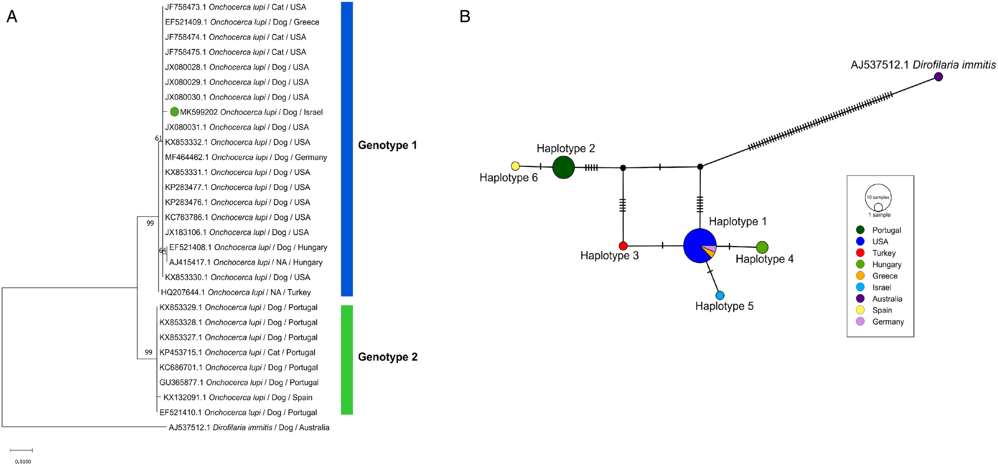
The 570 bp cox1 sequence from the Israeli dog nematode (GenBank accession number MK599202) was 99% identical to O. lupi collected from a dog in the USA (JX080030) (Labelle et al., Reference Labelle, Maddox, Daniels, Lanka, Eggett, Dubielzig and Labelle2013). The ML phylogram of O. lupi sequences showed two genotypes clustered according to the geographical location of the specimens (Fig. 1a): genotype 1 included 18 specimens from dogs, cats and a human in the USA, Greece, Hungary, Germany, Turkey and Israel (obtained herein) and genotype 2 included seven sequences from dogs and cats from Portugal and Spain. Five cox1 haplotypes were predicted, four of them belonging to genotype 1 and one to genotype 2 (Fig. 1b). The sequences obtained from the worms from Israel, Hungary and Turkey were closely related to the sequences from the USA and Greece. However, the sequences of haplotype 2 and 6 belonging to worms from Portugal and Spain were separated by 10 hypothetical haplotypes from the Turkish samples and 12 hypothetical haplotypes from the USA samples. In addition, cox1 sequences of genotype 1 differed from genotype 2 and 6 by 1.8 to 2.1% in their nucleotide pairwise distances. The difference within sequences of genotype 1 ranged from 0.2 to 0.4%, whereas the sequences from genotype 2 were all identical.
Analysis of a 570 bp cox1 gene sequences of O. lupi from different geographical locations and hosts. (A) Maximum likelihood tree with bootstrap values shown above each branch. The hosts, locations and GenBank accession numbers are indicated in each taxon, and the Israeli isolate is denoted with a green circle. Genotypes 1 and 2 are represented as coloured blue and green bars, respectively. (B) Haplotype network of the cox1 sequences of O. lupi. Each circle represents a haplotype, circle size is proportional to the number of sequences belonging to the same haplotype. The black circles and tick marks denote hypothetical sequences connecting each haplotype.

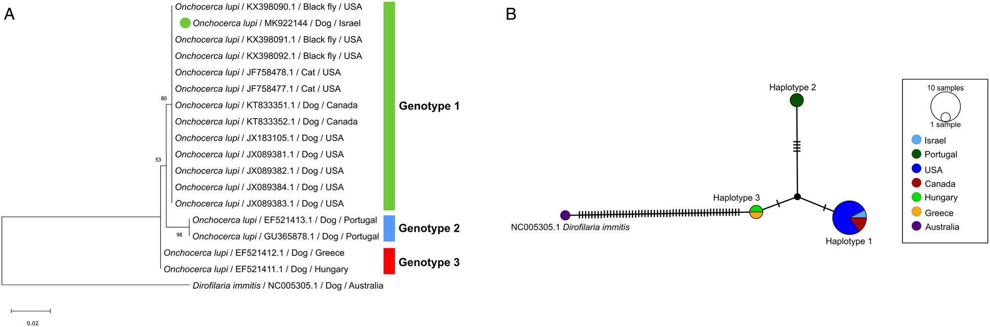
The 393 bp nad5 sequence of the Israeli specimen (MK922144) was 100% identical to O. lupi collected from a dog in the USA (JX089381) (Labelle et al., Reference Labelle, Maddox, Daniels, Lanka, Eggett, Dubielzig and Labelle2013). The haplotype analysis revealed the presence of three separate groups, as supported by the ML tree (Fig. 2a) and haplotype network (Fig. 2b): genotype 1 included the sequences from dogs, cats and black flies collected in Canada, USA and Israel, genotype 2 included the sequences from dogs of Portugal, and genotype 3 contained the sequences obtained from Hungarian and Greek dogs. Furthermore, the intergenotypic distances between genotypes 1 and 2, genotypes 1 and 3, and genotypes 2 and 3 were of 1.5%, 0.6% and 1.5%, respectively. All analysed sequences from the same genotype were identical between them, therefore, the intragenotypic nucleotide distances were zero for the three genotypes.
Analysis of a 393 bp nad5 gene sequences of O. lupi from different geographical locations and hosts. (A) Maximum likelihood tree with the hosts, locations and GenBank accession numbers indicated in each taxon. The Israeli isolate is denoted with a green circle. (B) Haplotype network of the ND5 sequences of O. lupi. Each circle represents a haplotype, circle size is proportional to the number of sequences belonging to the same haplotype. The black circles and tick marks denote hypothetical sequences connecting each haplotype.

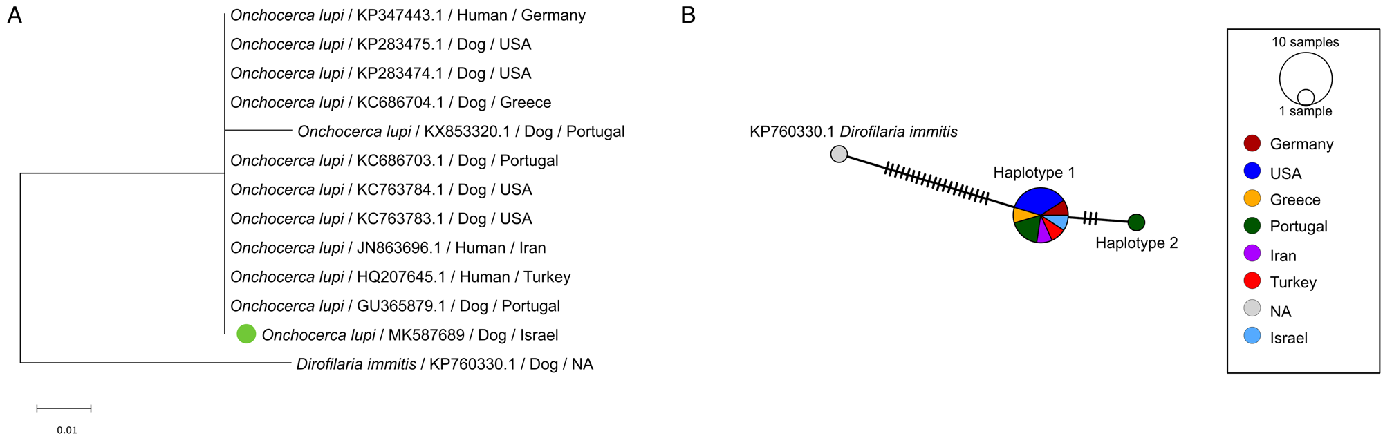
Similarly, the 248 bp 12S sequence obtained in the current study (MK587689) was 100% identical to O. lupi from ocular human cases from Iran (JN863696.1) (Mowlavi et al., Reference Mowlavi, Farzbod, Kheirkhah, Mobedi, Bowman and Naddaf2014) and Germany (KP347443.1) (Bergua et al., Reference Bergua, Hohberger, Held, Muntau, Tannich and Tappe2015), and a canine case from the USA (KC763784) (Hassan et al., Reference Hassan, Bolcen, Kubofcik, Nutman, Eberhard, Middleton, Wekesa, Ruedas, Nelson, Dubielzig, De Lombaert, Silverman, Schorling, Adler, Unnasch and Beeler2015). The ML tree confirmed that the analysed 12S sequences from the USA, Germany, Greece, Iran, Turkey, Israel and two from Portugal were identical to each other. Only one sample from a dog from Portugal was different from the ones mentioned previously (GenBank accession number KX853320.1) (Lefoulon et al., Reference Lefoulon, Giannelli, Makepeace, Mutafchiev, Townson, Uni, Verocai, Otranto and Martin2017) (Fig. 3a). Two haplotypes were found in the O. lupi 12S sequences, the first grouped with the sequences collected from Portugal, Greece, Turkey, Israel and the USA and the second was represented by sequence KX853320.1 from Portugal (Fig. 3b).
Analysis of a 248 bp 12S gene sequences of O. lupi from different geographical locations and hosts. (A) Maximum likelihood tree with bootstrap values shown above each branch. The hosts, locations and GenBank accession numbers are indicated in each taxon, and the Israeli isolate is denoted with a green circle. Genotypes 1 and 2 are represented as coloured blue and green bars, respectively. (B) Haplotype network of the 12S sequences of O. lupi. Each circle represents a haplotype, circle size is proportional to the number of sequences belonging to the same haplotype. The black circles and tick marks denote hypothetical sequences connecting each haplotype.

Discussion
This constitutes the first report of an autochtonous O. lupi ocular infection in a dog from Israel. The specimen analysed herein was genetically similar to specimens collected from the USA and belonged to genotype 1 along with other nematodes from Canada, Greece and Hungary.
The dog presented acute severe scleral conjunctival inflammation and its clinical condition deteriorated after removing the worms from the affected tissue. Intraocular damage might have been associated with the elicited host inflammatory response to degenerating worms or the mechanical damage that occurred during larvae or adult nematode migration, as described with the causative agent of human onchocercal keratitis, Onchocerca volvulus (Pearlman and Hall, Reference Pearlman and Hall2000). Additionally, the severe ocular inflammation and consequent loss of eye functionality might have worsened due to bacterial contamination or the release of antigens from the worm or its endosymbiont Wolbachia after dismembering the specimens during surgical intervention, as previously suggested (McLean et al., Reference McLean, Newkirk and Adema2017).
The dog described here lived in the Jerusalem region continuously since its adoption from an animal shelter in Central Israel at the age of 2-months. At least seven species of black flies of the family Simuliidae were reported in Israel (Freidberg, Reference Freidberg, Yom-Tov and Tchernov1988): Simulium aureum Fries, 1824, Simulium rubzovianum Sherban, 1961, Simulium ruficorne Macquart, 1838, Simulium buxtoni Austen, 1923, Simulium golani Beaucornu-Saguez, 1977, Simulium pseudequinum Seguy, 1921, and Simulium paraequinum Puri, 1933. Based on clinical reports from non-endemic countries, O. lupi displays a very long lifespan ranging from 3 years (Verocai et al., Reference Verocai, Conboy, Lejeune, Marron, Hanna, MacDonald, Skorobohach, Wilcock, Kutz and Gilleard2016) to 8 years (Hodzic et al., Reference Hodzic, Hinney, Konig, Naucke, Duscher and Joachim2018). Therefore, the dog could have become infected at any moment during its lifetime.
Different O. lupi genotypes were distinguished and confirmed in the phylogenetic and haplotype network analyses according to the cox1 and nad5, and further supported by high inter-genotypic nucleotide distances. The sequences from the USA, Greece, Turkey, Hungary and Israel constituted different haplotypes and clustered in the same monophyletic group with strong support and low genetic distance, according to the cox1 analysis, as previously observed by Otranto et al. (Reference Otranto, Dantas-Torres, Giannelli, Latrofa, Papadopoulos, Cardoso and Cortes2013c). In addition, the nad5 gene analysis showed that the Israeli sequence clustered with the sequences from the USA and kept the Portuguese sequences in a separate genotype. The low genetic variation found within these cox1 and nad5 genotypes might be due to the introduction of O. lupi from the Old World to the USA, as previously suggested (Otranto et al., Reference Otranto, Giannelli, Latrofa, Dantas-Torres, Trumble, Chavkin, Kennard, Eberhard and Bowman2015). In addition, the fact that the Israeli haplotype was differentiated from the other cox1 haplotypes in genotype 1 may strengthen the notion of an autochtonous source for this infection as the same haplotype was not described from other countries. Interestingly, all cox1 and nad5 sequences from Portugal obtained from different hosts, i.e. domestic dogs, cats and humans (Sreter-Lancz et al., Reference Sréter-Lancz, Széll and Sréter2007; Mutafchiev et al., Reference Mutafchiev, Dantas-Torres, Giannelli, Abramo, Papadopoulos, Cardoso, Cortes and Otranto2013; Faisca et al., Reference Faisca, Morales-Hojas, Alves, Gomes, Botelho, Melo and Xufre2010; Maia et al., Reference Maia, Annoscia, Latrofa, Pereira, Giannelli, Pedroso and Otranto2015; Lefoulon et al., Reference Lefoulon, Giannelli, Makepeace, Mutafchiev, Townson, Uni, Verocai, Otranto and Martin2017), belonged to the same haplotype and genotype, which confirms previous findings that a common O. lupi strain is circulating among Portuguese hosts (Maia et al., Reference Maia, Annoscia, Latrofa, Pereira, Giannelli, Pedroso and Otranto2015). Finally, the high inter-genotypic distance in the cox1 and nad5 genes might reflect the divergence of O. lupi due to geographical isolation, use of different intermediate host species or low prevalence within each geographical location, which may lead to genetic structure and speciation (Huyse et al., Reference Huyse, Poulin and Théron2005).
The analysed 12S gene region of O. lupi was highly conserved in the studied sequences. All available 12S sequences, except for one from Portugal, were identical between them. Therefore, the genotypes predicted by the cox1 and nad5 genes were not replicated with the 12S gene. This might be explained by the intraspecific nucleotide conservation of the analysed 12S gene region, which does not allow the separation of genotypes, as observed also for D. immitis (Huang et al., Reference Huang, Wang, Yang, Zhang, Wang, Yang, Luo, Liu, Lan and Huang2009).
Clinical manifestations elicited by specimens from genotype 1 and 2 suggest that the Iberian genotype produces a subclinical to mild infection, as observed in dogs from Spain (Miro et al., Reference Miro, Montoya, Checa, Galvez, Minguez, Marino and Otranto2016) and Portugal (Otranto et al., Reference Otranto, Dantas-Torres, Giannelli, Latrofa, Papadopoulos, Cardoso and Cortes2013c; Maia et al., Reference Maia, Annoscia, Latrofa, Pereira, Giannelli, Pedroso and Otranto2015). An exception to this observation comes from a Portuguese dog that showed hyperemic conjunctiva and oedematous cornea along with purulent discharge in the upper eyelid (Faisca et al., Reference Faisca, Morales-Hojas, Alves, Gomes, Botelho, Melo and Xufre2010), and the specimens collected from the case clustered in Genotype 2 according to the cox1 and nad5 phylogenetic analyses. In contrast, most cases reported from the domestic canine, feline and human hosts belonging to genotype 1, i.e. USA, Canada, Greece, Hungary, Germany and the reported herein in Israel, have in common the appearance of severe clinical signs including a nodular mass, conjunctival hyperemia, retinal detachment, blepharospasm, corneal oedema, and chemosis which may lead to loss of sight (Labelle et al., Reference Labelle, Daniels, Dix and Labelle2011; Otranto et al., Reference Otranto, Sakru, Testini, Gurlu, Yakar, Lia, Dantas-Torres and Bain2011; Otranto et al., Reference Otranto, Dantas-Torres, Cebeci, Yeniad, Buyukbabani, Boral, Gustinelli, Mounir, Mutafchiev and Bain2012; Otranto et al., Reference Otranto, Giannelli, Latrofa, Dantas-Torres, Trumble, Chavkin, Kennard, Eberhard and Bowman2015; Mowlavi et al., Reference Mowlavi, Farzbod, Kheirkhah, Mobedi, Bowman and Naddaf2014; McLean et al., Reference McLean, Newkirk and Adema2017; Hodzic et al., Reference Hodzic, Hinney, Konig, Naucke, Duscher and Joachim2018). Altogether, the delimitation of two genotypes according to the cox1 and nad5 phylogenetic trees and the difference in the clinical signs reported in cases from the Iberian Peninsula compared to elsewhere may indicate a potential differential virulence in both genotypes.
In conclusion, we report the first O. lupi infection in Israel. This filarioid should be recognized as a differential diagnosis for periocular and ocular lesions in humans and carnivores in Israel and its neighbouring countries. In addition, phylogenetic analyses confirmed the circulation of an O. lupi genotype in the Old and New Worlds, and a separate Iberian genotype. Further research is warranted to investigate the local transmission dynamics of this infection and its vectors, which could also provide insight into its yet undeciphered transmission globally.
Supplementary material
The supplementary material for this article can be found at https://doi.org/10.1017/S0031182020001560
Acknowledgements
The authors thank the University of Costa Rica for a partial stipend granted to AR. Also, the authors are grateful to Drs. Yaron Hershkovitz and Etai Kahana of the National Center for Aquatic Ecology, The Steinhardt Museum of Natural History, Tel Aviv University, Israel for sharing published and unpublished information regarding Simuliidae prevalence in Israel.
Financial support
The research was done using internal funds from Prof. Baneth's laboratory.
Conflict of interest
The authors have no financial conflicts of interest.
Ethical standards
Not applicable