Milk is the only food for the newborn during the first period of life. The composition of milk is complex and varies according to different factors including stage and number of lactations, and the maternal diet (Jensen, Reference Jensen and Jensen1995). The major milk proteins are well known as they were isolated and characterized long ago and have been widely studied: these include lactoferrin (Farnaud & Evans, Reference Farnaud and Evans2003), casein, α-lactalbumin (Farrell et al. Reference Farrell, Brown and Kumosinski1993; Permyakov & Berliner, Reference Permyakov and Berliner2000) and immunoglobulins (Larson, Reference Larson and Fox1993). However, new functions have been attributed to well characterized proteins, like α-la, for which antitumoral properties have been proposed in recent years (Shah, Reference Shah2000).
Furthermore, active peptides derived from milk proteins have been identified (Schlimme & Meisel, Reference Schlimme and Meisel1995). Peptides corresponding to digestion fragments have been obtained by casein hydrolysis. Some of these peptides have been characterized and different biological functions attributed to them (Silva & Malcata, Reference Silva and Malcata2005). In addition, growth factors have been identified in milks of different species (Campana & Baumrucker, Reference Campana, Baumrucker and Jensen1995), and also growth inhibitors which could have an important role in protecting newborn and mammary gland tissues from cancer (Gill & Cross, Reference Gill and Cross2000).
Human and bovine milk have been reported as cell growth promoters (Ichiba et al. Reference Ichiba, Kusuda, Itagane, Fujita and Issiki1992; Aranda et al. Reference Aranda, Sánchez, Pérez and Calvo1996) and bovine colostrum and milk have been proposed as replacers for foetal calf serum in cell culture (Sereni & Baserga, Reference Sereni and Baserga1981). The growth promoting activity of milk secretions has been attributed to different molecules, such as epidermal growth factor (EGF) in human milk, and insulin-like growth factor-I and II (IGF-1, IGF-2) in bovine and human milk (Campbell & Baumrucker, Reference Campbell and Baumrucker1989). On the other hand, some milk factors have been considered to be growth inhibitors for certain cells. These include mammary-derived growth inhibitor (MDGI) (Brandt et al. Reference Brandt, Pepperle, Otto, Kraft, Boehmer and Grosse1988), proteolytic fragments of lactoferrin (Yoo et al. Reference Yoo, Watanabe, Koike, Mitobe, Shimazaki, Watanabe and Azuma1997) and a new form of α-la (Hakansson et al. Reference Hakansson, Zhivotovsky, Orrenius, Sabharwal and Svanborg1995). This form of α-la, called HAMLET (human alpha-lactalbumin made lethal to tumour cells), was obtained from human casein and was shown to inhibit the growth of tumoural cells, but was not harmful for differentiated cells (Svanborg et al. Reference Svanborg, Agerstam, Aronson, Bjerkvig, Düringer, Fischer, Gustafsson, Hallgren, Leijonhuvud, Linse, Mossberg, Nilsson, Pettersson and Svensson2003). α-La has been reported to change its native form into the apoptosis-inducing form after removal of calcium normally bound to it. This causes unfolding of the protein into the apoptotic form, which is stabilized by incorporating oleic acid (Gustafsson et al. Reference Gustafsson, Hallgren, Mossberg, Pettersson, Fischer, Aronsson and Svanborg2005).
The gastrointestinal tract is the first target of milk bioactive molecules. Therefore, the identification of milk fractions that affect proliferation or migration of epithelial cells may have potential applications in promoting gastrointestinal development and health. The pharmaceutical, biotechnological and nutraceutical industries thus show great interest in finding novel bioactive components in available biological fluids (Shah, Reference Shah2000).
In the present work we have evaluated the activity of several samples of human and bovine milk and their corresponding whey and casein fractions on the growth of two cell lines, MDCK and Caco-2. We have studied the type of effect exerted by milk on the cells and we have also made a first approach at characterizing the nature of the factor responsible for the inhibitory activity found in human milk.
Materials and Methods
Materials
Human milk samples were kindly supplied by healthy donors belonging to Via Lactea Association (Zaragoza, Spain) who were in a non-colostral period. Pooled bovine milk samples were provided by Nuprosa (Zaragoza, Spain) and mature milk samples from individual cows by Tauste Ganadera (Tauste, Spain). All milk samples were immediately frozen after milking and maintained at −20°C until processed. Milk samples were not subjected to any heat treatment.
Minimum essential medium Eagle (MEM), Dulbecco's modified Eagle medium (DMEM), L-glutamine, non-essential amino acids (NEAA), fetal calf serum (FCS), penicillin-streptomycin, trypsin and EDTA were from Biological Industries (Kibbutz Beit Haemek, Israel). Pyruvic acid, glucose, HEPES, naphthol blue-black grade II (NBB) and bisbenzimide H 33342 trihydrochloride (Hoechst 33342) were obtained from Sigma (Poole, England).
Cell culture
MDCK, a canine kidney cell line, was grown in monolayers in 25 cm2 flasks and maintained with MEM supplemented with 2 mm-L-glutamine, 1% NEAA, 10% FCS, 50 000 IU penicillin-streptomycin/l and 1 mm-pyruvic acid. Caco-2 TC-7 clone, a human adenocarcinoma cell line, was grown in monolayers in 25 cm2 flasks and maintained with DMEM supplemented with 2 mm-L-glutamine, 1% NEAA, 20% FCS, 25 mm-glucose, 2·5% HEPES and 50,000 IU penicillin-streptomycin/l.
Isolation of casein
Casein was obtained by acid precipitation according to Svensson et al. (Reference Svensson, Sabharwal, Hakansson, Mossberg, Lipniunas, Leffler, Svanborg and Linse1999) and lyophilized. The resulting whey was neutralized to the original pH of milk, dialyzed using a membrane of 12 000–14 000 MWCO against 30 volumes of distilled water for 24 h at 4°C, and finally lyophilized.
Measurement of cell growth
Cells were grown in 25 cm2 cell culture flasks and near to confluence were subcultured and seeded in 96-well plates at a concentration of 3·6×104 cells/ml for MDCK and 2·9×104 cells/ml for Caco-2. Cell cultures were maintained at 37°C in a humidified atmosphere containing 5% CO2. After 48 h of culture, cells were rinsed with PBS, and then with cell culture medium without FCS. The samples to be tested were then added to wells in medium without FCS. The control wells contained medium without FCS. After 30 min, FCS was added at the optimal concentration for each cell line. Plates were then incubated for 4.5 h under the conditions described above.
Cell viability and cell growth were determined by trypan blue exclusion and by a protein staining assay, respectively. The protein staining assay was performed according to Everitt & Wohlfart (Reference Everitt and Wohlfart1987) using 0·1% of NBB. The absorbance measured at 620 nm corresponds to the total cellular protein content and it can therefore be directly related to cell number. The NBB method was chosen for all the assays as it allowed a higher number of replicates per experiment. The results obtained by this method were comparable to those obtained by trypan blue.
Determination of lactoferrin and α-la levels in human milk samples
The determination of lactoferrin and α-la concentrations in human milk samples was performed by radial immunodiffusion, according to Sánchez et al. (Reference Sánchez, Peiró, Castillo, Pérez, Ena and Calvo1992) using polyclonal rabbit antisera. Lactoferrin standard was isolated from human whey by Blue Cibacron chromatography according to Mata et al. (Reference Mata, Sánchez, Headon and Calvo1998). The concentrations used in standards were 0·05, 0·10, 0·15 and 0·20 mg/ml and milk samples were diluted 10−1 in distilled water and analysed in duplicate. α-La standard was obtained from human whey by chromatography on Sephadex G-100 according to Barbana et al. (Reference Barbana, Pérez, Pocovi, Sánchez and Wehbi2008). The concentrations of α-la standards were 50, 25, 12·5 and 10 μg/ml and milk samples were diluted 2·10−2 in distilled water and analysed in duplicate.
Treatments of milk caseins
Human casein samples at a concentration of 10 mg/ml in culture medium without FCS, and a control consisting of medium without FCS were heated at 80°C for 10 min in a temperature controlled bath (±0·1°C). Activity of each sample, with and without heat treatment, on cell growth was tested in quintuplicate in at least two independent experiments.
Human and bovine casein fractions were digested by trypsin as described by McDonagh & FitzGerald (Reference McDonagh and FitzGerald1998).
Morphological examination of cell cultures
Cells were stained with Hoechst 33342, a DNA binding stain, to study their morphology. After incubation with milk samples for 4·5 h, the cells were fixed with 2% paraformaldehyde in PBS for 10 min at 4°C. Cells were then incubated for 20 min with 20 mg Hoechst 33342/ml. After washing with PBS, cells were observed under a fluorescence microscope.
DNA fragmentation analysis
DNA was extracted from 4·106 cells by incubation in 75 mm-KCl lysis solution at 37°C for 30 min. The cell lysate was centrifuged at 2000 g for 5 min at 4°C. The nuclei were recovered from the pellet and incubated overnight at 50°C in 0·5% Triton X-100/0·5 mm-EDTA containing 25 mg proteinase K/ml. The hydrolyzed proteins were precipitated by adding saturated NaCl and centrifuged at 4000 g for 20 min. The supernatant was transferred into another microcentrifuge tube containing 2·2 volumes of 100% ethanol. DNA was pelleted by centrifugation at 13 000 g for 20 min and washed once in 70% ethanol. The supernatant was removed and the DNA pellet was allowed to dry for 3 min at room temperature. The DNA was then dissolved in 10 mm-Tris buffer, pH 8, containing 0·1 mm-EDTA (TE) and 4 mg RNAse/ml, and incubated for 1 h at 37°C.
DNA was loaded on to 1·8% agarose gels in 89 mm-Tris buffer, 89 mm-boric acid, pH 8, containing 0·5 m-EDTA (TBE), and electrophoresed at a constant voltage (100 V). The gels were stained with ethidium bromide and photographed under a Gel Doc 2000, Bio Rad UV transilluminator.
Results and discussion
Effect of human skimmed milk and casein on MDCK and Caco-2 cells
The cell lines employed in this work have been widely used in cellular studies, MDCK as a well established epithelial cell line of reference (Reveneau et al. Reference Reveneau, Paumelle, Deheuninck, Leroy, De Launoit, Fafeur and Diederich2003; Shoeib et al. Reference Shoieb, Elgayyar, Dudrick, Bell and Tithof2003) and Caco-2 as a human tumoural cell line used in studies related to cancer therapeutics (Sternhagen & Allen, Reference Sternhagen and Allen2001; Hallgren et al. Reference Hallgren, Gustafsson, Irjala, Selivanova, Orrenius and Svanborg2006).
The inhibitory effect of human milk had been previously reported on different cell lines (Hakansson et al. Reference Hakansson, Zhivotovsky, Orrenius, Sabharwal and Svanborg1995) and this activity had been associated with α-la bound to casein. In subsequent work, the active variant of α-la was characterized and its effect on cells described (Hakansson et al. Reference Hakansson, Andréasson, Zhivotovsky, Karpman, Orrenius and Svanborg1999; Svensson et al. Reference Svensson, Sabharwal, Hakansson, Mossberg, Lipniunas, Leffler, Svanborg and Linse1999). We intended to investigate the existence of a similar activity in bovine milk, as the previous studies were not very conclusive. For this purpose, we assayed the activity of some human milk samples from different donors, and found that the activity was very different according to the sample.
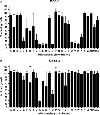
Milk samples from 29 healthy women were collected and fractionated into skimmed milk, whey and casein. The effect of skimmed milk on the growth of MDCK and Caco-2 cells is shown in Fig. 1A and 1B, respectively. On the cell line MDCK, eight of 28 skimmed milk samples at a 10−1 dilution decreased the growth below 20% with respect to the control. Cell growth was between 20 and 80% of that obtained with the control for nine samples of skimmed milk and more than 80% for the other eleven samples. On the cell line Caco-2, only one of the 28 samples of skimmed milk, assayed at a dilution of 10−1 reduced growth to 20% respect to control cells. The growth of Caco-2 cells was between 20 and 80% for five skimmed milk samples and more than 80% for the other 22 samples. The samples with inhibitory activity on Caco-2 cell growth were also inhibitory on MDCK cells.
Effect of skimmed human milk (28 samples) at a dilution of 10−1 on the cell lines MDCK (A) and Caco-2 (B). Cell growth, determined by the NBB method, is expressed as percentage of cell growth obtained in controls (considered as 100%). Error bars represent SD. Samples are represented by a letter and each corresponds with milk from one healthy donor in a non-colostral stage of lactation. Each sample was analyzed five times in at least two independent experiments.

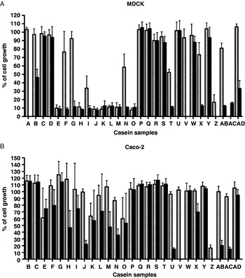
Whey obtained from human milk samples after casein separation was also tested on MDCK and Caco-2 cells and no effect was observed on their growth (data not shown). Fractions of casein were assayed on the two cell lines and the results are shown in Fig. 2A and 2B. Casein fractions from 25 human milk samples were tested on the cell line MDCK at two concentrations, 5 and 10 mg/ml, and 4 samples only at one concentration (Fig. 2A). For these cells, 16 of the 29 casein samples decreased the growth below 20% in at least one of the two concentrations tested and the cell growth did not differ by more than 8% between the two concentrations used in seven of the samples. The rest of the casein samples decreased cell growth to a higher degree when added at 10 mg/ml. The effect of caseins obtained from 27 human milk samples on Caco-2 cell growth was also studied (Fig. 2B). For 12 samples the effect on cell growth was very similar at the two concentrations studied, 5 and 10 mg/ml, the difference between their activity being no higher than 9%. The majority of the other casein samples decreased cell growth to a higher degree at 10 mg/ml than at 5 mg/ml. The growth of Caco-2 cells decreased below 20% only for four samples of human casein and between 20 and 80% for eleven samples.
Effect of casein fraction from human milk samples (casein samples originate from milks with the same letter) at the concentrations of 5 mg/ml (hatched bars) and 10 mg/ml (solid bars) on the cell lines MDCK (A) and Caco-2 (B). Cell growth, determined by the NBB method, is expressed as percentage of cell growth obtained in controls (considered as 100%). Error bars represent SD. Each sample was analyzed five times in at least two independent experiments.

These results show that the inhibitory activity of human milk on cell growth remains associated with casein and not with whey. For MDCK cells the percentage of active casein samples was the same as that found for the corresponding non-fractionated milks. However, although the majority of the samples that were active as non-fractionated milk were also active as casein, we found 10% of samples that did not follow this behaviour. For Caco-2, the percentage of samples with cytotoxic activity increased considerably when isolated casein was used instead of non-fractionated milk, 55% and 20%, respectively. Given that the susceptibility of Caco-2 cells to the inhibitory activity of human milk increases when casein is added, it is possible that the isolation of this fraction may exert a kind of concentration effect on the putative ‘inhibitory factor’.
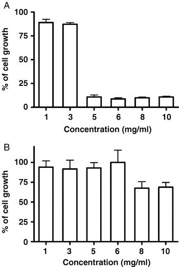
The previous studies by Hakansson et al. (Reference Hakansson, Zhivotovsky, Orrenius, Sabharwal and Svanborg1995) reported that casein from human milk showed inhibitory activity at 10 mg/ml, reducing the growth of different human carcinoma cell lines (A549, NCI, A-498 and J 82) to 0%. In the same study, MDCK and Caco-2 cells were used to evaluate the activity of a fraction isolated from active casein, though the effect that non-fractionated milk or casein exerted on those cells were not reported. Later, Svensson et al. (Reference Svensson, Sabharwal, Hakansson, Mossberg, Lipniunas, Leffler, Svanborg and Linse1999) and Pettersson et al. (Reference Pettersson, Mossberg and Svanborg2006) found human casein inhibitory at 0·5 mg/ml on the cell line L1210 which seemed to be more sensitive to the ‘inhibitory factor’ than the lines previously assayed. In preliminary experiments, we studied the effect of casein isolated from several active human milks on MDCK and Caco-2 cells at concentrations between 1 μg/ml and 10 mg/ml and found an inhibitory effect only above 5 mg/ml. Fig. 3A and 3B show the effect of the concentration of an active casein, from 1 to 10 mg/ml, on the growth of MDCK and Caco-2 cells. MDCK cells were found to be more sensitive than Caco-2 cells to active casein. No clear effect of casein concentration on its inhibitory activity was observed, since above a certain value, 5 mg/ml for MDCK cells and 8 mg/ml for Caco-2 cells, the cytotoxic effect did not further increase. Therefore, concentrations of 5 mg/ml and 10 mg/ml were used when screening human milk samples.
Effect of casein concentration on cell lines MDCK (A) and Caco-2 (B). Casein (H) was added at concentrations from 1 to 10 mg/ml to cell culture. Cell growth, determined by the NBB method, is expressed as percentage of cell growth obtained in controls (considered as 100%). Error bars represent SD. Each sample was analyzed five times in at least two independent experiments.

The high concentration at which casein is inhibitory would suggest that casein is not active by itself, as the effective concentrations of growth and inhibitory factors are usually in the range of nanograms per ml (Thompson et al. Reference Thompson, Farrell, Sanjeeva, Liu, Kidwell, Bansal, Cook, Medina, Kotts and Bano1992; Zavizion et al. Reference Zavizion, Politis and Gorewit1993). It is possible that the conditions we used for the fractionation of milk favoured the association of the ‘inhibitory factor’ with casein. However, this hypothesis would raise the question of whether the different activity exerted by milk from different donors is due to the presence or absence of the ‘inhibitory factor’ or to various degrees of association of the factor to casein during its isolation.
Effect of bovine skimmed milk and casein on MDCK and Caco-2 cells
Similar experiments to those performed with human milk were carried out with bovine milk. Firstly, pooled milk was tested on MDCK and Caco-2 cell lines. The fractions studied were: skimmed milk at dilutions 10−1 and 50−1, whey and casein at 5 mg/ml and 10 mg/ml. None of the fractions had any effect on the growth of the cells studied (results not shown). Similar experiments were also performed with 10 samples of bovine milk from individual animals and none had any effect on either of the cell lines.
In previous work by Aranda et al. (Reference Aranda, Sánchez, Pérez and Calvo1996) we studied the activity of bovine lacteal secretions on a fibroblast cell line and found that bovine colostrum promoted its growth, while the corresponding whey showed differing activity according to the isolation method. Whey obtained from colostrum by ultrafiltration did not exert growth promoting activity. However, when whey was isolated by ultracentrifugation, the activity was similar to that of colostrum. Shing & Klagsbrun (Reference Shing and Klagsbrun1984) also showed that the mitogenic activity of bovine and human colostrum was higher for the whole secretion than for the corresponding whey.
The presence of inhibitory factors has been reported in bovine milk, including mammary derived growth inhibitor (MDGI), which has been shown to inhibit proliferation of epithelial cells of the bovine mammary gland, thus participating in its development (Brandt et al. Reference Brandt, Pepperle, Otto, Kraft, Boehmer and Grosse1988; Kurtz et al. Reference Kurtz, Vogel, Funa, Heldin and Grosse1990; Zavizion et al. Reference Zavizion, Politis and Gorewit1993). This factor is present in bovine whey in trace levels and it has been isolated from the lactating bovine mammary gland and from the milk fat globule membrane (Yang et al. Reference Yang, Spitzer, Kenney, Zschiesche, Li, Kromminga, Müller, Spener, Lezius, Veerkamp, Smith, Salomon and Grosse1994). A MDGI-related polypeptide has been characterized in the pregnant bovine mammary gland (Brandt & Grosse, Reference Brandt and Grosse1992) which may suggest post-translational MDGI-processing related to its activity in cell differentiation. We have not detected any activity similar to that of MDGI when we studied the effect of bovine milk and their fractions on MDCK and Caco-2 cells. It is possible that the activity of that factor may be directed only to mammary cells and that the cells we have used are not sensitive to it.
Lactoferrin and α-la levels in human milk samples
Some whey proteins have antitumoural properties, for example lactoferrin, which has been reported to reduce the incidence of oxidant-induced carcinogenesis in rats (Gill & Cross, Reference Gill and Cross2000), and also α-la (Thompson et al. Reference Thompson, Farrell, Sanjeeva, Liu, Kidwell, Bansal, Cook, Medina, Kotts and Bano1992). We therefore measured the concentrations of lactoferrin and α-la to check that their levels were not abnormally high in active samples. We found that their concentrations were within the normal range (from 0·64 to 1·88 mg/ml1 for lactoferrin, and from 1·04 to 6·83 mg/ml for α-la) as reported by Sánchez et al. (Reference Sánchez, Aranda, Pérez and Calvo1988) and Pérez et al. (Reference Pérez, Sánchez, Aranda, Ena, Oria and Calvo1990), respectively. Furthermore, no correlation was found between the inhibitory activity of human milk samples and the corresponding concentrations of lactoferrin and α-la.
Effect of heat treatment of casein on cell growth
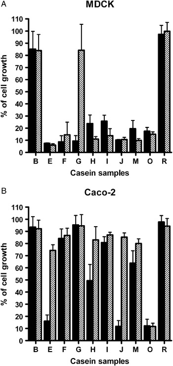
In order to know if the ‘inhibitory factor’ present in human milk was a proteic molecule, casein samples were subjected to 80°C for 10 min, a treatment that usually denatures most proteins (Ford et al. Reference Ford, Law and Marshall1977; Dannenberg & Kessler, Reference Dannenberg and Kessler1988). Nine samples of human casein showing inhibitory activity (B, E, F, G, H, I, J, M, O) and sample R with no inhibitory activity were tested at a 10 mg/ml concentration on MDCK and Caco-2 cells, before and after being subjected to heat treatment. Heating only suppressed the cytotoxic activity in one of the casein samples when evaluated on MDCK cell line and in three samples when tested on Caco-2 cells (Fig. 4A and 4B). These results suggest that the ‘inhibitory factor’ of human milk is a very thermoresistant protein, a polypeptide or a lipidic component.
Effect of heat treatment on the activity of human casein samples on the cell lines MDCK (A) and Caco-2 (B). Different samples of active casein (letters are the same than those used for the corresponding milks) were evaluated at a concentration of 10 mg/ml before (solid bars) and after being subjected to heating at 80°C for 10 min (hatched bars). Cell growth, determined by the NBB method, is expressed as percentage of cell growth obtained in controls (considered as 100%). Error bars represent SD. Each sample was analyzed five times in at least two independent experiments.

Effect of hydrolysis on casein activity on cell growth
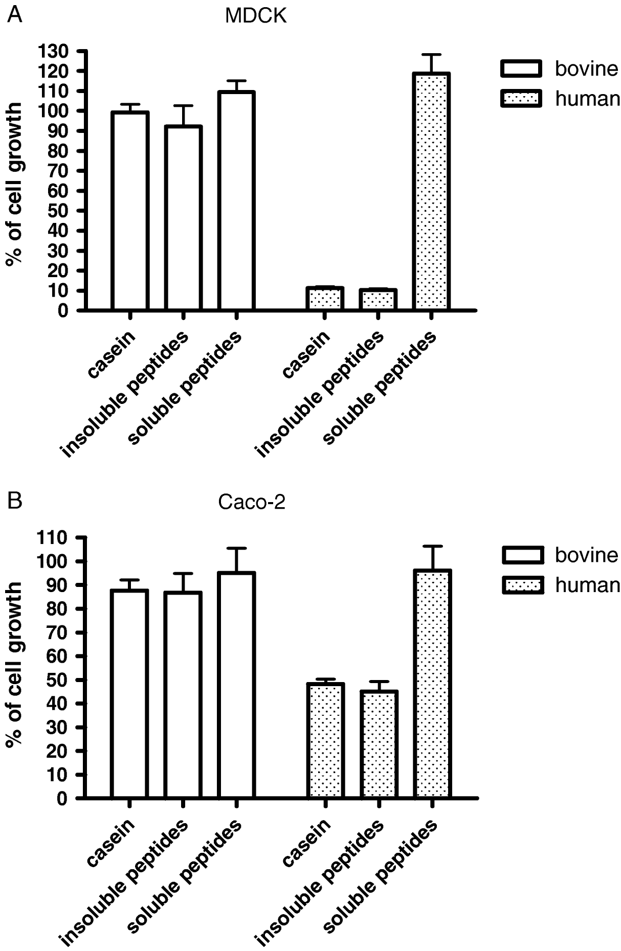
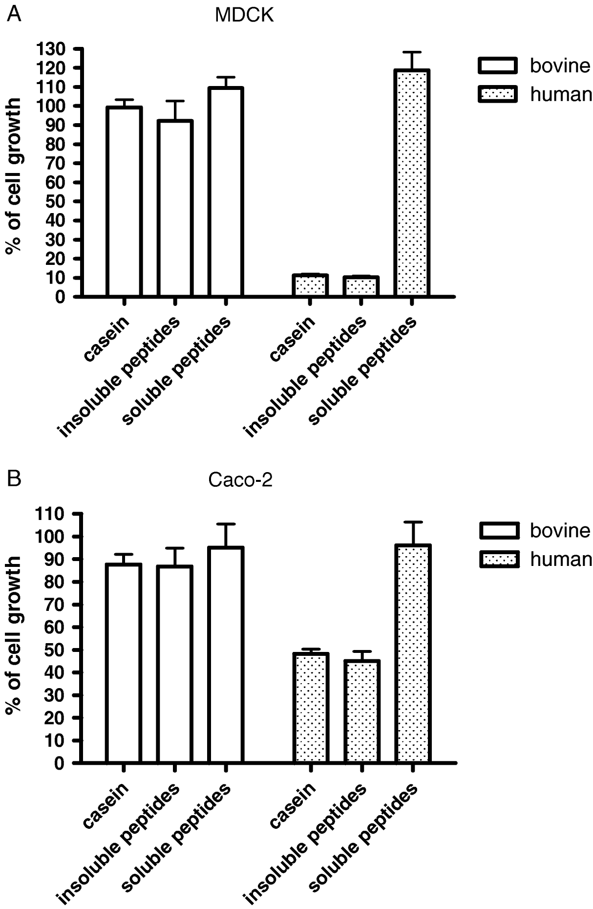
The effect of trypsin on the activity of human and bovine casein on MDCK and Caco-2 cell growth was studied and the results are shown in Fig. 5. The inhibitory activity remained intact in the insoluble peptide fraction after hydrolysis of human casein, while the soluble peptides did not show any activity on the growth of both cell lines. These results show that the factor responsible for the inhibitory activity of human casein must be strongly associated with this fraction, as the activity of trypsin was not able to release it to the soluble fraction. This finding would indicate that the factor could be a small peptide or a lipid, as both are resistant to proteolysis. In the case of bovine casein, proteolysis did not produce any fragment with either inhibitory or stimulatory activity on cellular growth, as has also been observed for some growth factors that increase their activity after trypsin digestion (Thaler & Michalopoulos, Reference Thaler and Michalopoulos1985).
Effect of trypsin hydrolysis on the activity of human and bovine casein samples on the growth of the cell lines MDCK (A) and Caco-2 (B). Fractions assayed were: whole human casein (sample H), whole bovine casein, soluble and insoluble peptides obtained after casein digestion with trypsin. Cell growth, determined by the NBB method, is expressed as percentage of cell growth obtained in controls (considered as 100%). Error bars represent SD. Each sample was analyzed five times in at least two independent experiments.
Analysis of nuclear morphology and DNA fragmentation
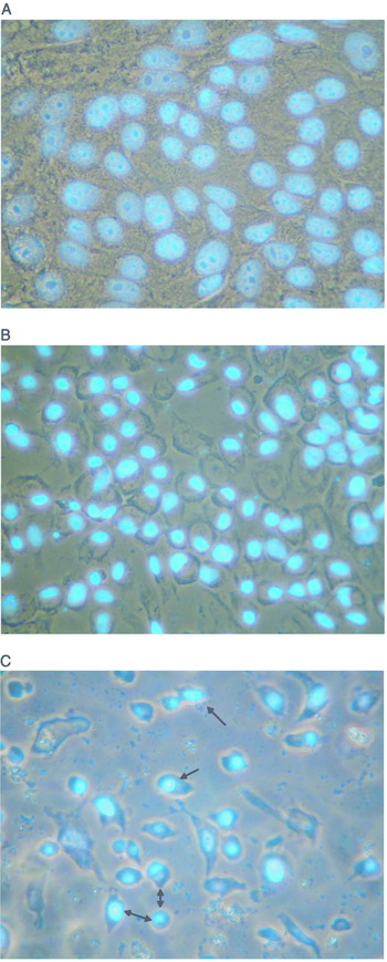
The cytotoxic effect of active milk samples was studied by evaluating the morphology of cells stained with Hoechst 33342, under fluorescence microscopy. Morphological examination of MDCK and Caco-2 cells cultured with samples of active casein revealed features compatible with apoptosis, such as cell shrinkage, membrane blebbing, nuclear condensation and chromatin aggregation (Kerr et al. Reference Kerr, Wyllie and Currie1972), as shown in Fig. 6B for MDCK cells still adhering to the plate. Cells that had detached from the plate due to the inhibitory effect of active casein samples were collected, stained and examined. These cells showed similar morphological features to those shown by cells attached to the plate (Fig. 6C). Cells incubated with samples of non-active caseins in the same conditions retained their normal morphology and size, with relaxed chromatin in the nuclei (Fig. 6A).
Morphological appearance of MDCK cells under fluorescence microscope (×100) after cell staining with Hoechst 33342. (A) Control, (B) cells incubated with active casein at a concentration of 10 mg/ml, (C) cells recovered from the supernatants of cultures incubated with 10 mg/ml active casein. Apoptotic features, such as cell shrinkage and membrane blebbing are marked with arrows in Fig. 6C.

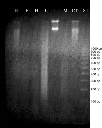
Agarose gel electrophoresis of DNA extracts from MDCK cells incubated with active casein samples at a concentration of 10 mg/ml. Lanes are labelled with the letter that corresponds to the casein used in the experiment of the effect on cell growth, control (CT) and standard (ST).

DNA was isolated from cells with abnormal morphology and from normal cells and subjected to electrophoresis (Fig. 7). Control cells and those incubated with non-active samples showed intact DNA. The majority of cells incubated with active milks showed internucleosomal fragmentation of DNA. The absence of fragmentation in some cells incubated with active milks (E and J) could be due to the fact that this feature only appears at a relatively advanced stage of apoptosis, which may not have been reached in those cultures. The fact that the inhibitory factor associated with casein is an inducer of apoptosis rather than necrosis may be relevant to understanding the mechanisms related to mammary gland tissue remodelling, with development of neonatal tissues, and also with its possible application. However, many unanswered questions remain about the nature of the factor responsible for the high cytotoxic activity of some human milk samples and more studies are in progress.
We thank Dr. Javier Naval for his advice on cell morphology analysis and Dr. Luis Monteagudo for the help with DNA analysis. This work was financially supported by grants HF 2001-0077 and AGL 2001-1814 from CICYT and also by a grant obtained by Coloma Pocoví from MEC (Spain).