History
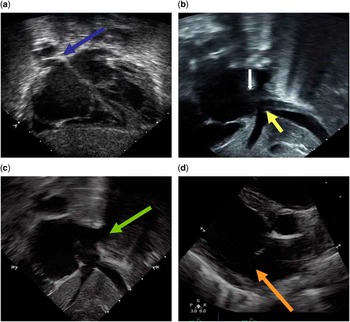
A 2-year-old girl presented to our institution after a recent umbilical hernia repair for respiratory distress and an umbilical abscess. The patient’s vital signs revealed a heart rate of 181 beats/minute, respiratory rate of 80, and oxygen saturation of 90% on room air that improved to 95% on 3 L of oxygen via the nasal cannula. Owing to difficulty in weaning her off oxygen, a chest X-ray was performed that demonstrated mild cardiomegaly, right atrial enlargement, and pulmonary interstitial oedema. These findings prompted a cardiac evaluation, which identified a grade 1/6 low-pitched systolic murmur at the upper left sternal border. Her electrocardiogram showed peaked P-waves consistent with right atrial enlargement and tall R (20 mm)-waves in V1, consistent with right ventricular hypertrophy. The echocardiogram demonstrated severe right atrial dilatation, malposition of the septum primum to the left, inferior sinus venosus defect, and possible anomalous drainage of all of the right-sided pulmonary veins to the right atrium (Fig 1a–d).
( a ) Sub-xiphoid long-axis view demonstrating septum primum malposition defect and anomalous drainage of the right upper pulmonary vein (blue arrow). ( b ) Sub-xiphoid short-axis view demonstrating overriding of the inferior caval vein (yellow arrow) and the absence of the septal tissue at the mouth of the inferior caval vein. The atrial septum is marked by a white arrow. ( c ) Sub-xiphoid sagittal view illustrating the anomalous drainage of the right lower pulmonary vein (green arrow). ( d ) Parasternal short-axis view illustrating a defect in the posteroinferior part of the atrial septum (orange arrow).

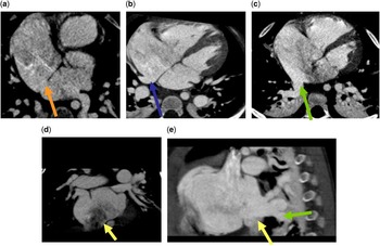
Because of this unusual anatomy and to better define the anomalous pulmonary venous drainage, a cardiac CT scan was performed, which confirmed the absence of the septum secundum, inferior sinus venosus defect, and septum primum malposition defect. In addition, it further delineated the pulmonary venous anatomy, demonstrating anomalous drainage of the right upper pulmonary vein to the right atrium and the right lower pulmonary vein at the level of inferior sinus venosus defect, having normal connection to the left atrium (Fig 2a–e). We have elected to allow our patient to grow and recover from her illness before referring for surgical repair.
( a ) Cardiac CT demonstrating a defect in the posteroinferior part of the atrial septum (orange arrow). ( b ) Cardiac CT illustrating septum primum malposition and anomalous drainage of the right upper pulmonary vein to the right atrium (blue arrow). ( c ) Cardiac CT illustrating septum primum malposition and the anomalous right lower pulmonary vein (green arrow) to the right atrium. ( d ) Cardiac CT demonstrating overriding of the inferior caval vein and a large defect at the mouth of inferior caval vein. ( e ) Cardiac CT illustrating the anomalous drainage of the right lower pulmonary vein into the right atrium (green arrow), just above the entrance of the inferior caval vein (yellow arrow).

Discussion
We report a rare case of inferior sinus venosus defect associated with the absent septum secundum and septum primum malposition defect, resulting in an anomalous venous drainage of the right upper and lower pulmonary veins to the right atrium, with normal pulmonary venous connection to the left atrium. Inferior sinus venosus defect and septum primum malposition defects are both rare cardiac malformations and their association is extremely rare and only two cases have been reported to date.Reference Valente, Sena, Powell, Del Nido and Geva 1
Inferior sinus venosus defect is a rare veno-atrial communication located posteriorly and inferiorly in the mouth of the inferior caval vein along with anomalous right pulmonary venous drainage to the inferior caval vein, while the pulmonary veins retaining normal connection to the left atrium.Reference Ettedgui, Siewers, Zuberbuhler and Anderson 2 , Reference Attenhofer Jost, Connolly and Danielson 5 It accounts for about 3% of atrial septal defectsReference Crystal, Al Najashi, Williams, Redington and Anderson 3 and 5% of sinus venosus defects.Reference Muñoz-Castellanos, Espinola-Zavaleta, Kuri-Nivón, Ruiz and Keirns 4
In one study, it was found that 50% of the patients with inferior sinus venosus defect were misdiagnosed as secundum atrial septal defect by echocardiography preoperatively, but the authors found that the surgical outcomes were not affected by this misdiagnosis.Reference Attenhofer Jost, Connolly and Danielson 5 Another study comparing the outcomes of secundum atrial septal defect and inferior sinus venosus defect found that 64% of the patients with inferior sinus venosus defects were misdiagnosed by preoperative echo imaging and 40% intraoperatively, but the authors demonstrated preoperative diagnostic accuracy improving over time.Reference Banka, Bacha, Powell, Benavidez and Geva 6 This study had disparate outcomes from the first, demonstrating worse surgical outcome, prolonged cardiopulmonary bypass time, longer ICU stay, and increased rate of reintervention as compared with the secundum atrial septal defect group.Reference Banka, Bacha, Powell, Benavidez and Geva 6
Accurate preoperative diagnosis is important to improve the perioperative outcome and decrease the incidence of reintervention for residual defects. With the current technological advancement, transthoracic echocardiography can accurately diagnose the defect in majority of children with sub-xyphoid and parasternal short-axis views, demonstrating overriding of the inferior caval vein with the absence of the septal tissue at the mouth of the inferior caval vein;Reference Ettedgui, Siewers, Zuberbuhler and Anderson 2 , Reference Attenhofer Jost, Connolly and Danielson 5 , Reference Plymale, Kolinski, Frommelt, Bartz and Tweddell 7 however, it may be difficult in obese and older patients because of poor sub-xyphoid windows. In such patients, alternative imaging modalities such as transoesophageal echocardiogram, cardiac CT, or cardiac MRI can be helpful to clearly delineate the defect and coexisting anomalous pulmonary venous drainage.Reference Plymale, Kolinski, Frommelt, Bartz and Tweddell 7 – Reference Beerbaum, Parish, Bell, Gieseke, Körperich and Sarikouch 10 Cardiac MRI has an additional benefit of volumetric analysis and shunt quantification using phase-contrast flow measurements.Reference Prompona, Muehling, Naebauer, Schoenberg, Reiser and Huber 8
Excellent surgical results have been reported utilising various surgical approaches, including closure of the inferior sinus venosus defect by displacement of the posterior edge of the atrial septum and suturing it to the right of the right pulmonary veinsReference Sturm and Ankeney 11 and reimplantation of the anomalous pulmonary vein along with direct suture closure of the defect,Reference Perna and Alajmo 12 closure of the defect with a patch or construction of a baffle to redirect the pulmonary venous flow to the left atrium.Reference Attenhofer Jost, Connolly and Danielson 5
Septum primum malposition defects have been speculated to result from displacement of the septum primum as a result of underdevelopment of the septum secundum, which allows anomalous drainage of half or all of the pulmonary veins to the right atrium, but having normal connection to the left atrium.Reference Van Praagh, Carrera, Sanders, Mayer and Van Praagh 13 It has been shown to be associated with visceral heterotaxy in majority of patients, of which 92% had polysplenia and 87% had rhythm disturbances.Reference Van Praagh, Carrera, Sanders, Mayer and Van Praagh 13 Our patient is unusual in that she had neither of these findings. In one study, it was demonstrated that one was able to accurately delineate the defect with two-dimensional echocardiography with sub-xyphoid coronal, parasternal short-axis, and apical four-chamber views.Reference Tomar, Radhakrishnan and Shrivastava 14 Anatomic repair was achieved by atrial septal incision at the posterior edge and repositioning to redirect pulmonary venous flow to the left atrium and excellent results have been reported.Reference Hiramatsu, Takanashi and Imai 15
Conclusion
Septum primum malposition defects and inferior sinus venosus defect are rare cardiac malformations, which are associated with anomalous pulmonary venous drainage to the right atrium and normally connected to the left atrium. To date, the association of septum primum malposition defect with inferior sinus venosus defect has been reported only in two cases. Transthoracic echocardiography is an excellent non-invasive imaging modality to accurately diagnose both defects and associated anomalous pulmonary venous drainage in a majority of patients. We recommend additional imaging, such as cardiac CT or MRI in patients with doubtful diagnosis and to clearly delineate pulmonary venous anatomy. A high index of suspicion is essential for accurate preoperative diagnosis, which can significantly improve perioperative outcome and reduce the need for reintervention.
Acknowledgement
The authors would like to thank Jennifer Staley for her assistance with literature search.
Financial Support
This research received no specific grant from any funding agency, commercial or not-for-profit sectors.
Conflicts of Interest
None.