Introduction
Human strongyloidiasis, a soil-transmitted helminthiasis and one of the most neglected tropical diseases, is a deleterious gastrointestinal disease mainly caused by Strongyloides stercoralis infection (Olsen et al., Reference Olsen, van Lieshout and Marti2009; Schär et al., Reference Schär, Trostdorf, Giardina, Khieu, Muth, Marti, Vounatsou and Odermatt2013). Estimates of the number of people infected vary, but are as high as 370 million worldwide (Olsen et al., Reference Olsen, van Lieshout and Marti2009; Bisoffi et al., Reference Bisoffi, Buonfrate and Montresor2013; Nutman, Reference Nutman2017). Humans are infected by the skin penetration of Strongyloides infective third-stage larvae (Grove, Reference Grove1996). The disease occurs in communities with low standards of sanitation, and among travellers, immigrants from poor areas, immunocompromised individuals and people exposed to soil (Beknazarova et al., Reference Beknazarova, Whiley and Ross2016). Strongyloides stercoralis has been successfully transmitted from humans to experimental dogs and is generally considered a zoonotic nematode (Deplazes et al., Reference Deplazes, Eckert, Mathis, von Samson-Himmelstjerna and Zahner2016; Jariwala et al., Reference Jariwala, Redding and Hewitt2017; Thamsborg et al., Reference Thamsborg, Ketzis, Horii and Matthews2017). However, since different genotypes have been previously identified in different host species, it has been assumed that host-specialized populations of S. stercoralis may exist, and that the risk of zoonotic transmission might be overstated (Ramachandran et al., Reference Ramachandran, Gam and Neva1997; Takano et al., Reference Takano, Minakami, Kodama, Matsuo and Satozono2009; Hasegawa et al., Reference Hasegawa, Sato and Fujita2010; Thamsborg et al., Reference Thamsborg, Ketzis, Horii and Matthews2017). Overall, previous studies strongly support the hypothesis that dogs are a potential source for human S. stercoralis infection and that dogs should be treated, along with their owners, in settings where people are exposed to dog excrement (Jaleta et al., Reference Jaleta, Zhou, Bemm, Schär, Khieu, Muth, Odermatt, Lok and Streit2017; Nagayasu et al., Reference Nagayasu, Aung and Hortiwakul2017).
People in rural communities often share their premises closely with their dogs. That is also the case in north-east Thailand where the prevalence of human strongyloidiasis is high (Laoraksawong et al., Reference Laoraksawong, Sanpool, Rodpai, Thanchomnang, Kanarkard, Maleewong, Kraiklang and Intapan2018). To our knowledge, evidence of zoonotic transmission of S. stercoralis between humans and pet animals has never been reported in Thailand. To address this question, we sequenced mitochondrial and nuclear DNA of individual Strongyloides isolated from humans and dogs at the same time and location in rural areas. The possibility of zoonotic transmission of S. stercoralis is discussed based on the analysis of these sequences.
Materials and methods
Study areas and populations
Faecal samples were collected from humans and dogs of the same households in two villages of the Kalasin province in north-east Thailand: (1) Nong Bua sub-district in Nong Kung Si district (16.716733°N, 103.383900°E); and (2) Phu Din sub-district in Mueang Kalasin district (16.643328°N, 103.517948°E). Most participants were agriculturists and defecated into the surrounding environment while working on their farms. In addition, soil around their houses was generally damp, and they had one or more pets (primarily dogs).
Stool examination and sample collection
Strongyloides free-living adults and larvae were detected using an agar-plate culture (APC) (Arakaki et al., Reference Arakaki, Hasegawa, Asato, Ikeshiro, Kinjo, Saito and Iwanaga1988). A 3-g portion of each faecal sample from humans (n = 220) and dogs (n = 120) was placed on a nutrient agar plate in the field. Strongyloides rhabditiform larvae and free-living adults were observed and collected after three days of culture at room temperature. Parasite stages harvested were fixed in 70% ethanol, transported to the laboratory and kept at −20°C until used for molecular identification. From each positive sample, one adult male free-living worm was used for DNA extraction, amplification and sequencing. Strongyloides species were differentiated from hookworm larvae using morphological criteria (Garcia & Bruckner, Reference Garcia and Bruckner2001). Participants and pet dogs infected with Strongyloides were treated according to the standard guidelines.
DNA extraction, polymerase chain reaction (PCR) and sequencing
Each free-living adult worm selected for sequencing was crushed with a disposable polypropylene pestle (Bellco Glass, Vineland, NJ, USA) and DNA was extracted using the Nucleospin Tissue kit (Macherey-Nagel GmbH & Co, Duren, Germany). PCR primers used were specific for a part of the Strongyloides 18S rRNA gene (hyper-variable region IV (HVR-IV) corresponding to nucleotides between positions 843 and 1410; GenBank number AF279916) and partial cox1 gene (positions 5600–6435; GenBank number AJ558163). PCR conditions were as previously described (Laymanivong et al., Reference Laymanivong, Hangvanthong and Insisiengmay2016). DNA sequencing was done using the Applied Biosystems 3730×I DNA Analyzer and ABI Big Dye Version 3.1 (Applied Biosystems, Foster City, CA, USA) in both directions, using the PCR primers as sequencing primers. The gene sequences were analysed using a nucleotide BLAST search via NCBI (National Centre for Biotechnology Information). New sequences of both regions were aligned with reference sequences from the NCBI. Alignment lengths were 552 bp for cox1 of S. stercoralis and 331 bp for the 18S rRNA region of S. stercoralis after trimming the primer sequences and reducing the alignment to the length of the shortest published sequence using BioEdit (Hall, Reference Hall1999).
Alignment and phylogenetic analysis
All sequences were aligned using ClustalW (Thompson et al., Reference Thompson, Higgins and Gibson1994). Phylogenetic relationships were inferred using the maximum likelihood method implemented in MEGA version 6.0 (Tamura et al., Reference Tamura, Stecher, Peterson, Filipski and Kumar2013). The Hasegawa–Kishino–Yano model (Hasegawa et al., Reference Hasegawa, Kishino and Yano1985), with non-uniformity of evolutionary rates among sites modelled by using a discrete Gamma distribution (+G), was found to be the best model for 18S rRNA and cox1 sequences. Support for groupings within the tree was evaluated by bootstrapping with 1000 pseudoreplications. DnaSP v5 was used to infer haplotype composition and haplotype diversity (Librado & Rozas, Reference Librado and Rozas2009). A median-joining network was constructed using the Network software version 5.0.0.0 (Fluxus Technology Ltd., www.fluxus-engeneering.com).
Results
A total of 220 human study participants, 120 from Nong Bua sub-district and 100 from Phu Din sub-district, were enrolled in the study. Twenty three (23/220; 10.45%) of the participants were found to be positive for Strongyloides infection according to APC. We obtained rectal faecal samples from 120 dogs. Five dogs were positive for Strongyloides by APC. In the same community, some dog owners were infected, while their dogs were not, and vice versa. The 23 infected owners had, between them, 45 uninfected dogs. All five infected dogs belonged to uninfected owners.
Each of the 28 adult worms sequenced from humans (n = 23) and dogs (n = 5) possessed an 18S sequence identical (100% similarity and 100% query coverage) to GenBank accessions of S. stercoralis (KU962182 and KY081221), as reported from Lao PDR and Thailand. These sequences correspond to HVR-IV haplotype A of Jaleta et al. (Reference Jaleta, Zhou, Bemm, Schär, Khieu, Muth, Odermatt, Lok and Streit2017). Phylogenetic analysis placed all these sequences in a clade with S. stercoralis, distinct from other species (supplementary fig. S1)
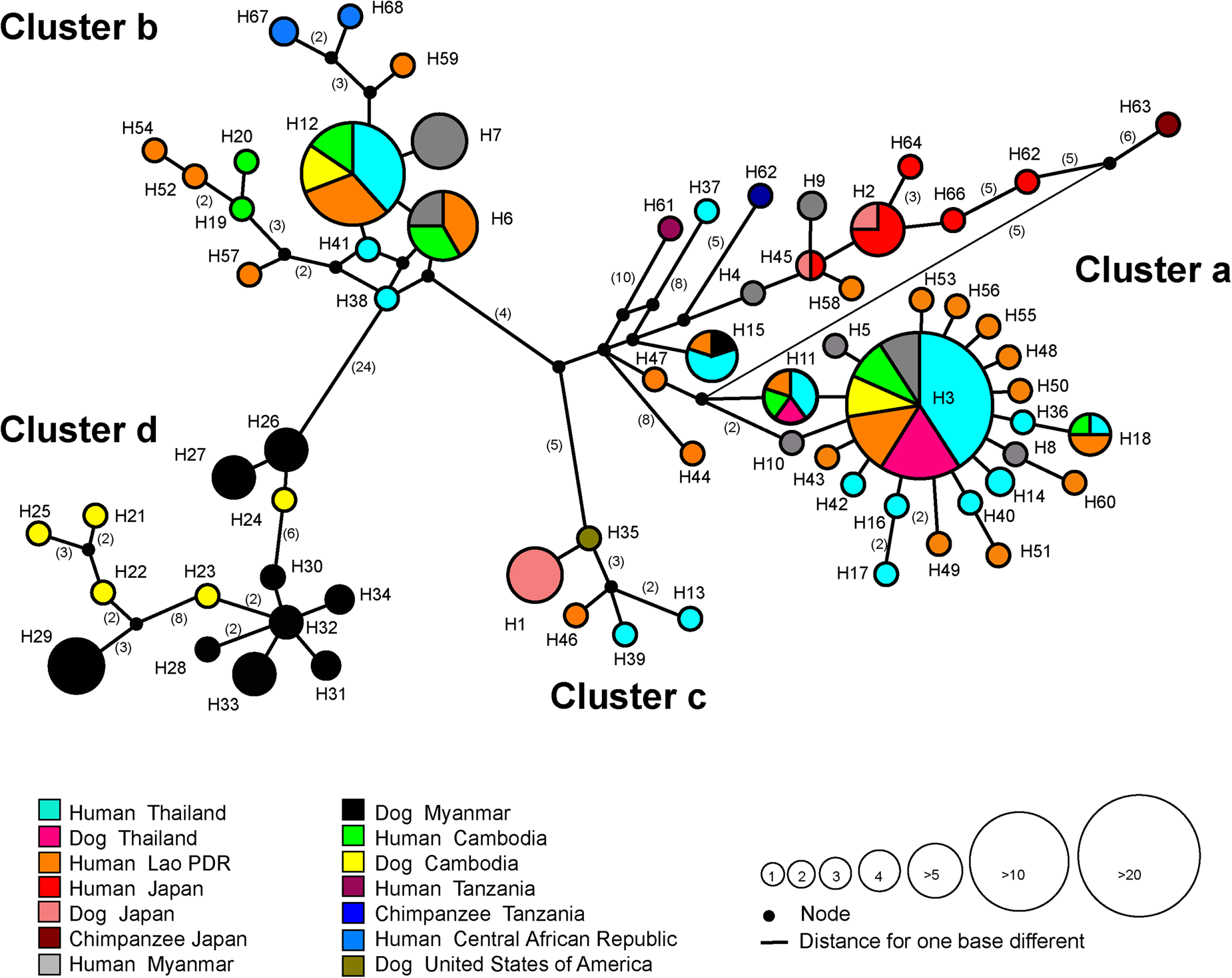
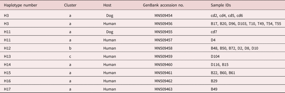
All 28 cox1 sequences from humans and dogs were clustered with reference sequences of S. stercoralis from GenBank (n = 150). In a phylogenetic tree (supplementary fig. S2) and a median-joining network (fig. 1), our new sequences were located in S. stercoralis clusters a, b and c of Thanchomnang et al. (Reference Thanchomnang, Intapan and Sanpool2017), with previously reported sequences from Thailand (KY081237–KY081241), Lao PDR (KU962164–KU962166) and Myanmar (LC179139–LC179147). In total, the 178 sequences represented 68 haplotypes. Our new sequences fell into eight distinct haplotypes (fig. 1), most of which were placed in cluster a (21 sequences from humans and dogs belonging to haplotypes 3, 11, 14–17). Fewer new sequences belonged to cluster b (six sequences from humans; haplotype 12) and cluster c (one sequence from a human; haplotype 13), while cluster d comprised sequences from dogs in Cambodia and Myanmar. Strongyloides stercoralis sequences from dogs (this study) represented haplotypes 3 and 11 shared with humans from Thailand, Lao PDR, Myanmar and Cambodia (fig. 1, table 1).
Median-joining haplotype network for the mitochondrial cox1 gene. Each circle represents one haplotype. The size of the circle is proportional to the number of samples with that haplotype. Numbers in parentheses indicate numbers of nucleotide differences inferred as having occurred between nodes. The colours represent the different host species and geographical origin (country).

Haplotypes of Strongyloides stercoralis cox1 sequences from the present study deposited in GenBank.

Discussion
We have presented the molecular identification and phylogeny of S. stercoralis infection in humans and dogs from the two rural communities in Thailand. Phylogenetic analysis of the 18S rRNA gene (HVR-IV haplotype A) identified the causative agent as S. stercoralis (supplementary fig. S1). We observed that HVR-IV haplotype A is associated with strains infective both to humans and dogs, while HVR-IV haplotype B (not found in this study) is restricted to strains that are only infectious to dogs. The same result was also discovered in recent studies of S. stercoralis from humans and dogs in the US, Italy, Australia, Lao PDR and Cambodia (Jaleta et al., Reference Jaleta, Zhou, Bemm, Schär, Khieu, Muth, Odermatt, Lok and Streit2017; Barratt et al., Reference Barratt, Lane and Talundzic2019; Beknazarova et al., Reference Beknazarova, Barratt, Bradbury, Lane, Whiley and Ross2019), where two genetically distinguishable S. stercoralis populations were identified based on the HVR-IV region. Worms with haplotype A were infective to dogs and humans, while those with HVR-IV haplotype B were specific for dogs (Jaleta et al., Reference Jaleta, Zhou, Bemm, Schär, Khieu, Muth, Odermatt, Lok and Streit2017; Barratt et al., Reference Barratt, Lane and Talundzic2019; Basso et al., Reference Basso, Grandt, Magnenat, Gottstein and Campos2019). All worms from dogs that we sequenced presented the HVR-IV haplotype A variant, suggesting a zoonotic potential. The result is consistent with previous reports on possible zoonotic transmission in Australia, Cambodia and Myanmar (Jaleta et al., Reference Jaleta, Zhou, Bemm, Schär, Khieu, Muth, Odermatt, Lok and Streit2017; Nagayasu et al., Reference Nagayasu, Aung and Hortiwakul2017; Beknazarova et al., Reference Beknazarova, Barratt, Bradbury, Lane, Whiley and Ross2019). The whole amplified sequence including HVR-IV of the parasites in this study (accession numbers MN498129–MN498130) showed a 100% identity (529/529 bp) with sequences of S. stercoralis (GenBank accession numbers AF279916) that cannot distinguish between human and dogs by these sequences. Whereas the HVR-I is highly conserved within many nematode species, it appears to be more variable in S. stercoralis, and its usefulness to distinguish dog- and human-adapted strains should be further clarified (Nagayasu et al., Reference Nagayasu, Aung and Hortiwakul2017).
Our median-joining network based on cox1 sequences revealed that S. stercoralis haplotypes obtained in this study were shared by both human and dog hosts (H3 and H11; cluster a). Despite this, there was no correlation between S. stercoralis infection in dogs and the dogs’ owners. This is probably because dogs usually roam freely, depositing contaminated faeces anywhere in the village. A similar pattern was seen in a study of the zoonotic transmission of S. stercoralis between dogs and their owners in Cambodia (Jaleta et al., Reference Jaleta, Zhou, Bemm, Schär, Khieu, Muth, Odermatt, Lok and Streit2017). Taken together, these results suggest that dogs may play a far greater role in the zoonotic transmission of human strongyloidiasis than do other animals (i.e. cats) in rural Thailand. Recently, the role of dogs in the zoonotic transmission of S. stercoralis to humans has been discussed in the literature. Some molecular studies suggest the persistence of different host-adapted variants, subspecies or even species (Ramachandran et al., Reference Ramachandran, Gam and Neva1997; Hasegawa et al., Reference Hasegawa, Hayashida, Ikeda and Sato2009, Reference Hasegawa, Sato and Fujita2010; Jaleta et al., Reference Jaleta, Zhou, Bemm, Schär, Khieu, Muth, Odermatt, Lok and Streit2017; Thamsborg et al., Reference Thamsborg, Ketzis, Horii and Matthews2017). Based on polymorphisms of the nuclear 18S (HVR-I and IV) rDNA sequences and mitochondrial cox1 gene, two genetically different populations occurred in dogs living in an endemic region for human and canine strongyloidiasis in Cambodia. One population was restricted to dogs (‘dog-adapted’) while the other was found both in dogs and humans (‘dog–human shared’) (Jaleta et al., Reference Jaleta, Zhou, Bemm, Schär, Khieu, Muth, Odermatt, Lok and Streit2017). These results would support the existence of a zoonotic species (S. stercoralis) and a dog-adapted species (Strongyloides canis) (Jaleta et al., Reference Jaleta, Zhou, Bemm, Schär, Khieu, Muth, Odermatt, Lok and Streit2017). Similarly, Nagayasu et al. (Reference Nagayasu, Aung and Hortiwakul2017) recently reported the existence of two genetically different lineages of S. stercoralis, mainly characterized on the basis of cox1 sequence data. While worms belonging to their S. stercoralis type A were isolated from both humans and dogs from different countries (i.e. humans from Japan, Myanmar, Thailand, Lao PDR, Uganda, Central Africa and dogs from Japan and Myanmar), those belonging to type B were isolated exclusively from dogs from Myanmar. These authors suggested that type B worms represented an ancestral canid-adapted S. stercoralis line, from which type A parasites evolved, expanding their host spectrum to infect humans (Nagayasu et al., Reference Nagayasu, Aung and Hortiwakul2017). Our cox1 sequences from humans and dogs (clusters a, b and c in fig. 1) all fell within the type A clade of Nagayasu et al. (Reference Nagayasu, Aung and Hortiwakul2017), in which both human and dog S. stercoralis isolates were located, suggesting their zoonotic potential. We did not find any cox1 sequences belonging to cluster d (clade II and type B of Nagayasu et al., Reference Nagayasu, Aung and Hortiwakul2017 and Jaleta et al., Reference Jaleta, Zhou, Bemm, Schär, Khieu, Muth, Odermatt, Lok and Streit2017, respectively). Members of this cluster are known only from dogs in Myanmar and Cambodia.
In conclusion, zoonotic transmission between dogs and humans in rural communities in Thailand appears likely. These results should prompt public health workers to be mindful of the risk of transmission of harmful parasites, such as S. stercoralis, between reservoir hosts and humans. Nationwide assessment and campaigns to control S. stercoralis infection should be conducted to reduce the prevalence and improve prevention and control measures.
Supplementary material
To view supplementary material for this article, please visit https://doi.org/10.1017/S0022149X1900107X
Acknowledgements
We thank Hiroshi Yamasaki for his scientific suggestions. We would also like to thank David Blair for his valuable suggestions and assistance with the presentation of this paper through Khon Kaen University Publication Clinic.
Financial support
This work was supported by a Distinguished Research Professor Grant, Thailand Research Fund (P.M.I. and W.M., grant number DPG 6280002) and the Faculty of Medicine, Mahasarakham University (T.T.).
Conflicts of interest
None.
Ethical standards
The study protocol was approved by the Khon Kaen University Ethics Committee for Human Research (HE601048) and institutional guides on the care and use of laboratory animals, and was approved by the Mahasarakham University Ethics Committee for Animal Research (0022/2560). Each participant was informed of the study methods, risks and benefits of the process. Before enrolment, written consent was obtained from all adult participants and from parents or legal guardians of minors.