Introduction
Strepsiptera is an order of endoparasitic insects known for its unique life cycle and extreme sexual dimorphism (Kathirithamby Reference Kathirithamby1989, Reference Kathirithamby2009, Reference Kathirithamby, Footit and Adler2018). Both sexes develop and feed from within their host, but only the adult males emerge and become free-living, except in the basal Mengenillidae, where the adult females are also free-living (Kathirithamby Reference Kathirithamby1989, Reference Kathirithamby2009, Reference Kathirithamby, Footit and Adler2018). In the other families, adult females remain inside their host’s gaster (abdomen) with only their cephalothorax extruding between the host’s abdominal segments. The presence of these parasites within a host has long been known to cause severe effects on the host’s anatomy, mainly by inhibiting the development of the reproductive organs in female hosts (Pérez Reference Pérez1886; Smith and Hamm Reference Smith and Hamm1914; LaBerge and Ribble Reference LaBerge and Ribble1972; LaBerge Reference LaBerge1973, Reference LaBerge1977, Reference LaBerge1980; Kathirithamby Reference Kathirithamby1989, Reference Kathirithamby2009, Reference Kathirithamby, Footit and Adler2018; Cappa et al. Reference Cappa, Manfredini, Dallai, Gottardo and Beani2014).
Stylops Kirby (Strepsiptera: Stylopidae) is a genus that parasitises bees of the genus Andrena Fabricius (Hymenoptera: Andrenidae). The host-seeking, first-instar larvae leave their mother’s body over the course of weeks (Balzer and Davis Reference Balzer and Davis2020) due to asynchronous development (Kathirithamby Reference Kathirithamby2009). First-instar larvae of Stylops presumably require transport on adult bees in order to transfer to the host’s nest where an immature bee can be detected and parasitised (Ulrich Reference Ulrich1956; Linsley and MacSwain Reference Linsley and MacSwain1957; Balzer and Davis Reference Balzer and Davis2019a). During the following spring, the stylopised females and all males of Andrena emerge from their respective nests around the same time (Linsley and MacSwain Reference Linsley and MacSwain1957; Straka et al. Reference Straka, Rezkova, Batelka and Kratochvíl2011; Balzer and Davis Reference Balzer and Davis2020). Manipulation of stylopised female Andrena to emerge earlier than normal allows for all free-living male Stylops to search out and mate with an endoparasitic female whose cephalothorax protrudes from an emerged bee (Straka et al. Reference Straka, Rezkova, Batelka and Kratochvíl2011).
Andrena, one of the largest bee genera, with an estimated 2000 species (Dubitzky et al. Reference Dubitzky, Plant and Schönitzer2010), is found throughout the Holarctic region and in parts of Africa and Asia (Michener Reference Michener2007). The importance of Andrena and other wild bees as pollinators has become more apparent over the last few decades (Kremen et al. Reference Kremen, Williams and Thorp2002; Memmott et al. Reference Memmott, Waser and Price2004; Morandin and Winston Reference Morandin and Winston2005; Fontaine et al. Reference Fontaine, Dajoz, Meriguet and Loreau2006; Garibaldi et al. Reference Garibaldi, Steffan-Dewenter, Winfree, Aizen, Bommarco and Cunningham2013; Winfree et al. Reference Winfree, Reilly, Bartomeus, Cariveau, Williams and Gibbs2018). Andrena milwaukeensis forages on flowers of many plant species representing several families, presumably pollinating them (LaBerge Reference LaBerge1980; Ascher and Pickering Reference Ascher and Pickering2020).
This study aims to examine the anatomical impact of neotenic females of Stylops advarians Pierce (identified using molecular techniques by Balzer et al. Reference Balzer, Thoroughgood, Diyes, Davis and Chilton2020) on Andrena milwaukeensis Graenicher, which in Saskatoon, Saskatchewan, Canada can be found foraging on Cotoneaster Medik (Rosaceae), Shepherdia Nuttall (Elaeagnaceae), and Syringa Linnaeus (Oleaceae) from early May to late June (Balzer and Davis Reference Balzer and Davis2020). During 2016–2018, the annual prevalence of this strepsipteran parasite on Andrena milwaukeensis was relatively high and consistent (22–24%; Balzer and Davis Reference Balzer and Davis2020). Accordingly, to broadly investigate the impact of the parasite on this native bee species, we have endeavoured to identify the adaptive morphology of its host-seeking first-instar larva (Balzer and Davis Reference Balzer and Davis2019a) and the free-living, winged adult male (Balzer and Davis Reference Balzer and Davis2019b). Eighteen percent of bees (n = 455) hosted a single mature parasite of S. advarians, whereas 3% and 1% of hosts were infected by two or three parasites, respectively (Balzer and Davis Reference Balzer and Davis2020). Here, our main objective was to report anatomical changes occurring in female bees of A. milwaukeensis when they are stylopised by an increasing parasite load – namely one, two, or three adult females of S. advarians – and to compare these results to the anatomy of nonstylopised bees. During this histological pursuit, we incidentally acquired data about viviparous development of the first-instar larvae within the bodies of their neotenic mothers. We include those observations in the present report.
Materials and methods
Study site and specimen collection
To collect female specimens of Stylops advarians, foraging females of Andrena milwaukeensis were sweep-netted from flowers throughout Cosmopolitan Park in Saskatoon, Saskatchewan, Canada (Balzer and Davis Reference Balzer and Davis2019a, Reference Balzer and Davis2020). Captured live bees were taken to the laboratory in vials, and their gasters were examined using an Olympus SZ-ST dissecting microscope (Olympus, Tokyo, Japan) to screen for the presence of the protruding cephalothorax of the Stylops advarians female. Nonstylopised bees were also collected in this same way (controls) for anatomical investigation.
Histological techniques
Four nonstylopised and 11 stylopised bees of A. milwaukeensis from early May to the third week of June (2016–2018) were chosen for anatomical investigation. Gasters were removed and immersed in fixative (2% glutaraldehyde in 25 mM sodium phosphate buffer) for up to a few weeks before processing for resin sectioning in a protocol modified from Benedeczky et al. (Reference Benedeczky, Tanács and Móczár1990) and Chapman and Abu-Eid (Reference Chapman and Abu-Eid2001), but without post-fixation nor processing for electron microscopy. Briefly, this fixative was exchanged three times with the buffer and then distilled water before specimens were dehydrated in an ascending series of ethanol (10, 30, 50, 70, 85, 95, and 100%). Each ethanol change lasted one hour, except for the 100% ethanol, which was introduced three times, the final change being left overnight. Specimens were then infiltrated gradually with a mixture of 100% ethanol and LR White resin (London Resin Company Limited, Basingstoke, United Kingdom). Finally, specimens were infiltrated with fresh resin three times, with the final change occurring overnight. Each gaster was transferred to a Beem capsule and polymerised in an incubator at 60 °C. Specimens were sectioned at two micrometres using a Reichert Ultracut microtome and glass knives and then stained using 1% toluidine blue 0 in a 1% sodium borate solution, a modification from Peinert et al. (Reference Peinert, Wipfler, Jetschke, Kleinteich, Gorb, Beutel and Pohl2016).
Four nonstylopised bees (controls) were sectioned to compare anatomically with stylopised bees. Gasters of nonparasitised bees were sectioned transversely (n = 2) and longitudinally (n = 2). Of the 11 bees stylopised by S. advarians, seven were sectioned transversely, three were sectioned longitudinally, and one was sectioned laterally. Most (n = 7) of the 11 stylopised bees investigated anatomically were infected by a single neotenic female, whereas three and one gasters possessed two and three strepsipterans, respectively. Actual collection dates of the sectioned bees are provided by Balzer (Reference Balzer2019). One stylopised bee collected on 3 May 2017 also was examined in detail for eggs, embryos, and first-instar larvae within a viviparous neotenic female.
To assist interpretation of the anatomy of female Stylops, we followed Peinert et al. (Reference Peinert, Wipfler, Jetschke, Kleinteich, Gorb, Beutel and Pohl2016). For specific histological features of the host’s digestive tract, we were guided by Snodgrass (Reference Snodgrass1910). Chapman and Abu-Eid (Reference Chapman and Abu-Eid2001), Serrão (Reference Serrão2005), Santos and Serrão (Reference Santos and Serrão2006), and Ceylan et al. (Reference Ceylan, Sevin and Özgenc2019).
Imaging
Sections were initially viewed and screened using an Olympus CH30 light microscope at 40–400X. Selected sections were imaged using a Zeiss AxioPlan microscope (Zeiss, Oberkochen, Germany) with an AxioCam105 (Zeiss) mounted to the top. Images captured (25–400X) with this camera were adjusted for contrast and colour using the software ZEN 2 (Zeiss, Jena, Thuringia, Germany).
Specimen deposition
Pinned bees with female parasites of S. advarians, as well as the anatomical sections prepared during this study, are housed in the Department of Biology, University of Saskatchewan, Saskatoon. One free-living male specimen (paratype) of S. advarians is stored in 70% ethanol as RSKM_ENT_E-216645 (Balzer and Davis Reference Balzer and Davis2019b) at the Royal Saskatchewan Museum in Regina, Saskatchewan.
Results
Nonstylopised versus stylopised bee anatomy
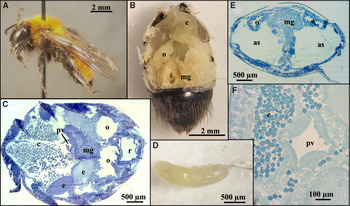
Examination of nonstylopised A. milwaukeensis (Fig. 1A) showed that the bee’s abdominal organs occupied the gaster fully (Fig. 1B). All three domains (foregut, midgut, hindgut) of the digestive tract were represented in the gaster. The foregut’s oesophagus extended through the thorax (not shown) and connected at its posterior to the expanded crop (honey sac) located anteriorly in the gaster (Fig. 1B – top, C – left). The major organs of the digestive tract, such as the crop, ventriculus (midgut), and rectum, resided anteriorly to posteriorly within the abdomen and were centralised medially (i.e., not shifted to the left or right side; Fig. 1C). The crop was often laden with pollen (Fig. 1C, F) and, when full, occupied a large portion of the abdomen (Fig. 1B, C). The proventriculus consisted of four lobes lining the narrow lumen (Fig. 1F) and terminated the foregut by connecting the crop to the midgut (Fig. 1C). The latter occupied the gaster posterodorsally (Fig. 1B, C, E). Via the ileum (not shown), the midgut was connected to a rectum sometimes found empty (Fig. 1C) – presumably simply reflecting a recent voiding of wastes – but was often filled with nondigested exines of pollen (Figs. 2C, 3B). Reproductively, nonstylopised bees were fertile. Eggs inside the elongate ovaries were present dorsally (Fig. 1B, E) upon dissection of several nonstylopised bees (Fig. 1D), and a bee’s pair of ovaries, each with two or more developing eggs, was detected in some sections of noninfested bees (Fig. 1C). Representing the gaster’s major sector of the respiratory system, two large air sacs resided anteriorly and ventrolaterally within the gaster, thus flanking the major components (crop, midgut) of the digestive tract, below the ovaries (Fig. 1E).
A, Nonstylopised female of Andrena milwaukeensis and B–F, photographs and light micrographs of the gasters of several nonstylopised A. milwaukeensis. B, An isolated gaster with several tergites removed to show the anterior of the abdominal digestive tract. The tergites covering the hindgut remain. C, Longitudinal section of the gaster showing the crop (containing hundreds of pollen grains) leading to the proventriculus and anterior portion of the ventriculus (midgut). Note two large eggs developing within each ovary. D, Isolated ovary with developing eggs; oviduct at right. E, Transverse section of the gaster depicting the centralised, dorsal placement of the midgut that intervenes the pair of ovaries and large air sacs, located laterally. F, Transverse section of the gaster showing the mid-region of the proventriculus, which leads from the pollen-filled crop to the anterior of the midgut. as, air sac; c, crop; e, egg; mg, midgut; o, ovary; pv, proventriculus; r, rectum.

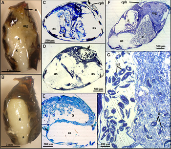
Gasters of female Andrena milwaukeensis infected with one adult female of Stylops advarians. A, Ventral view of host gaster following removal of several sternites, showing the neotenic parasitic female and the host’s organs beside and below her. B, Ventral view of 2A following removal of most of the host’s abdominal organs, depicting the full volume of the gaster occupied by the neotenic parasite in situ. The asterisk approximately corresponds to location of the asterisk in 2E. C, Transverse section showing the lateral location of the pair of air sacs near the posterior end of the host bee. At this plane of sectioning, the parasite is entirely external to the gaster, with its cephalothorax residing on the host’s tergite. Note the first-instar larvae (arrow) of S. advarians within the birth canal of their mother’s cephalothorax. The diamonds denote the ducts beside the rectal pads. D, Transverse section of gaster showing the female parasite’s body occupying upper region, above an air sac of the host. Note first-instar larvae (arrow) that have developed within their mother’s body. E, Tangential longitudinal section of the host gaster showing the neotenic female parasite residing dorsally and longitudinally along one side of her host. Hundreds of first-instar larvae, liberated from their mother’s ovarial matrix, are evident in her body cavity. Host’s air sac below the adult parasite in figure. This asterisk approximately aligns with the asterisk of Fig. 2B. F, Lateral section showing the female’s abdomen occupying the upper portion of her host, with her body supported by an air sac and the crop. G: Section highlighting the asynchronous development of first-instar larvae within an adult female parasite. Mature larvae at left have moved out of their mother’s haemolymph, whereas immature larvae (right) are still found within haemolymph. as, air sac; c, crop; cph, cephalothorax of female Stylops; il, immature first-instar larvae; mg, midgut; ml, mature first-instar larvae; o, host’s ovipositor (sting); S, female Stylops.

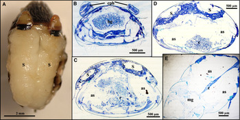
Gasters of female Andrena milwaukeensis infected with two adult females of Stylops advarians. A, Gaster with its anterior tergites removed, revealing the in situ location of the two adjacent, longitudinally oriented females, and B–D, transverse sections of two different hosts. B, Posterior end of a bee’s gaster showing the cephalothoraces of the two strepsipterans above the host’s tergites, not yet localised within its body. C, Middle of the gaster, showing the host’s midgut between the two air sacs, which each support a parasite above. D, Abdomens of the two female parasites, each above an air sac. Note the host’s crop, with an abundance of pollen, confined towards the gaster’s venter. E, Ventral tangential-longitudinal section through the gaster’s pair of air sacs below the two strepsipterans (not seen), showing the heavily reduced volume of the host’s crop. as, air sac; c, crop; cph, cephalothorax; hg, hindgut; mg, midgut; r, rectal sac; S, female Stylops.

When one adult female S. advarians inhabited the gaster of A. milwaukeensis, the host’s internal organs were no longer centralised, but were shifted to the side opposite from where the female parasite resided (Fig. 2A, E), as well as below the parasite (Fig. 2A, F). Figure 2B displays the region of the host’s gaster occupied by the female parasite after most of the host’s organs were removed. The posterior end of the host bee’s gaster was less affected than the anterior region because the female parasite’s body does not occupy the gaster beyond the host’s fourth segment (Fig. 2F). However, the host’s midgut and rectum were more tightly squeezed into the gaster’s posterior region (Fig. 2A, F). Near the bee’s posterior end, the parasitic female’s cephalothorax extruded externally above and along the tergite of her host (Fig. 2C, F). Where the female parasite’s body occupied her host, it was supported by one of her host’s air sacs (Fig. 2D, F). Neither ovaries nor eggs belonging to the host bee were discernible in hosts infected by a neotenic female of S. advarians (Fig. 2D–F).
When two female parasites of S. advarians presented in the same gaster of A. milwaukeensis, a slight difference was apparent in their body size, but they both protruded above the same tergite (Fig. 3A). The host’s crop and midgut were not shifted laterally away from one parasite, likely due to the presence of the second. Instead, these major organs of the bee’s digestive tract were compressed below the two parasites (Fig. 3A) and towards the posterior end of the bee’s abdomen (Fig. 3C–E). Expansion of the crop evidently was restricted severely by the presence of the two parasites and by the air sacs on either side of it (Fig. 3D, E). As a result, the crop occupied a ventral position in the gaster of the doubly infected host (Fig. 3D). The posterior end of the bee, however, remains relatively unaffected because the cephalothoraces and the first abdominal segments of the two female parasites were not contained distally within the host (Fig. 3A, B). Where the two parasites did occupy the host’s gaster, the two air sacs appeared to flank each side of the midgut (Fig. 3C) and likely supported the abdomens of the female parasites (Fig. 3C, D). No recognisable signs of reproductive structures or of eggs of the host bees were observed inside their gasters (Fig. 3C–E).
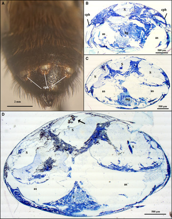
Abdominal organisation inside bees of A. milwaukeensis infected by three female parasites showed similar results to those infected by two. The third female parasite occupied the centre of the host’s gaster and was flanked by the other two females (Fig. 4A). Although all cephalothoraces extended from the same tergite of the host’s gaster, extrusion of the central occupant was staggered slightly compared to the two laterally residing parasites: its cephalothorax extended less posteriorad (Fig. 4A) or more posteriorad (Fig. 4B) than those of its neighbours. The three female parasites occupied a greater volume inside the host bee’s gaster than when two females were present, but the organs were similarly shifted beneath the abdomens of the three parasites (Fig. 4B, C). The gaster’s major anterior organs (crop, midgut) of the digestive system were reduced (Fig. 4C, D), similar to those in bees stylopised by one or two female parasites. Neither reproductive structures nor eggs of the host were observed inside the triply parasitised gaster (Fig. 4C, D). The females that occupied the left and right side of the bee’s gaster were supported by the host’s air sacs (Fig. 4C, D). The central female, however, was supported most directly by the females on either side of her (Fig. 4C, D).
Gasters of female Andrena milwaukeensis infected with three adult females of Stylops advarians. Photographs of the following: A, the gaster showing the slightly staggered extrusion of the cephalothoraces of the three female parasites protruding between the same tergites, and B–D, transverse sections at different planes of the same bee’s gaster. B, Section near gaster’s posterior end, showing staggered location of anterior body regions of the three adjacent parasites, dorsad to the host’s hindgut and abdominal air sacs. C, Section near mid-region of the gaster, showing the reduced and ventrally residing midgut, as well as the host’s air sacs below the three female parasites. D, Section near the middle of the gaster, showing the reduced crop towards the host’s venter. The two lateral-most female parasites are each supported by one of the host’s air sacs, whereas support for the central female is shared by the females adjacent. Note first-instar larvae (arrow) within body cavity of central parasite. as, air sac; c, crop; cph, cephalothorax; hg, hindgut; mg, midgut; S, female Stylops.

Observations of viviparous and asynchronous development of Stylops advarians
When sectioning gasters of A. milwaukeensis infected by neotenic females of S. advarians in situ, it became apparent that larval development was both viviparous and asynchronous. Fully developed first-instar larvae were observed alongside embryos within the parasite’s abdomen (Fig. 2G), whereas first-instar larvae simultaneously were evident within the brood canal and emerging from the birth opening (Peinert et al. Reference Peinert, Wipfler, Jetschke, Kleinteich, Gorb, Beutel and Pohl2016) of the female’s cephalothorax (Fig. 2C, D), illustrated by scanning electron microscopy in other specimens (Balzer and Davis Reference Balzer and Davis2019a). The bee shown in Fig. 2C–D was collected on 13 June 2017, almost three weeks after that year’s inaugural observation of first-instar larvae (24 May 2017) at the field site sampled. The specimen shown in Fig. 2G, which shows free mature larvae (left) within the mother’s haemolymph in proximity to immature larvae still enclosed within the haemocoel (right) of their mother’s abdomen, was collected on 8 June 2016, two weeks after first-instar larvae were first observed (25 May 2016).
Discussion
The most significant impact of female S. advarians on female A. milwaukeensis is the loss of the host’s reproductive organs. Strepsiptera have long been known to inhibit the development of the reproductive organs of their host (Pérez Reference Pérez1886; Smith and Hamm Reference Smith and Hamm1914; Kathirithamby Reference Kathirithamby1989, Reference Kathirithamby2009; Cappa et al. Reference Cappa, Manfredini, Dallai, Gottardo and Beani2014) and to impact host morphology (Pérez Reference Pérez1886; Pierce Reference Pierce1909; Smith and Hamm Reference Smith and Hamm1914; LaBerge Reference LaBerge1973, Reference LaBerge1977, Reference LaBerge1980), and we found the same results in stylopised A. milwaukeensis. Nonstylopised bees had distinct ovaries and eggs present, whereas female bees stylopised by even a single parasite – 82 of 99 (83%) of all infestations of A. milwaukeensis by S. advarians examined (Balzer and Davis Reference Balzer and Davis2020) – showed no sign of reproductive organs other than the ovipositor, which now functions only as a sting.
Only bees identified as female were examined anatomically in the present study, although stylopisation can cause morphological changes in Andrena (LaBerge Reference LaBerge1973, Reference LaBerge1977, Reference LaBerge1980). Further research is required on male Andrena, but Smith and Hamm (Reference Smith and Hamm1914) report little impact on the testes of stylopised males of A. nigroaenea Kirby. Presumably, the testes require fewer nutrients than the larger ovaries, and thus they are not affected by the presence of a Stylops (Smith and Hamm Reference Smith and Hamm1914). A similar result was found in stylopised male paper wasps, Polistes dominula Christ (Hymenoptera: Vespidae) (Cappa et al. Reference Cappa, Manfredini, Dallai, Gottardo and Beani2014), wherein the wasps were still observed to maintain their sexual behaviour, and development of their reproductive organs was not inhibited by the strepsipteran parasite (Cappa et al. Reference Cappa, Manfredini, Dallai, Gottardo and Beani2014). Additional studies are necessary to determine the impact of parasite load on male hosts – namely, whether the number of strepsipterans within the same gaster of a male bee or wasp affects development of the testes and sperm production.
Besides sterilisation of female A. milwaukeensis caused by a single neotenic female of S. advarians, the presence of one or more female Stylops likely impacted the amount of pollen and nectar stylopised bees could collect and store in their crops. When one female is present in a host, the crop apparently still has room to expand upwards and laterally to the side opposite the parasite. However, when two or three female parasites are present per host – as was detected in 17 of 99 (17%) stylopised bees (Balzer and Davis Reference Balzer and Davis2020) – the capacity of the crop’s expansion is heavily restricted; thus, food intake and storage appear limited. The midgut and rectum of stylopised bees, however, did not seem to be greatly affected. These digestive organs may become more compressed into the posterior end of the bee, but because the female parasite’s body is not inside her host’s in this region, these organs are likely still able to expand to close their normal size and complete their normal functions. Furthermore, the aforementioned loss of the singly stylopised female bee’s reproductive organs may provide additional space for the midgut and rectal sac of the hindgut to expand within the bee’s gaster.
Females of S. advarians were most often found dorsolaterally within the host’s gaster, with their cephalothoraces protruding between the fourth and fifth tergites (Balzer and Davis Reference Balzer and Davis2020). This positioning may allow the horizontally oriented abdomen of the female parasite to be supported most favourably inside her host, discovered in the present study to occur above the host’s air sacs. In the relatively rare occurrence (4%) of three parasites of S. advarians in a stylopised A. milwaukeensis (Balzer and Davis Reference Balzer and Davis2020), the third, central female is supported most directly along the host’s midline by the two female parasites flanking her rather than substantially by either of the bee’s two air sacs of the gaster.
Histological observations of neotenic females of S. advarians in situ revealed that the first-instar larvae within their mother exhibited both viviparous and asynchronous development (Kathirithamby Reference Kathirithamby2009; Balzer and Davis Reference Balzer and Davis2020). Therefore, the asynchronous production of primary larvae per neotenic female of S. advarians evidently extends the duration and hence distribution of these host-seeking larvae during the host’s foraging activities, leading to the birth of live young (ovoviviparity; Balzer and Davis Reference Balzer and Davis2019a). However, it is unclear from our anatomical observations, which involved destructive and hence one-time sampling of neotenic females of S. advarians, whether all eggs within the neotenic female eclose synchronously, with staggered development of the first-instar larvae then occurring, or whether eggs hatch at different times.
We show that female A. milwaukeensis are greatly impacted by the presence of one or more S. advarians female parasites within their gasters. Fortunately, A. milwaukeensis populations are able to withstand the loss of some reproductivity, as evidenced by their expansive range (LaBerge Reference LaBerge1980). More study is required into the biology of the many bee-infecting species of Strepsiptera and the bees that they infect to improve understanding of the impacts of these parasites on their hosts and to consider the results alongside the findings on pesticide use and habitat loss.
Acknowledgements
The authors appreciate the constructive comments by two anonymous reviewers of the original submission, plus thank the study’s funding sources, namely a University of Saskatchewan Graduate Scholarship, the Arthur R. Brooks Prize from the Entomological Society of Saskatchewan (ZSB), and an NSERC Discovery Grant (ARD) that made this study possible.