Introduction
Cells sense their microenvironment not only through biochemical signals, but also through physical and mechanical cues present in the extracellular matrix (ECM). These mechanical cues are mainly attributed to ECM stiffness, topography, dimensionality, and porosity,[Reference Pathak and Kumar1, Reference Dennis, Discher and Wang2] all of which are known to regulate fundamental cellular functions such as proliferation, migration, and differentiation.[Reference Balzer, Tong, Paul, Hung, Stroka, Boggs, Martin and Konstantopoulos3–Reference Saez, Ghibaudo, Buguin, Silberzan and Ladoux8] In recent years, growing evidence suggests that there is a direct link between various nuclear transcription factors and mechano-regulated cell activities.[Reference Sero, Sailem, Ardy, Almuttaqi, Zhang and Bakal9, Reference Hao, Zhang, Wang, Ye, Qiu, Zhao and Li10] In particular, Yes-associated-protein (YAP) is recognized as the classical sensor that transduces mechanical signals, including ECM stiffness and cell shape, into biologic outcomes.[Reference Dupont, Morsut, Aragona, Enzo, Giulitti, Cordenonsi, Zanconato, Le Digabel, Forcato, Bicciato, Elvassore and Piccolo11] These mechanosensitive functions of subcellular YAP localization are processed through the Hippo signaling pathway, which acts upstream of YAP. Upon activation of the Hippo pathway, large tumor suppressor homolog (LATS) gets phosphorylated, resulting into YAP phosphorylation.[Reference Zhou, Conrad, Xia, Park, Payer, Yin, Lauwers, Thasler, Lee, Avruch and Bardeesy12] The inactivated (phosphorylated) YAP is excluded from the nucleus and aggregated within the cytoplasm.[Reference Zhao, Wei, Li, Udan, Yang, Kim, Xie, Ikenoue, Yu, Li, Zheng, Ye, Chinnaiyan, Halder, Lai and Guan13] However, the inactivated form of Hippo pathway leads to de-phosphorylation of YAP and re-localization to nucleus to induce gene expression.[Reference Moroishi, Hansen and Guan14, Reference Zhao, Li, Lei and Guan15] Therefore, the shuttling of YAP between the nucleus and cytoplasm serves as a key mechanism through which mechanical cues are transduced to gene expression.[Reference Sun, Spill and Zaman16]
Several recent studies have demonstrated a direct relationship between ECM stiffness and the nuclear YAP activation in various cells types when cultured singly on two-dimensional (2D) substrates of variable stiffness.[Reference Dupont17–Reference Yang, Tibbitt, Basta and Anseth20] On stiff fibronectin-coated polyacrylamide (PA) gels, mammary epithelial cells (MECs) and human mesenchymal stem cells (hMSCs) showed predominantly nuclear YAP localization. However, on soft substrates, the cytoplasmic localization was the dominant phenotype.[Reference Dupont, Morsut, Aragona, Enzo, Giulitti, Cordenonsi, Zanconato, Le Digabel, Forcato, Bicciato, Elvassore and Piccolo11] The YAP activation is also associated with osteogenic differentiation of hMSCs on stiff substrates.[Reference Dupont, Morsut, Aragona, Enzo, Giulitti, Cordenonsi, Zanconato, Le Digabel, Forcato, Bicciato, Elvassore and Piccolo11, Reference Yang, Tibbitt, Basta and Anseth20] In fibrosis, higher matrix stiffness activates fibroblasts to enhance the rate of matrix synthesis, where YAP is known to be the key mediator of stiffness-dependent fibroblast activation in pulmonary fibrosis.[Reference Calvo, Ege, Grande-Garcia, Hooper, Jenkins, Chaudhry, Harrington, Williamson, Moeendarbary, Charras and Sahai21, Reference Liu, Lagares, Choi, Stopfer, Marinkovic, Vrbanac, Probst, Hiemer, Sisson, Horowitz, Rosas, Fredenburgh, Feghali-Bostwick, Varelas, Tager and Tschumperlin22] Moreover, the enhanced YAP localization on stiff substrates is correlated with the higher cell spreading area.[Reference Dupont, Morsut, Aragona, Enzo, Giulitti, Cordenonsi, Zanconato, Le Digabel, Forcato, Bicciato, Elvassore and Piccolo11] Notably, the restriction of cell spreading led to more cytoplasmic YAP localization.[Reference Wada, Itoga, Okano, Yonemura and Sasaki23] Although a direct correlation between substrate stiffness and cell shape with YAP activation has been identified for single cells, there is not enough evidence regarding the mechano-regulated YAP localization in epithelial clusters. Much of our understanding of mechano-regulated YAP localization stems from studies in 2D culture, where PA hydrogels conjugated with different ECM proteins have been commonly used for the independent control of ECM stiffness and biochemical ligand density.[Reference Dupont, Morsut, Aragona, Enzo, Giulitti, Cordenonsi, Zanconato, Le Digabel, Forcato, Bicciato, Elvassore and Piccolo11] In this study, we aim to advance our understanding of mechano-regulated YAP localization in three-dimensional (3D) microenvironments that better mimic the in vivo conditions. To study YAP activation in epithelial clusters due to 3D mechanical cues, we employed a PA-based microchannels platform that allows independent control over matrix stiffness and topography. Utilizing this platform, we aim to address three key questions regarding YAP activity in epithelial cell clusters – (1) How does the ECM stiffness influence YAP distribution in epithelial clusters? (2) How does the cell density in epithelial clusters affect YAP activation on 2D soft and stiff matrices? (3) How does ECM confinement regulate YAP activity in epithelial clusters in soft and stiff matrices? Given that epithelial cell clusters of irregular densities often reside in ECMs that vary in both stiffness and topography, it is crucial to understand how mechanosensitive YAP activity varies with the degree of confinement surrounding such cell clusters.
To understand how matrix stiffness regulates YAP localization in clustered epithelial cells, we cultured MCF-10A human MECs on collagen-coated flat PA gels of variable stiffness. Our results demonstrate for the first time that stiffness-dependent YAP activation holds true for epithelial cell clusters, similar to the effects previously observed for single cells.[Reference Dupont, Morsut, Aragona, Enzo, Giulitti, Cordenonsi, Zanconato, Le Digabel, Forcato, Bicciato, Elvassore and Piccolo11] While cell density is known to influence YAP activation, it is not clear how cell density affects the stiffness-dependent YAP localization in epithelial clusters. Our results indicate that the density of epithelial cells minimally affects YAP localization on soft ECMs. However, on stiff ECMs, the nuclear YAP localization rises with lower cell density. Next, to investigate the role of ECM confinement in regulating YAP activation in epithelial clusters, we need a device that permits independent control over matrix stiffness and topography. Here, we employed a microchannels-based matrix scaffold with tunable stiffness and confinement. We examined subcellular YAP localization for epithelial clusters trapped inside the channels of varying width and stiffness. We found that the nuclear YAP localization increased in narrower channels. Our results indicate that reduced cell roundness, due to increased confinement, accompanies YAP activation in narrower channels. Surprisingly, the enhanced nuclear YAP in more confined environments persisted even in soft ECMs, which were previously shown to have inhibitory effect on YAP activation. Taken together, our findings expand the understanding of ECM stiffness-dependent and cell-shape-dependent YAP activation in the context of clustered epithelial cells.
Materials and methods
PA gels and microchannels
To prepare flat hydrogels, 18 mm glass coverslips were plasma cleaned, treated with Bind-Silane (GE Healthcare), rinsed with ethanol, and air-dried. The PA precursor solutions were mixed by choosing monomer:crosslinker ratios based on previous stiffness characterizations of PA gels – acrylamide:bisacrylamide (A:B) percentages of 5%A:0.2%B and 15%A:1.2%B, corresponding to PA elastic moduli of 1 and 120 kPa.[Reference Pathak and Kumar5] To make flat PA gels, the precursor solution was sandwiched between a reactive coverslip and a glass slide coated with Sigmacote (Sigma-Aldrich), and let to polymerize for 30 min. To fabricate hydrogel-microchannel PA gels, a mixed solution was polymerized against the silicon wafers, made using a photolithography technique described previously.[Reference Nasrollahi and Pathak24] Polymerized gels and microchannels were soaked in phosphate-buffered saline (PBS) and stored at 4 °C until use. Gels surfaces were functionalized with 0.5 mg/mL Sulfo-SANPAH (Thermo Scientific Pierce) in HEPES buffer under 365 nm ultraviolet light for 10 min, and incubated at 4 °C overnight with 0.05 mg/mL of rat tail collagen I (Santa Cruz Biotechnologies).
Cell culture
Human mammary gland epithelial (MCF-10A) cells were cultured, in DMEM/F12 (Invitrogen), with 5% (v/v) horse serum (Invitrogen), 20 ng/mL epidermal growth factor (EGF, Miltenyi Biotec Inc.), 0.5 mg/mL hydrocortisone (Sigma-Aldrich), 100 ng/mL cholera toxin (Sigma-Aldrich), 10 ug/mL insulin (Sigma-Aldrich), and 1% (v/v) penicillin–streptomycin (Sigma-Aldrich). A colony of MCF10A cells was seeded inside the central chamber of a PA hydrogel–microchannel device, as described previously,[Reference Nasrollahi and Pathak24] and allowed to grow out of the chamber and enter into the channels. To culture cells on flat gels, media containing enough MCF-10A cells to generate low and high density of epithelial cells was added to each well and incubated for 3 days at 37 °C and 5% CO2. To inhibit F-actin, cells were treated with 1 µM of Latrunculin A (LatA) (Cayman Chemical) after 4 days of culture inside the PA hydrogel–microchannel devices. Samples were fixed with 4% paraformaldehyde (Santa Cruz Technologies) in PBS, followed by permeabilization of cell membrane with 0.5% Triton-X 100 (Sigma-Aldrich) and blocking with 1% BSA (bovine albumin serum) (EMD milipore). Samples were incubated with mouse monoclonal YAP antibody (Santa Cruz Biotechnology; diluted 1:100) overnight followed by incubation with Alexa Fluor 488-labeled goat anti-mouse antibody (Invitrogen; diluted 1:500) for 1 h. After thoroughly rinsing the substrates with PBS, 1:250 10 mg/mL DAPI (4',6-diamidino-2-phenylindole) (Santa Cruz) was added for 30 min at room temperature. Finally, samples were rinsed again with PBS and stored at 4 °C before imaging.
Confocal microscopy and quantification of YAP localization
Images were taken using a laser-scanning confocal microscope (Zeiss LSM 730; Carl Zeiss MicroImaging, Germany) at 20× objective, and confocal stacks were obtained. Captured z-stacks were imported to ImageJ (NIH), and the stacks were projected with the maximum intensity. To quantify the subcellular YAP activity, the average fluorescence intensity was measured in the nucleus and the cytoplasm. Next, the nuclear/cytoplasmic ratio of YAP expression was plotted. As an alternative method, cells were examined for the nuclear inclusion/exclusion and the percentage of each type of YAP localization calculated by finding the number of cells representing the corresponding YAP localization category (nuclear or cytoplasmic).[Reference Kim, Koh, Chen and Gumbiner25]
Statistical analysis
Images used for analysis were selected randomly from at least three different experiments for each condition. Results are reported as the mean± standard error (SE), unless stated otherwise. To identify the significant differences between experimental conditions, we used two-way ANOVA followed by Tukey–Kramer HSD (honestly significant difference) for pairwise comparisons in MATLAB (Mathworks). Differences were considered to be significant for P < 0.05.
Results
ECM stiffness regulates nuclear YAP localization in epithelial cell clusters
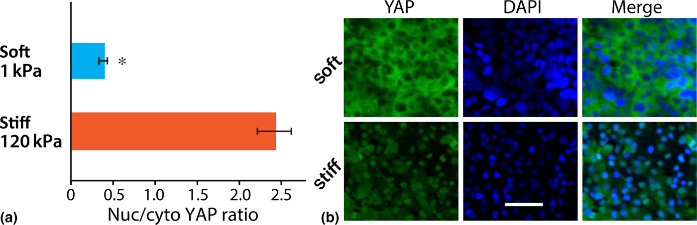
To test if YAP activity in clustered epithelial cells is regulated by ECM stiffness, we measured nuclear localization of YAP in MCF-10A cells grown on collagen-coated PA gels of either 1 or 120 kPa elastic modulus. Following 3 days of culture in these substrates, substrates were stained for the YAP antibody. Through the analysis of immunofluorescence images, we measured nuclear/cytoplasmic ratio of YAP expression for individual cells within epithelial cell sheets grown on soft or stiff substrates [Fig. 1(b)]. We found that the average nuclear localization of YAP, calculated as mentioned above, in the case of soft ECM was less than one-fifth of the value calculated for stiff ECM. These results indicate that ECM stiffness-dependent nuclear translocation of YAP that has been observed for single cells also holds true for multi-cellular epithelial sheets.
YAP activity in clustered cells on flat gels of varying stiffness. (a) Quantification of nuclear YAP localization in terms of nuclear/cytoplasmic ratio of YAP expression on soft (1 kPa) and stiff (120 kPa) flat PA gels. *P < 0.05 with respect to stiff ECM. N > 30 cells per condition, from at least three separate experiments. (b) Representative immunofluorescence images of YAP (green) and DAPI (blue) in MCF-10A epithelial sheet cultured on soft (top) and stiff (bottom) ECMs. Scale bar = 50 µm.

Stiffness-dependent YAP activity depends on the cell density of epithelial clusters
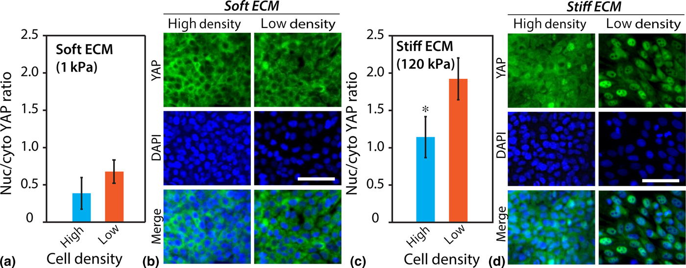
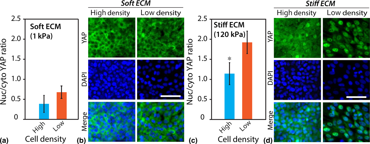
Cell density is known to regulate Hippo signaling and nuclear YAP accumulation.[Reference Wada, Itoga, Okano, Yonemura and Sasaki23] At low cell densities, weak Hippo signaling allows nuclear YAP accumulation, whereas culturing cells at high density induces strong Hippo signaling and the inhibition of nuclear YAP accumulation.[Reference Wada, Itoga, Okano, Yonemura and Sasaki23] However, it remains unknown whether such cell density-dependent YAP activity differs on soft and stiff matrices. To answer this question, we seeded MCF10A cell colonies at either low density of 25,000 cells/cm2 or high density of 75,000 cells/cm2 on both soft and stiff PA gels and imaged YAP expression [Fig. 2(b)]. Subsequently, we measured nuclear YAP localization in terms of the nuclear/cytoplasmic ratio of YAP expression, as described earlier. On soft substrates [Figs. 2(a) and (b)], the difference in nuclear YAP localization between low (2.5×10⁵ cells/cm2) and high (8.5×10⁵ cells/cm2) cell density conditions was small and statistically insignificant. However, on stiff ECMs [Figs. 2(c) and (d)], we observed an approximately 80% reduction in nuclear YAP localization in densely packed cells (5.5×10⁵ cells/cm2) compared with the cell clusters with lower density (1.5×10⁵ cells/cm2). On gels of an intermediate stiffness, 55 kPa, the YAP activity was similar to that observed on the stiff, 120 kPa, substrates (Fig. S1). Going forward, all measurements for “stiff” ECMs are conducted on the 120 kPa gels. In all measurements, the numbers corresponding to cell densities are average densities evaluated by counting the number of cells from the DAPI images for each condition. In both soft and stiff cases, the final “high” cell density is at least three times the “low” density. Note that the final cell densities on soft and stiff ECMs are different because we found a sparser distribution of cells on the stiffer ECM, which could be due to increased mechanosensitive spreading and EMT.[Reference Nasrollahi and Pathak24, Reference Das, Fischer, Pan and Waterman26, Reference Pathak27]. These results indicate that the density of cells in an epithelial cluster significantly influenced the mechanosensitive YAP localization when epithelial cells were grown on stiff substrates.
Regulation of YAP activity by cell density. (a) On soft ECM, nuclear/cytoplasmic ratio of YAP expression in cells within the low-density (250,000 cells/cm2) or high-density (850,000 cells/cm2) epithelial clusters. (c) On stiff ECM, nuclear/cytoplasmic ratio of YAP expression in cells within the low-density (150,000 cells/cm2) or high-density (550,000 cells/cm2) epithelial clusters. *P < 0.05 with respect to the low cell density case. N > 30 cells per condition, from at least three separate experiments. Confocal immunofluorescence images of YAP expression (green) and nuclei (blue) for MCF-10A cell clusters of varying cell densities, cultured on (b) soft and (d) stiff substrates. Scale bar = 50 µm.
Matrix confinement regulates YAP localization through changes in cell morphology
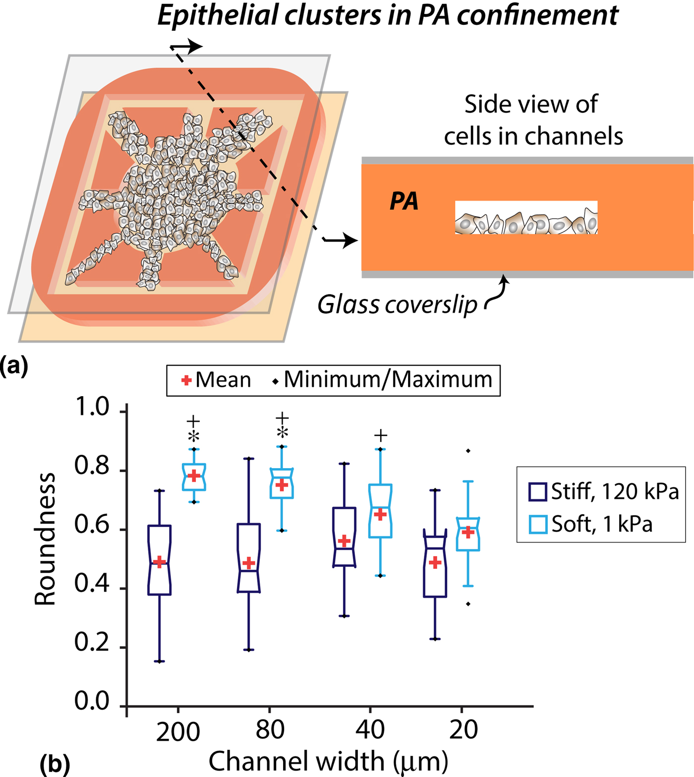
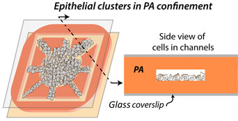
To study the independent roles of stiffness and confinement on YAP activation in epithelial clusters, we fabricated a PA microchannel-based matrix platform for cell culture [Fig. 3(a)], as we have done previously.[Reference Nasrollahi and Pathak24] Briefly, we combined photolithography techniques and PA hydrogel synthesis to construct microchannels of varying width, embedded in PA gels of specified stiffness. In this system, an epithelial colony is seeded in the central chamber and allowed to grow beyond the central chamber to facilitate the entry of cell clusters into channels of widths ranging between 20 and 200 µm. Thus, our platform allows independent control over confinement and stiffness around epithelial clusters.
Cell morphology and YAP activity in microchannels. (a) Schematic representation describing epithelial cell clusters grown inside the polyacrylamide (PA)–microchannels based platform. (b) Cell roundness for single cells in epithelial clusters inside channels of varying widths made of soft and stiff substrates. *P < 0.05 with respect to stiff ECM. +P < 0.05 with respect to narrow (20 µm) channels. N > 20 cells per condition, from at least three separate experiments.

Cells with flat and spread morphology have been shown to exhibit nuclear YAP localization, while rounded cell morphology favors YAP cytoplasmic localization.[Reference Wada, Itoga, Okano, Yonemura and Sasaki23] Given that cell morphology is associated with the subcellular YAP localization, we hypothesized that morphological adaptation of epithelial cells to confinement might also influence YAP distribution. First, to measure the effect of confinement on cell morphology, we quantified roundness of individual cells in epithelial clusters in channels of varying confinement and stiffness. As Fig. 3(b) shows, the roundness parameter is decreased with increasing confinement (decreasing channel width). We also found that the cells on soft substrate were rounder compared with the ones on stiff substrate regardless of the channel width. These results demonstrate that confining of epithelial clusters within narrow channels reduces the roundedness of individual cell that make up the epithelial cluster [Fig. 3(b)].
Greater nuclear YAP localization in stiffer and more confined ECMs
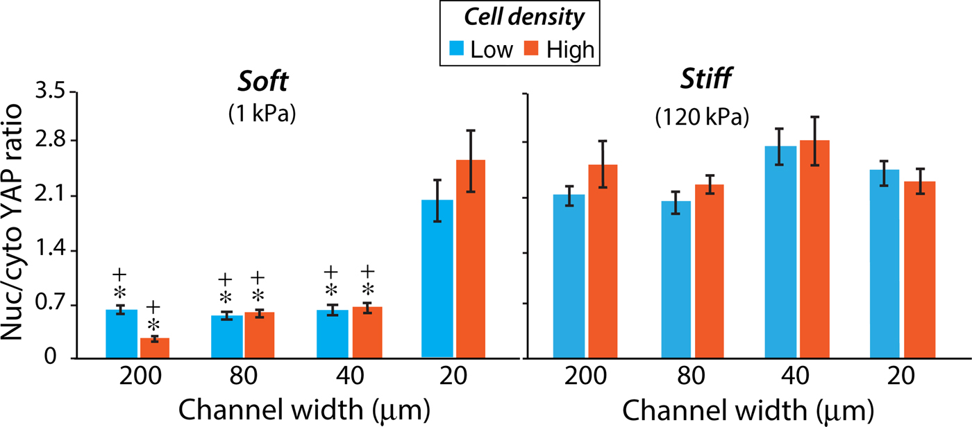
Given the previously described dependence of YAP localization on cell morphology[Reference Wada, Itoga, Okano, Yonemura and Sasaki23] and our measurements of the influence of channel width on cell morphology [Fig. 3(b)], we examined the effect of confinement on YAP distribution inside the channels made of soft or stiff ECMs. Since cell density influenced mechanosensitive YAP activity on flat 2D substrates (Fig. 2), we first posited whether a similar density-dependence could occur in confined ECMs. Surprisingly, we found that nuclear YAP localization did not change significantly between low and high cell density conditions for any given channel width or ECM stiffness (Fig. 4). These results indicate that YAP activity in epithelial clusters in confinement is less sensitive to cell density as compared with the epithelial colonies on flat substrates (compare Figs. 2 and 4).
YAP activity in cell clusters cultured in channels of varying width and stiffness. Nuclear/cytoplasmic ratio of YAP expression in cells within the low- or high-density epithelial clusters cultured in channels of varying width made of soft (left) or stiff (right) PA gels. In soft channels, the average low and high cell densities are 500,000 and 1,500,000 cells/cm2, respectively. In stiff channels, the average low and high cell densities are 250,000 and 750,000 cells/cm2, respectively. +P < 0.05 with respect to stiff ECM. *P < 0.05 with respect to narrow (20 µm) channels. N > 30 cells per condition, from at least three separate experiments.

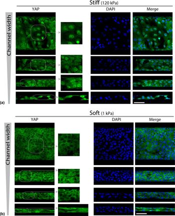
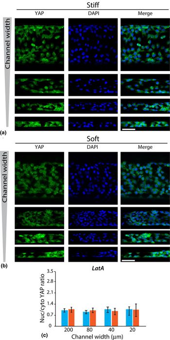
Inside soft channels, we found that nuclear YAP localization increased with decreasing channel width (for either cell density), which indicated a rise in YAP activation in more confined ECMs [Figs. 4 and 5(b)]. The epithelial clusters confined inside narrow (20 µm) channels showed an approximately threefold increase in nuclear YAP localization compared with ones located in the wide (200 µm) channels (Fig. 4). Next, to examine the effect of ECM stiffness on YAP activation in confinement, we repeated these experiments in channels made of stiff (120 kPa) PA gels. As expected, the cell clusters inside the stiff channels showed relatively high nuclear localization regardless of the channel width [Figs. 4 and 5(a)]. Overall, in stiffer channels, we found higher nuclear YAP compared with soft substrates for any given channel width. Thus, our results demonstrate that higher matrix elasticity regulates nuclear YAP localization even for epithelial clusters trapped inside channels, which is in agreement with earlier observations on flat PA gels.[Reference Dupont, Morsut, Aragona, Enzo, Giulitti, Cordenonsi, Zanconato, Le Digabel, Forcato, Bicciato, Elvassore and Piccolo11] YAP-regulated mechanosensation is mediated through the actin cytoskeleton, which determines cell morphology.[Reference Dupont, Morsut, Aragona, Enzo, Giulitti, Cordenonsi, Zanconato, Le Digabel, Forcato, Bicciato, Elvassore and Piccolo11, Reference Wada, Itoga, Okano, Yonemura and Sasaki23] Therefore, we hypothesized that disruption of the integrity of the actin fibers may hinder the morphological adaptation of clustered epithelial cells and consequently inhibit a confinement-induced nuclear YAP localization. We treated MCF10A cells with LatA, which is a known inhibitor of actin polymerization. Analysis of YAP subcellular localization demonstrated that such pharmacological disruption of F-actin polymerization significantly reduces nuclear YAP localization in epithelial clusters, regardless of the ECM stiffness and confinement (Fig. 6), compared with untreated cells (Fig. 5). After F-actin inhibition, the YAP activity in cells did not show any sensitivity to ECM confinement or stiffness [Fig. 6(c)].
Enhanced YAP nuclear localization in stiffer and narrower channels. Representative immunofluorescence images of YAP (green; insets showing higher resolution images), nuclei stained with DAPI (blue), and merged for epithelial cells embedded inside (a) stiff and (b) soft channels of varying width. Scale bar = 50 µm.

Reduced YAP nuclear localization in stiffer and narrower channels after disrupting F-actin polymerization. Representative immunofluorescence images of YAP (green), nuclei stained with DAPI (blue), and merged for LatA-treated epithelial cells embedded inside (a) stiff and (b) soft channels of varying width. Scale bar = 50 µm.

Discussion
Mechanical signals are abundantly present in the microenvironment that surrounds the living cells. In recent years, there has been an increasing appreciation that the mechanical properties of the ECM, such as stiffness and matrix topography, impact fundamental cellular functions through cell–ECM interactions. In cancer metastasis, epithelial clusters escape from the primary tumor and pass through ECMs of varying mechanical properties before arriving at the secondary sites. It is now known that the elasticity and microstructure of the ECM surrounding the epithelial clusters can independently prepare these cells for malignant transformation.[Reference Nasrollahi and Pathak24, Reference Paszek, Zahir, Johnson, Lakins, Rozenberg, Gefen, Reinhart-King, Margulies, Dembo, Boettiger, Hammer and Weaver28, Reference Wei, Fattet, Tsai, Guo, Pai, Majeski, Chen, Sah, Taylor, Engler and Yang29] Specifically, on stiffer substrates, higher actomyosin contractility and stronger cell–ECM adhesions lead to the dissolution of cell–cell adhesions, resulting in epithelial-to-mesenchymal transition (EMT).[Reference Paszek, Zahir, Johnson, Lakins, Rozenberg, Gefen, Reinhart-King, Margulies, Dembo, Boettiger, Hammer and Weaver28] Recent studies, including our work, have shown that MCF10A MECs cultured on stiff collagen-coated PA gels undergo EMT and attain invasive phenotype.[Reference Nasrollahi and Pathak24, Reference Wei, Fattet, Tsai, Guo, Pai, Majeski, Chen, Sah, Taylor, Engler and Yang29] Additionally, we have shown that ECM topography can perpetuate mesenchymal transformation independently of matrix stiffness.[Reference Nasrollahi and Pathak24] We also found that the elongated cell morphology in narrower channels was associated with a confinement-sensitive reduction in E-cadherin localization at cell–cell junctions and induction of EMT, even in softer ECMs.[Reference Nasrollahi and Pathak24]
In this study, we hypothesized that the ECM-dependent response of clustered epithelial cells[Reference Nasrollahi and Pathak24] due to the mechanical cues presented by the ECM are processed by signaling pathways transmitted through the nucleus. Since YAP is known as the classical transducer of ECM stiffness,[Reference Dupont, Morsut, Aragona, Enzo, Giulitti, Cordenonsi, Zanconato, Le Digabel, Forcato, Bicciato, Elvassore and Piccolo11, Reference Wada, Itoga, Okano, Yonemura and Sasaki23] we shortlizted it as a potential candidate for instructing ECM-sensitive responses in epithelial clusters. While it is known that ECM stiffness and cells shape can regulate YAP distribution in single cells, it is not yet clear how these parameters might affect YAP localization in epithelial cells that typically grow in clusters with intact cell–cell junctions. Given that the disassembly of tight junctions is known to enable ZO2-mediated translocation of YAP to the nucleus, it would be important to understand the regulation of YAP activity during ECM-dependent EMT.[Reference Oka, Schmitt and Sudol30] Current understanding of stiffness-dependent YAP activation has mainly been derived from experiments of cells cultured on flat surfaces. This gap in knowledge persists mainly because of an absence of matrix platforms that permit an orthogonal control over confinement and stiffness of the ECM around the epithelial clusters. In this work, we addressed this challenge by utilizing a matrix platform for culturing cell clusters in PA channels of varying width and stiffness.[Reference Nasrollahi and Pathak24] Our soft microchannels-based platform allows new measurements of YAP distribution in epithelial clusters in ECMs of defined stiffness and confinement.
We have shown for the first time that matrix confinement alters the dependence of YAP activation on ECM stiffness. Although previous studies have indicated that YAP remains inactive on soft substrates,[Reference Dupont, Morsut, Aragona, Enzo, Giulitti, Cordenonsi, Zanconato, Le Digabel, Forcato, Bicciato, Elvassore and Piccolo11, Reference Liu, Lagares, Choi, Stopfer, Marinkovic, Vrbanac, Probst, Hiemer, Sisson, Horowitz, Rosas, Fredenburgh, Feghali-Bostwick, Varelas, Tager and Tschumperlin22] our results demonstrate that confining the epithelial clusters inside the narrow channels can lead to nuclear YAP localization even on soft ECMs. On stiff substrates, most cells in the cluster showed maximal YAP activation regardless of the channel width. Here, cells confined within narrow channels were forced to become less round, which was similar to the cellular elongation and simultaneous nuclear YAP localization observed on flat stiff substrates.[Reference Wada, Itoga, Okano, Yonemura and Sasaki23] It is possible that cells near the channel walls intimately interact with the wall surface and align themselves along the walls. This rise in spreading and elongation along the channel walls may enhance cellular mechano-activation, as we have argued in previous studies,[Reference Pathak and Kumar4, Reference Pathak and Kumar5, Reference Nasrollahi and Pathak24, Reference Pathak27] and thus lead to higher YAP activation. Thus, increased YAP activity may arise due to either stiffer or more confined ECM, both of which are also associated with increased cell elongation. The results presented in this study, along with our previous findings,[Reference Nasrollahi and Pathak24, Reference Pathak27] have revealed that both ECM stiffness and confinement can independently induce EMT and enhance YAP activity in epithelial clusters.
We have also shown that nuclear YAP localization increases in sparser epithelial clusters cultured on 2D substrates, which is in agreement with another recent study.[Reference Das, Fischer, Pan and Waterman26] Notably, we report that denser epithelial clusters exhibit lower YAP activity even on stiff ECM. However, our measurements for epithelial clusters inside channels of varying width show that the density-dependent YAP activity does not hold true in confined ECM settings. It should be noted that the presented framework of ECM- and density-dependent YAP activity in epithelial clusters has only been tested for an immortalized epithelial cell line (MCF-10A) and could potentially differ in other cell lines and primary cultures. It is likely that the influence of ECM confinement on YAP activity in clustered cells might also regulate cellular behaviors other than EMT, such as migration and differentiation, for a variety of cell types and matrices that have not been covered thus far.
Supplementary Material
The supplementary material for this article can be found at https://doi.org/10.1557/mrc.2017.87.
Acknowledgments
This work was in part supported by grants to A.P. from the National Science Foundation (CAREER Award 1454016) and the Edward Mallinckrodt, Jr. Foundation (New Investigator Award). Lithography facilities were provided by the Institute of Materials Science & Engineering (IMSE) at Washington University in St. Louis.