
Introduction
The development of scaffolds that are able to mimic extracellular matrices and instinctive biologic functions is one of the goals of the tissue engineering.[ Reference Choi, Zhang and Xia 1 , Reference O'Brien 2 ] Having large specific surface area, small diameter and interconnected pores enable fiber scaffolds to provide a suitable cellular environment for cell proliferation, attachment, transport of nutrients and waste.[ Reference Erencia, Cano, Tornero, Macanás and Carrillo 3 ] Among currently available fiber fabrication techniques, electrospinning is a widely used method. However, the electrospinning needs high voltage and is limited to a set of solvents. It also yields low fibers.[ Reference Sarkar, Gomez, Zambrano, Ramirez, de Hoyos, Vasquez and Lozano 4 ] However, electrospun scaffolds require considerable additional steps like manual cell seeding and incubation, which reduces cell infiltration by blocking pores.[ Reference Sarkar, Gomez, Zambrano, Ramirez, de Hoyos, Vasquez and Lozano 4 ] As well, these seeded cells on the fiber scaffold are two-dimensional, not three-dimensional. Therefore, this process is not economical and is time-consuming. Thus, electrospun scaffolds have limited demand in biologic and clinical applications.
Therefore, cell electrospinning technology is developed to incorporate living cells into fiber scaffolds without a separate cell seeding process and it uses electrostatic force to fabricate fiber scaffolds.[ Reference Townsend-Nicholson and Jayasinghe 5 , Reference Jayasinghe 6 ] The cells distribute homogenously, survived well and scattered uniformly within the fiber scaffolds. Cell electrospinning showed different three-dimensional shapes, i.e., a product that has vessel-like architecture.[ Reference Townsend-Nicholson and Jayasinghe 5 ] The addition of cells shows cell aggregations in the fibers and the fibers prepared by cell electrospinning are not in uniform shape and thickness.[ Reference Townsend-Nicholson and Jayasinghe 5 , Reference Jayasinghe 6 ]
On the other hand, Forcespinning® (FS) was developed based on the use of centrifugal force where it can offer a high production rate of the fibers, with uniform shape and thickness and can be used in a broad range of materials.[ Reference Sarkar, Gomez, Zambrano, Ramirez, de Hoyos, Vasquez and Lozano 4 , Reference Padron, Fuentes, Caruntu and Lozano 7 ] FS is an eco-friendly technique to produce fibers from conductive and nonconductive polymers. The fiber production rate is more than 1 g/min per nozzle at the laboratory scale unit. The surface morphology of fibers is mainly dependent on solution parameters and device configurations such as orifice diameter, solution/melt viscosity, and fiber collection methods. Several synthetic biopolymer-based fibers have been fabricated and used in tissue engineering, surgical sutures, drug delivery, and wound dressing.[ Reference Jeong, Krebs, Bonino, Khan and Alsberg 8 ] However, solely biopolymer or synthetic polymer solutions are not sufficient for the preparation of fibers or could not provide unique mechanical and biocompatible properties. Several fibers have been fabricated by using the combination of biopolymers (gelatin) and synthetic polymers.[ Reference Nguyen, Ko, Oh, Heo, Jeon, Park, Choi, Choi and Jung 9 ]
Gelatin is a hydrolysis product of collagen with repeated α-amino acid (arginine-glycine-aspartic acid) (RGD) residues that helps in the cell attachment.[ Reference Liu and Chan-Park 10 ] Gelatin has better advances over collagen in terms of less antigenicity and it has been widely used in tissue engineering and pharmaceutical fields.[ Reference Wu, Liu, Li, Wen, Zhang, Long, Wang, Guo, Xing and Gao 11 ] Gelatin fibers have been developed using several synthetic biopolymers. For example, gelatin and polymer (polylactide-co-glycolide, chitosan, and polycaprolactone) composites were used as potential artificial nerve scaffolds in nerve tissue engineering.[ Reference Jafari, Emami, Samadikuchaksaraei, Bahar and Gorjipour 12 – Reference Cheng, Deng, Yang, Gong, Zhao and Zhang 14 ] PCL/gelatin fibers were developed by disc-electrospinning and studied their cell proliferation with fibroblast and bone marrow stromal cells.[ Reference Li, Chen, Sun, Li, Wu, Ke, Huang, Ei-Hamshary, Al-Deyab and Mo 15 ] Sodium alginate/gelatin fibers were also developed and used in blood vessel endothelial applications.[ Reference Jiang, Jiang, Huang, Wang, Li and Turng 16 ] Even though several investigations have succeeded in the development of gelatin-based fibers using electrospinning. However, some issues such as poor mechanical properties, cell binding, and viability to control the microarchitecture of ECM need further enhancements. Poly(epichlorohydrin-co-ethylene oxide) (PECO) is an eminent blending elastomer to enhance the durability and crystallization of fragile biodegradable polymers.[ Reference Soto-Oviedo and De Paoli 17 – Reference Juliana and Felisberti 19 ] PECO has been used in various applications, including solid electrolytes, air canals, fuel pipes, and Lithium polymer batteries.[ Reference Zhang, Ran, Wang, Han, Han, Wen, Zhuang and Dong 20 ]
Diclofenac is a non-steroidal anti-inflammatory drug (NSAID) used as an anti-rheumatic analgesic.[ Reference Souza, Sakamoto and Mattoso 21 ] Diclofenac reduces prostaglandin synthesis by blocking the cyclooxygenase expression, which can lead to gastrointestinal bleeding and renal insufficiency.[ Reference Cooper and Harirforoosh 22 ] The side effects of gastrointestinal lesions and renal damage are associated with orally delivered NSAIDs have been reduced when encapsulated into polymer-based nanoparticles.[ Reference Italia, Yahya, Singh and Ravi Kumar 23 ] In addition, previous reports suggest that NSAIDs such as diclofenac may facilitate transdermal, oral, colon-targeted, and ophthalmic delivery.[ Reference Souza, Sakamoto and Mattoso 21 – Reference Kim, Khil, Kim, Lee and Jahng 25 ]
Due to the high surface area-to-volume ratio, fibers can serve as good carriers for therapeutic agents such as anti-inflammatory, antifungal, anti-bacterial, and antimicrobials. Therefore, there is a clear and unmet need to develop novel gelatin-based fibers, which could be utilized to deliver therapeutics with admirable biocompatibility. For example, polycaprolactone fibers having antibiotics (biteral) are used in wound healing to prevent the post-surgery abdominal adhesions.[ Reference Bolgen, Vargel, Korkusuz, Menceloglu and Piskin 26 ] PMMA and hydrophilic polymer fibers are used for the sustained release of Ciprofloxacin.[ Reference Liu, Lin, Fang, Yao, Zhao, Dodson and Wang 27 ] Berberine chloride loaded electrospun collagen/zein fibers are developed for wound healing and controlled release of berberine.[ Reference Lin, Li, Zhao, Hu and Zhang 28 ] In addition, dexamethasone bearing peptide amphiphile-dexamethasone fiber gels are synthesized and studied in vivo inflammatory response.[ Reference Webber, Matson, Tamboli and Stupp 29 ]
In the present study, first time biocompatible GL : PECO is synthesized and fibers are fabricated using FS. The blending of PECO into gelatin solution enhances the tensile strength and elastic properties of fibers. Water contact angle, porosity, degradation, and cell viability properties of GL : PECO fibers are evaluated and improved results are observed. Prolonged release of diclofenac is also studied.
Experimental section
Materials
Unless stated otherwise, all chemicals were purchased from Sigma-Aldrich, USA and used without further purification. Cell culture consumables and supplements were Sigma Aldrich, sterile, and used as provided.
Methods
Synthesis of GL : PECO (1 : 1) composite
Gelatin (3.0 g) was dissolved in trifluoroacetic acid (TFA) at room temperature followed by addition of PECO (3.0 g) and diclofenac (0.5 g). The final solution was stirred at 60°C for 24 h to get a clear homogeneous solution.
Synthesis of GL : PECO (2 : 1) composite
Gelatin (4.0 g) was dissolved in TFA at room temperature followed by addition of PECO (2.0 g) and diclofenac (0.5 g). The final solution was stirred at 60°C for 24 h to get a clear homogeneous solution. Similarly, GL : PECO (3 : 1) and GL : PECO (4 : 1) are also prepared. Then, each homogeneous solution was fed in a dual orifice spinneret of FS. The optimum speed and time were found to be 9000 rpm and 30–40 s, respectively. Similarly, GL : PECO (3 : 1) and GL : PECO (4 : 1) are also prepared. (Please see the supporting information for the characterization of fibers, drug release, and cell study experimental condition).
Results and discussion
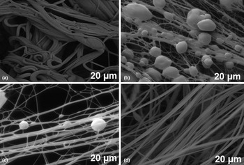
Fibers made of biocompatible and biodegradable polymers are of great interest for skin tissue engineering.[ Reference O'Brien 2 ] To obtained forcespun GL : PECO fibers, TFA is used as a solvent. Figure 1 shows SEM images of fibers from GL : PECO blend with different weight ratios of gelatin. In these cases, the total concentration of each solution is kept to be 30% (w/v). As showed in Fig. 1(a), 1 : 1 ratio of GL : PECO is provided elastic micro fibers at 9000 rpm. These fibers are very dense and attached on the collector. In contrast, 2 : 1 ratio of GL : PECO is produced fibers with many beads [Fig. 1(b)]; whereas 3 : 1 ratio of GL : PECO is produced fibers with few beads [Fig. 1(c)]. When a 4 : 1 ratio of GL : PECO is used, it could be forcespun effectively into bead-free fibers with homogeneous morphology and smaller fiber diameters. With the increase of gelatin concentration from 1 : 1 to 4 : 1, the diameter of the fibers is decreased to several hundreds of nanometers and improved uniformity of the fibers. Therefore, 3 : 1 and 4 : 1 of GL : PECO are selected as the optimized ratio for the rest of the studies.
Optimization of gelatin concentration in PECO blend; (a) GL : PECO (1 : 1), (b) GL : PECO (2 : 1), (c) GL : PECO (3 : 1), and (d) GL : PECO (4 : 1). All fiber scaffolds produced at 9000 rpm.

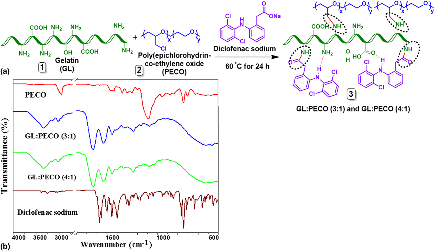
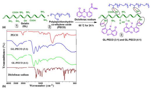
The GL : PECO (3 : 1 and 4 : 1) composites are synthesized, then characterized by Fourier transform infrared spectroscopy (FTIR) and the results are presented in Fig. 2. It is hypothesized that during the reaction, PECO underwent the chloride substitution reaction with a free amine of gelatin and forms N–C bonding as showed in Fig. 2(a) (inside dotted regions of GL : PECO). On the other hand, diclofenac can generate amide bonding from a free amine of gelatin (inside dotted regions of GL : PECO). Secondary amine of diclofenac can form hydrogen bonds with an amine of gelatin (dotted red lines of GL : PECO). As seen in Fig. 2(b), the FTIR spectrum of pure PECO has absorption bands from C to H stretching of aliphatic groups at 2930 and 2865/cm and –CH2 bending peaks at 1245–1424/cm. Furthermore, C–H wagging peaks were observed in the range of 1300–1150/cm; whereas C–Cl stretching peaks were detected in the range of 850–550/cm. However, fibers of GL : PECO (3 : 1) and GL : PECO (4 : 1) showed different absorption peaks at 3281/cm for –NH2 and –OH stretching vibration, 3075–2900/cm for C–H aliphatic groups, stretching vibration (3075/cm for alkenyl C–H stretching and 2900/cm for CH2 asymmetrical stretching).
(a) Synthesis of GL : PECO (3 : 1 and 4 : 1) composites, (b) FTIR spectra of PECO, GL : PECO (3 : 1 and 4 : 1) fiber scaffolds and diclofenac sodium.
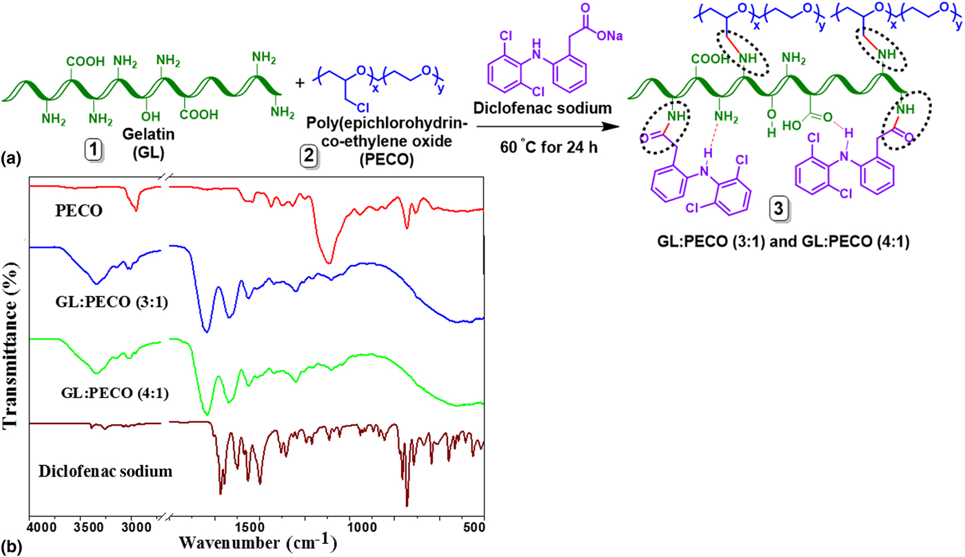
Further, 1628/cm for amid I (C=O) stretching vibration, 1538/cm for amid II (N–H) bending vibration of GL : PECO (3 : 1) and GL : PECO (4 : 1). CH2 bending peaks of pure PECO was shifted from 1300 to 1446/cm. In both gelatin composites, around at 1334/cm, a (C–N) stretching vibration peak was observed for amid III and –C–N stretching at 1235/cm. In addition, C–Cl stretching peaks of pure PECO at 850–550/cm was vanished in the both GL : PECO fibers. This indicates that there could be a chloride substitution reaction between PECO and gelatin. FTIR data showed that there might be weak interactions between the backbone of PECO and gelatin. In addition, there was an amide peak observed at 1692/cm, which indicates the amide bonding between diclofenac and gelatin.
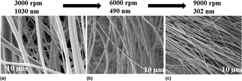
After synthesis of GL : PECO composites, GL : PECO (4 : 1) fibers were produced using FS. Continuous aligned fibers with diameters ranging from 300 to 1030 nm are obtained as showed in Figs. 3(a)–3(c). By increasing the rotation speed of the spinneret from 3000 to 9000 rpm, the fiber diameter dropped from 1030 to 300 nm [Figs. 3(a)–3(c)]. Therefore, the rotation speed was kept constant while varying the polymer concentration.
Fiber morphology and diameter distribution of GL : PECO (4 : 1) solution forcespun at different rotation speeds, (a) 3000 rpm, (b) 6000 rpm, and (c) 9000 rpm.

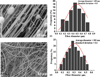
Figure 4 shows a SEM images of fibers produced at rpm 9000 by the solution of GL : PECO (3 : 1) and GL : PECO (4 : 1) (see S1.1., Supplementary Information). The solution of GL : PECO (3 : 1) produced some beads with an average diameter of 490 nm; whereas, at the same speed (rpm), GL : PECO (4 : 1) solution produced beads free homogenous fibers with an average diameter of 300 nm. It was observed that the fiber morphology and diameter were influenced by the concentration of polymer solution and the rotational speed of the spinneret (rpm). Increasing the concentration of gelatin from (3 : 1) to (4 : 1) wt.% led to a drop in the average fiber diameter (from 490 to 300 nm). Figures 4(c) and 4(d) exhibit the diameter distributions of GL : PECO (3 : 1) and GL : PECO (4 : 1) fibers, respectively. These results suggest that by increasing the rotation speed of the spinneret, the polymer liquid travels faster and stretches as the solvent is evaporated. This could control the polymer liquid jet and resists surface tension-induced bead formation.
SEM images of GL : PECO fibers; (a) GL : PECO (3 : 1) and (b) GL : PECO (3 : 4) respectively. (c) and (d) Diameter distribution of GL : PECO (3 : 1) and GL : PECO (4 : 1) fibers at rpm 9000, respectively.

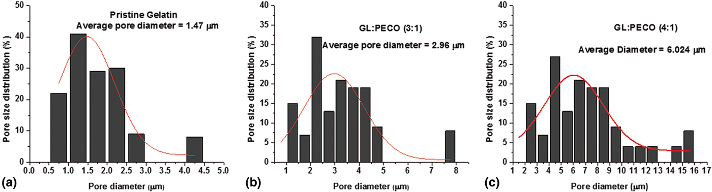
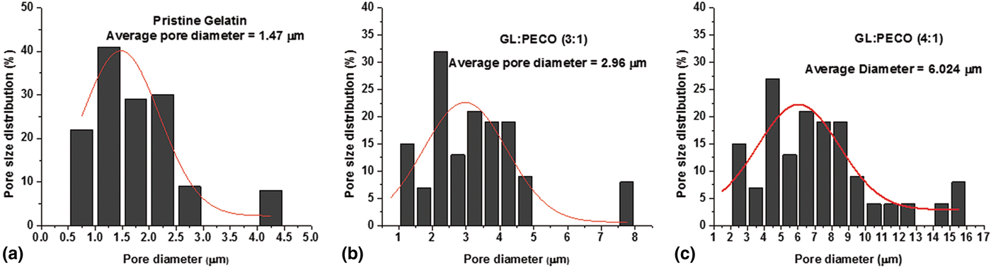
As showed in Fig. 5, the pore size distribution of gelatin and GL : PECO fibers was measured (see S1.1., Supplementary Information). It can be seen in pure gelatin fibers that the diameter of pores underwent a distribution ranging from 0.5 to 4.0 µm and average pore diameter was 1.4 µm. This size is due to the densely packed fibers produced in a compact structure, reducing the pore size. GL : PECO (3 : 1) fibers were showed the average diameter 2.9 µm; the diameter of pores was distributed in the range of 1.2–7.9 µm [Fig. 5(b)]. In contrast, GL : PECO (4 : 1) showed highest pore size and the diameter of the pore underwent a broad distribution ranging from 2.3 to 15.8 µm and the average diameter was 6.0 µm. This could be due to the loosely distributed microfibers [Fig. 5(c)]. Overall, the pore size of fibers was gradually increased with the concentration of gelatin.
Pore size distribution of fibers; (a) gelatin, (b) GL : PECO (3 : 1), and (c) GL : PECO (4 : 1) scaffolds, respectively. The average pore diameter for gelatin, GL : PECO (3 : 1) and GL : PECO (4 : 1) was 1.4, 2.9, and 6.0 µm, respectively.
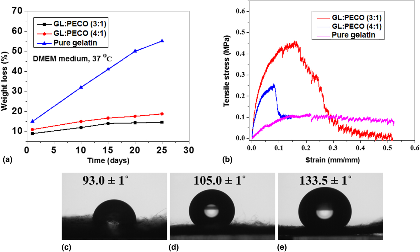
In vitro degradation results of pure gelatin and GL : PECO (3 : 1 and 4 : 1) fibers are shown in Fig. 6(a) (see S1.1., Supplementary Information for the conditions). The fibers were degraded over the time. Based on the blending ratio of gelatin, the degradation rate of GL : PECO (3 : 1 and 4 : 1) fibers was varied. Particularly, the degradation in 25 days reached approximately 14.7% for GL : PECO (3 : 1) fibers and 18.8% for GL : PECO (4 : 1) fibers, respectively; whereas pure gelatin fibers showed approximately 55.1% of degradation profile after 25 days. The degree of the degradation rate was decreased when the content of PECO increased in the composites. This could be due to stronger mechanical properties and the hydrophobic nature of GL : PECO (3 : 1) and GL : PECO (4 : 1) fibers.
(a) In vitro degradation, (b) tensile curve, (c–e) water contact angles of gelatin, GL : PECO (3 : 1) and GL : PECO (4 : 1) fibers, respectively.

Pristine gelatin and GL : PECO solutions were forcespun into ~1.0 mm thick scaffolds to measure mechanical properties. Representative stress/strain tensile curves of the scaffolds are shown in Fig. 6(b). The results reveal that the tensile strength and elongation at break of GL : PECO scaffolds were enhanced compared with neat gelatin scaffolds. Particularly, GL : PECO (3 : 1) fibers showed 0.45 MPa of the tensile strength and 15.75 MPa of Young's modulus. Whereas, GL : PECO (4 : 1) fibers showed 0.24 MPa the tensile strength and 3.35 MPa of Young's modulus. The tensile results showed that increasing the concentration of PECO in the blend solution strengthens the fibers. Mechanical properties of GL : PECO (3 : 1 and 4 : 1) fibers are compared with pure gelatin fibers and improved results are observed. This could be due to the chloride substation reaction or intermolecular hydrogen bonding between gelatin, PECO, and diclofenac. It is known that the hydrophilic/hydrophobic fibers can influence the cell adhesion and their proliferation to a higher rate.[ Reference Zhang, Ran, Wang, Han, Han, Wen, Zhuang and Dong 20 ] Therefore, water contact angle of fibers was measured and shown in Figs. 6(c)–6(e). By decreasing the amount of gelatin, the water contact angle was increased from 105.0° to 133.5°. Particularly, 93.0°, 105.0° and 133.5° water contact angles are observed in gelatin, GL : PECO (4 : 1) and GL : PECO (3 : 1), respectively. These results demonstrate that with an increase of PECO concentration, the surface wettability of fibers was decreased. That could be due to a hydrophobic and elastic nature of PECO.
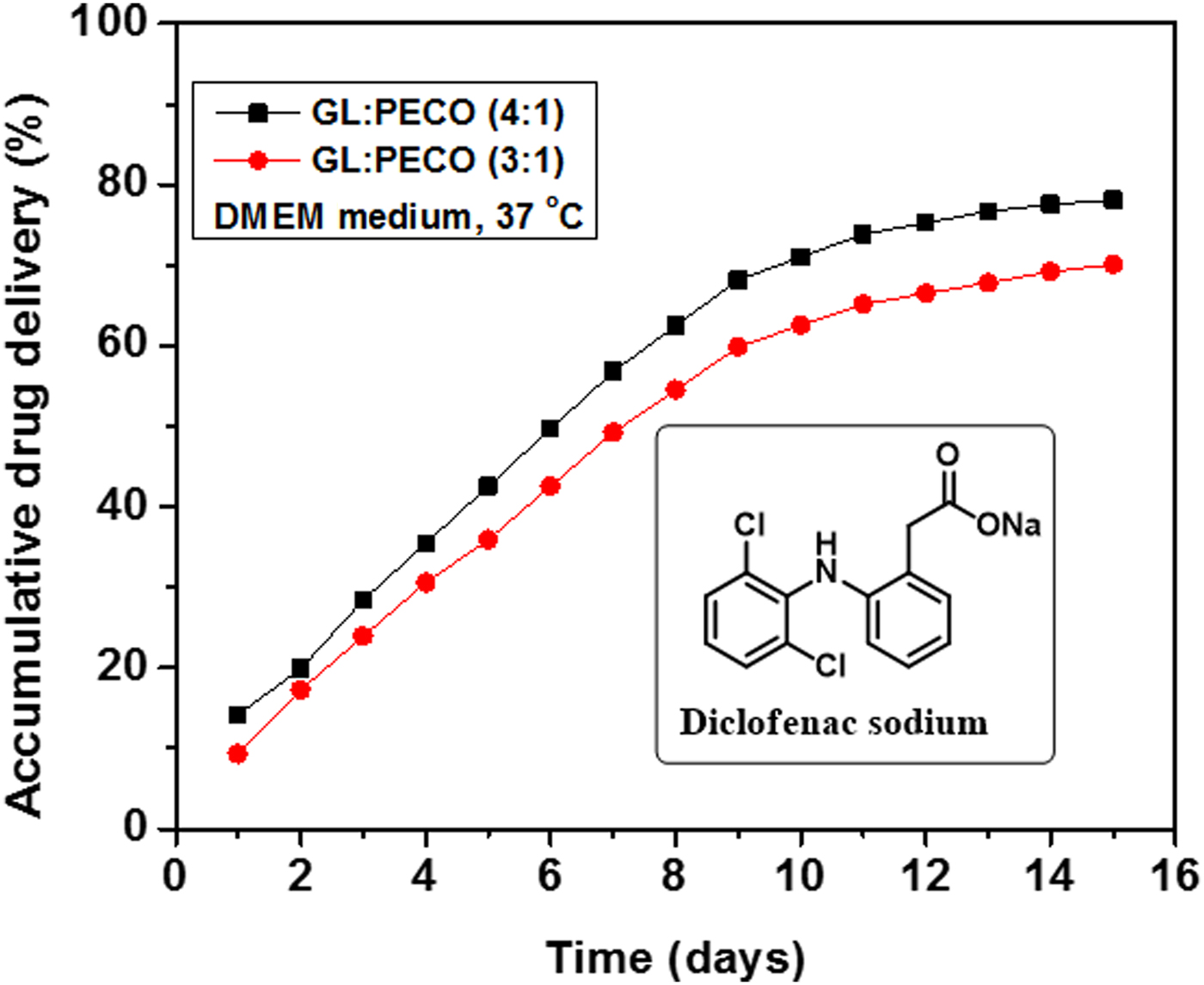
In vitro cumulative drug release of fibers was studied (see S1.2., Supplementary Information). Figure 7 shows the relation between the cumulative drug release percentage and release time. GL : PECO (3 : 1) and GL : PECO (4 : 1) fibers displayed minimal burst release and zero-order release kinetics with the sustained release over 15 days. By the end of this time, approximately 70–77% of the total diclofenac was released from GL : PECO fibers. As expected, a greater burst release at onset and also a faster drug release rate occurred. The prolonged release of diclofenac from GL : PECO (3 : 1) and GL : PECO (4 : 1) confirms the formation of amide linkage (between gelatin and diclofenac sodium). Therefore, GL : PECO (3 : 1) and GL : PECO (4 : 1) fibers can be useful to deliver a low and consistent dose of diclofenac for prolonged times.
In vitro drug release profiles of diclofenac from fiber scaffold of GL : PECO (3 : 1) and GL : PECO (4 : 1).

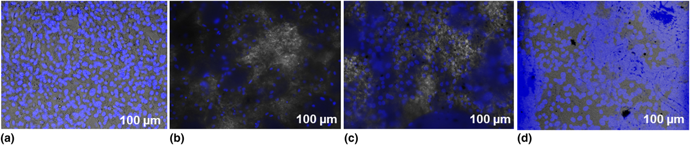
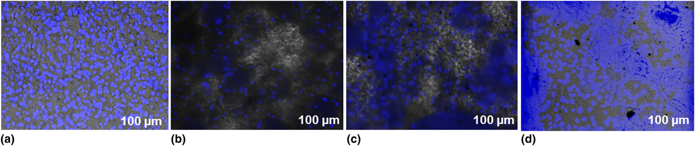
The cell attachment and proliferation on pristine gelatin, GL : PECO (3 : 1) and GL : PECO (4 : 1) fibers are viewed by nuclear staining (Hoechst 33258) (see S1.3., Supplementary Information). These images of stained nuclei indicate the distribution, attachment, and proliferation of cells on the surface of fibers (Fig. 8). Among all fibers, GL:PECO (4 : 1) fibers were supported more cell attachment on the surface, followed by GL : PECO (3 : 1) and pristine gelatin. It indicates that GL : PECO fibers are biocompatible and suitable for biomedical applications.
Fluorescence microscopic images of Hoechst 33258 stained NIH/3T3 cells on fiber scaffolds (after 24 h): (a) control (without fibers), (b) pristine gelatin fibers, (c) GL : PECO (3 : 1) and (d) GL : PECO (4 : 1) fibers.
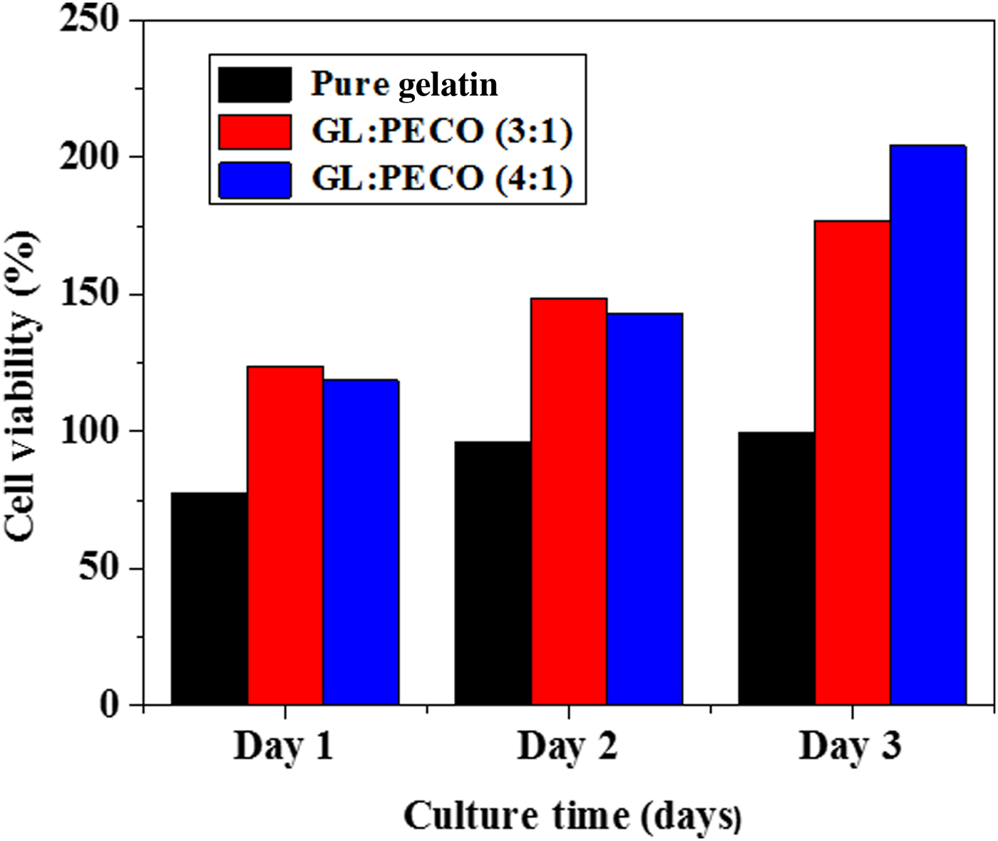
Cell proliferation, adhesion, migration, and spreading on the fibers are important issues to evaluate the suitability for tissue regeneration. To determine the proliferation on the GL : PECO (3 : 1) and GL : PECO (4 : 1) fibers, NIH/3T3 cells are cultured for 1, 2, and 3 days.
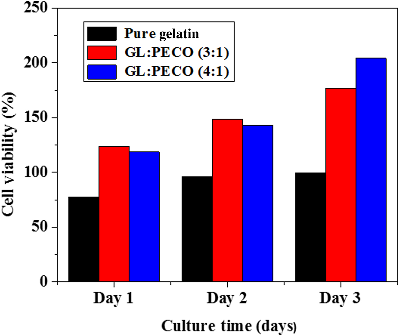
CellTiter96®AQueous One Solution cell proliferation assay is used to measure the cell viability (see S1.3. in Supplementary Information for the experimental conditions). Initially, GL : PECO (3 : 1) showed a higher cell proliferation rate (on day one) than pristine gelatin and GL : PECO (4 : 1) fibers. After 2 days, the proliferation rate was very similar in GL : PECO (3 : 1) and GL : PECO (4 : 1) fibers; however, the proliferation rate was much higher than pure gelatin fibers. This could be due to the high degradation, high surface wettability and lower pore size of gelatin fibers. In addition, after 3 days of cell seeding, cell viability significantly increased in GL : PECO (4 : 1) fibers; the proliferation rate was higher than that for pristine gelatin and GL : PECO (3 : 1) fibers (Fig. 9). This may be caused by large pore size, moderate surface wettability and degradation properties of GL : PECO (4 : 1) fibers. The pores size of the fibers is ideal for cell migration, as well as for diffusion of nutrients.[ Reference Jeong, Krebs, Bonino, Khan and Alsberg 8 ] It is reported that cell attachment and growth could be promoted by fiber stiffness and pore size.[ Reference Sisson, Zhang, Farach, Chase and Rabolt 30 ] As highest porosity bearing GL : PECO (4 : 1) fibers (see Fig. 4) showed higher cell viability on the third day.
Cell viability of NIH/3T3 is seeded on pure gelatin, GL : PECO (3 : 1) and GL : PECO (4 : 1) fiber scaffolds.
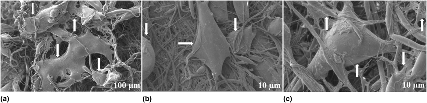
SEM images of cell interactions with GL : PECO (4 : 1) fibers are shown in Fig. 10 (conditions are shown in S1.3., Supplementary Information). Fibroblast cells inclined to form clusters on the surface of fibers on the first day and growth of clusters was observed on the second day. At longer cell culturing time, fibroblast cells started to form cellular extensions [Figs. 10(a)–10(c)], which were extended in many directions (white arrows). The length and number of these extensions were improved along with time. The cell mass, cellular extensions, and colonization were improved along with time. Few cells were found to grow in between the fibers and below the surface [Fig. 10(c)]. This could be due to the porous structure of the fibers. Similar penetrations of cells were observed in different fibers. SEM examination at two magnifications of fibroblast cultures [Figs. 10(a)–10(c)] showed good attachment and spreading of NIH/3T3 cells on the GL : PECO (4 : 1) fibers. These observations demonstrate the enhanced properties of GL : PECO (4 : 1) fibers for cell attachment and proliferation.
The morphology and cell interactions of fibroblast cells cultured on GL : PECO (4:1) fiber scaffolds (a, b) after 2 days and (c) after 3 days.

Conclusions
First time, gelatin, PECO, and diclofenac composites are synthesized. The use of FS provides biocompatible scaffolds from gelatin and PECO. FTIR results show the chemical reactions between gelatin, PECO, and diclofenac. The fiber diameter, morphology, surface wettability, mechanical properties, in-vitro degradability, cell adhesion and proliferation can be manipulated with the blending ratio of gelatin, PECO and process parameters of FS. The incorporation of PECO is helpful for the improvement of forcespinnability, flexibility, and tensile strength, whereas, the addition of gelatin is useful for the enhancement of surface wettability, in vitro degradability, cell adhesion, and proliferation. GL : PECO (4 : 1) fibers show a prolonged drug release, and cell viability. These biocompatible fibers are useful for drug delivery, wound healing, and skin regenerative scaffolds.
Supplementary material
The supplementary material for this article can be found at https://doi.org/10.1557/mrc.2017.117
Acknowledgments
The authors would like to acknowledge the financial support from the Consejo Nacional de Ciencia y Tecnología de México (CONACyT), Project Number 242269 and they thank Mónica Arreola Flores for her review of this paper. They also thank Dr. Alan Osiris Sustaita Narváez for the water contact angle.