Advanced glycation end products (AGE) are a group of heterogeneous compounds produced through nonenzymatic reactions between proteins, lipids or nucleic acids and reducing sugars(Reference Rowan, Bejarano and Taylor1). These reactions are irreversible, resulting in the accumulation of AGE in the blood and tissues over time. AGE can be endogenously formed in the body or exogenously derived from food(Reference Vistoli, De Maddis and Cipak2). Endogenous AGE are generated through metabolic processes, such as reactions between reducing sugars and proteins under conditions of hyperglycaemia or oxidative stress, while exogenous AGE are primarily derived from high-temperature cooked foods (e.g. grilled or fried) via the Maillard reaction. These pathways contribute differently to the overall AGE burden in the body(Reference Phuong-Nguyen, McNeill and Aston-Mourney3). The representative AGE include Nε-carboxymethyl-L-lysine (CML), Nε-(1-carboxyethyl) lysine (CEL), Nδ-(5-hydro-5-methyl-4-imidazolon-2-yl)-ornithine (MG-H1) and pentosidine, with CML, CEL and pentosidine as lysine-derived AGE and MG-H1 as arginine-derived AGE, respectively. These AGE are commonly found in foods, particularly those subjected to high-temperature processing methods. For example, CML and CEL are abundant in processed meats, baked goods and dairy products, while MG-H1 is frequently detected in high-fat and high-sugar foods. These food-derived AGE can be absorbed into the bloodstream through dietary intake and may contribute to the body’s overall AGE pool, which has been mechanistically linked to oxidative stress, chronic inflammation and insulin resistance via receptor for AGE (RAGE)-mediated pathways, thereby exacerbating the progression of diabetes mellitus complications, CVD and neurodegenerative disorders(Reference Zhao, Sheng and Wu4). AGE accumulation in the body could be assessed by the quantification of representative AGE via ELISA or chromatography and also via the measurement of skin autofluorescence(Reference Gelzinsky, Mayer and Seidlerová5).
Most of the existing evidence suggests that AGE accumulation in the body might be a risk factor for obesity. For example, Amin et al. (Reference Amin, Mosa and El-Shishtawy6) reported a positive association between CML and obesity measurements including BMI and waist:hip ratio (WHR) in Egyptian participants. Cordova et al. (Reference Cordova, Knaze and Viallon7) demonstrated that high dietary intake levels of energy-adjusted CML, CEL and MG-H1 were associated with greater weight gain over a 5-year follow-up of European adults, with a one-standard deviation (sd) increment in CEL intake associated with 0·11 kg (95 % CI: 0·09, 0·14) additional weight gain, while the corresponding additional weight gain for CML and MG-H1 was 0·07 kg (95 % CI: 0·04, 0·09) and 0·03 kg (95 % CI: 0·01, 0·06), respectively. Likewise, via randomised clinical trial, Goudarzi et al. (Reference Goudarzi, Sedaghat and Hedayati8) reported that 8 weeks of AGE restricted diet in overweight patients with metabolic syndrome significantly reduced body weight and waist circumference and HOMA-IR and inflammatory markers. In contrast, in a cross-sectional study of 265 aged 18–75 years from Iranian adults, dietary AGE intake had a negative association with obesity and body composition, which is independent from energy intake(Reference Ghorbaninejad, Djafarian and Babaee9).
Besides adipose tissue, skeletal muscle is another organ highly susceptible to AGE accumulation(Reference Semba, Nicklett and Ferrucci10). In a systematic review of observational studies, Granic et al. (Reference Granic, Hurst and Dismore11) reported that circulating AGE were negatively associated with muscle strength and physical functioning in older adults aged ≥ 65 years. In a relatively large cross-sectional study recruiting 9203 participants (mean age, 57·8 years) in Japan, accumulation of AGE measured via skin autofluorescence was negatively associated with low skeletal muscle index (SMI)(Reference Tabara, Ikezoe and Yamanaka12). In contrast, Debora et al. (Reference Silva, Queiroz and Freitas13) demonstrated that serum AGE level measured via ELISA were not associated with muscular strength in hemodialysis patients. While skeletal muscle impacts are debated, the relationship between AGE and adiposity remains equally contentious, particularly given methodological limitations in obesity assessment.
Previous studies investigating the links between AGE accumulation and obesity have predominantly relied on BMI and WHR as measures of adiposity. However, these indices suffer from significant methodological limitations. BMI, for instance, does not differentiate between fat mass and lean body mass, which is particularly problematic in older adults who often experience sarcopenia and changes in body composition with ageing(Reference Han, Tajar and Lean14,Reference Merchant, Seetharaman and Au15) . Moreover, the association between BMI and body fatness weakens with age, as evidenced by studies showing that BMI becomes less accurate in reflecting adiposity in older populations due to increasing percentage of body fat (PBF) and decreasing lean body mass(Reference Jeong, Lee and Rezende16). These limitations underscore the need for more precise body composition markers such as PBF and fat mass index (FMI), which provide a more accurate assessment of adiposity by directly quantifying fat mass relative to total body weight.
In this study, we addressed these methodological shortcomings by employing PBF and FMI, alongside traditional measures like BMI and WHR, to explore the association between AGE accumulation and obesity. Our study not only overcomes the limitations of prior research but also contributes novel insights into the relationship between AGE and body composition. We hypothesised that PBF and FMI will show a stronger association with AGE accumulation compared with BMI and WHR, given their superior ability to accurately reflect adiposity. Findings from this study could inform development of targeted interventions aimed at reducing obesity-related health risks in aging populations.
Methods
Study population
This cross-sectional study recruited participants aged ≥ 40 years via simple random sampling from two sources between April and December 2021: (1) community-dwelling adults randomly selected from resident registries of three Xiangcheng District towns, invited to local health centers for assessments and (2) hospital participants were recruited from outpatient clinics during routine visits. All surveys and examinations, including anthropometric measurements and blood sample collection, were conducted in real time by trained graduate students or medical personnel at the recruitment sites. During the initial contact, the purpose of the study, procedures, potential risks and benefits were explained in detail. Among the 1865 individuals initially invited, 1149 completed both the surveys and clinical inspections, resulting in a response rate of 61·6 %. The primary reasons for non-participation included lack of interest, time constraints and health issues. This study was conducted in accordance with the Declaration of Helsinki, and all procedures involving human participants were approved by the ethical committee of Soochow University (approval no. SUDA20200601H01& SUDA20210923H03). After excluding participants for lacking sex (n 1), and those with BMI < 18·5 kg/m2 (n 9), which represents the lower extreme of BMI values, a total of 1139 participants were finally included in the analysis. Written informed consent was obtained from all participants.
Measurement of plasma free and combined advanced glycation end products level via ultra-high performance liquid chromatography-tandem MS
Fasting blood samples were collected in the morning, transported within 1 h and processed for plasma. Plasma was stored at −80°C until AGE measurement within 1 month. Plasma free AGE (CML, CEL and MG-H1) and combined AGE (CML and CEL) were measured via ultra-high performance liquid chromatography-tandem MS(Reference Scheijen, Clevers and Engelen17,Reference Zhang, Zhang and Ren18) . Detailed information for the AGE measurements was shown in supplementary methods. Briefly, for free AGE, a 50 μl plasma sample was taken and a 25 μl internal standard was added for pretreatment, for combined AGE, 25 μl plasma was mixed adequately with 500 μl 0·5 M PH 9·2 borate buffer and 100 µl 1 M sodium borohydride, the cap was loosened and left to stand at 4°C for 16h for pretreatment. After preprocessing, the samples were detected in ESI positive multiple reaction monitoring (MRM) mode using a Xevo TQ MS (Waters TQD, Milford, USA). Quantification of CML, CEL and MG-H1 was performed by calculating the ratio of the peak area of each nominal peak area to the peak area of the internal standard region of the response. This approach, known as the internal standard method, is widely used in MS-based quantification to correct for variations in sample preparation and instrument response. The limits of detection was calculated at signal-to-noise (S/N) of 3. Missing data for advanced glycation end products (AGE) were handled by imputing values based on the limits of detection.
Anthropometric and body composition measurement
Health examinations were performed in the morning by trained medical staff. Height (cm), weight (kg), waist circumference (cm), hip circumference (cm), fat mass related index including body fat mass (BFM, kg) and BFM of trunk and legs (kg), skeletal muscle-related index including skeletal muscle mass (SMM, kg) were measured by a bioelectrical impedance analyser (Inbody770; Bio space Co., Ltd, Seoul, Korea), which can estimate body composition with small individual error(Reference McLester, Nickerson and Kliszczewicz19). The participants were instructed to stand upright and to grasp the handles of the analyser, thereby providing contact with a total of eight electrodes.
The other indices, including BMI, WHR, FMI, PBF, the ratio of trunk fat to legs fat (trunk fat/legs fat), and fat free mass (FFM), fat free mass index (FFMI) and skeletal muscle index (SMI), were calculated using the following equations.
 $${\rm{BMI }}\left( {{\rm{kg/}}{{\rm{m}}^{\rm{2}}}} \right){\rm{ = weight }}\left( {{\rm{kg}}} \right){\rm{ / heigh}}{{\rm{t}}^{\rm{2}}}\left( {\rm{m}} \right)$$
 $${\rm{WHR }}\left( {\rm{\% }} \right){\rm{ = waist \,measurement }}\left( {{\rm{cm}}} \right){\rm{ / hip \,measurement }}\left( {{\rm{cm}}} \right)$$
 $${\rm{FMI }}\left( {{\rm{kg/}}{{\rm{m}}^{\rm{2}}}} \right){\rm{ = BFM }}\left( {{\rm{kg}}} \right){\rm{ / heigh}}{{\rm{t}}^{\rm{2}}}\left( {\rm{m}} \right)$$
 $${\rm{PBF }}\left( {\rm{\% }} \right){\rm{ = BFM }}\left( {{\rm{kg}}} \right){\rm{ / weight }}\left( {{\rm{kg}}} \right){\rm{ \times 100\% }}$$
 $$\eqalign{& {\rm{Trunk \,Fat/Legs \,Fat = BFM \,of \,trunk \,fat}}\left( {{\rm{kg}}} \right){\rm{ / (BFM \,of \,right \,leg}} \cr & {\rm{ + BFM \,of \,left \,leg) }}\left( {{\rm{kg}}} \right) \cr} $$
 $${\rm{FFM = weight }}\left( {{\rm{kg}}} \right){\rm{ - BFM }}\left( {{\rm{kg}}} \right)$$
 $${\rm{FFMI = FFM }}\left( {{\rm{kg}}} \right){\rm{ / heigh}}{{\rm{t}}^{\rm{2}}}\left( {\rm{m}} \right)$$
 $${\rm{SMI = SMM }}\left( {{\rm{kg}}} \right){\rm{ / heigh}}{{\rm{t}}^{\rm{2}}}\left( {\rm{m}} \right)$$
Covariates
Continuous variables included age (in years) and HbA1c level (in %). Categorical variables included sex (male, female), marital status (married, others), occupation status (work in enterprises and institutions, business and self-employed work), average monthly household income (< 5000 yuan, 5000–10 000 yuan, ≥ 10 000 yuan), smoking status (yes or no), drinking status (yes or no) and chronic diseases (yes or no), where chronic diseases included hypertension, diabetes, CVD (coronary heart disease or stroke), chronic respiratory diseases (chronic obstructive pulmonary disease or asthma) and other physician-confirmed chronic conditions. Covariates were selected based on published literature, and there was no collinearity between selected variates.
Statistical analysis
SAS 9.4 (SAS Institute Inc.) and R (version 4.3.1) were used for data analysis. Characteristics of the study population were presented as mean (standard deviation (sd)) for continuous variables and the number of instances (n) and frequency (%) for categorical variables.
The associations between co-exposure to the five AGE, as well as individual AGE and body composition related parameters, were explored. First, according to previous findings(Reference Liu, Nie and Deng20,Reference Zhang, Dong and Hu21) , the joint effects of co-exposure to free CML, free CEL, free MG-H1, combined CML and combined CEL on fat-related and skeletal muscle-related indices were considered simultaneously via the weighted quantile sum regression model(Reference Gennings, Curtin and Bello22), in conjunction with multivariable linear regression model. The weighted quantile sum regression model was chosen over other models due to its ability to handle complex mixtures of exposures and address multicollinearity among correlated variables. Unlike traditional regression models, weighted quantile sum regression assigns weights to each component of the mixture, allowing for the identification of the most influential factors contributing to the overall effect. This approach is particularly suitable for assessing the cumulative impact of multiple AGE, as it provides a single index (the weighted sum) that represents the joint effect of the mixture, making the results more interpretable and actionable. The complete model specification, including full mathematical formulation and parameter definitions, is provided in the supplementary material (Equation S1).
Second, multivariable linear regression model was also used to explore the associations between specific AGE and fat-related and skeletal muscle-related indices. The final results were expressed as beta coefficients (β) (se) and 95 % CI. Subgroup analyses stratified by age (median cut-point: 61 years), sex, chronic disease and HbA1c level (ADA prediabetes threshold: 5·7 %) were conducted. Interaction terms were included in the multivariable regression models, and P interaction values were derived from the significance of these terms. These analyses were conducted to explore potential effect modification by key variables that may influence the relationship between AGE and body composition. Additionally, participants were stratified by sex (male, female) and the presence of chronic diseases (yes, no). This approach allowed us to examine potential differences in the associations between AGE and body composition parameters across subgroups with varying metabolic risk profiles(23). P < 0·05 for a two-sided test was considered statistically significant.
Results
Basic characteristics of recruited participants
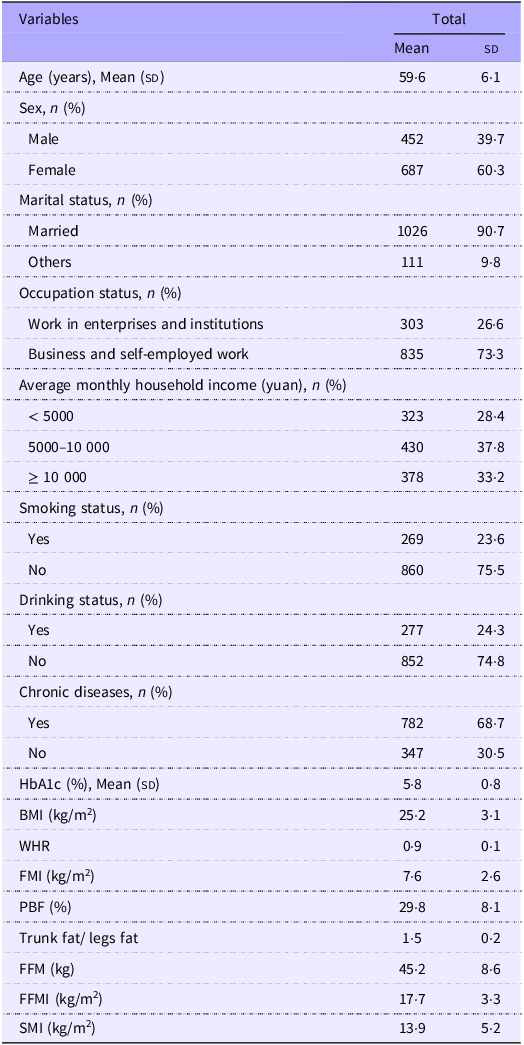
The baseline characteristics of the recruited participants are presented in Table 1. The study population had an average age of 59·6 years (sd = 6·1), with more females and married participants. Most participants were engaged in business or self-employed work (73·3 %), and household income was evenly distributed across three categories (< 5000 yuan: 28·4 %; 5000–10 000 yuan: 37·8 %; > 10 000 yuan: 33·2 %). Chronic diseases were prevalent (68·7 %), while current smoking (23·6 %) and drinking (24·3 %) were less common. The average HbA1c of the study population was 5·8 % (sd = 0·9). The average value for BMI, WHR, FMI, PBF, trunk fat/legs fat, FFM, FFMI and SMI were 25·2 kg/m2, 0·9, 7·6 kg/m2, 29·8 %, 1·5, 45·2 kg, 17·7 kg/m2 and 13·9 kg/m2, respectively.
Baseline characteristics of recruited participants

HbA1c, glycosylated hemoglobin; WHR, waist:hip ratio; FMI, fat mass index; PBF, percentage of body fat; FFM, fat free mass; FFMI, fat free mass index; SMI, skeletal mass index.
Missing data: marital status (n 2), occupation status (n 1), average monthly household income (n 8), smoking status (n 10), drinking status (n 10) and chronic diseases (n 10).
Associations between co-exposure of plasma advanced glycation end products and individuals advanced glycation end products and body composition index
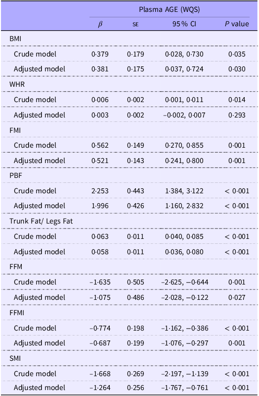
The estimated weights of each AGE on body composition-related parameters were presented in Supplementary Figure 1. As shown in Table 2, in adjusted model, with regard to fat mass-related parameters, total exposure of plasma AGE was positively associated with BMI (β (95 % CI): 0·381 (0·037, 0·724), P = 0·030), FMI (β (95 % CI): 0·521 (0·241, 0·800), P = 0·001), PBF (β (95 % CI): 1·996 (1·160, 2·832), P < 0·001), trunk fat/legs fat (β (95 % CI): 0·058 (0·036, 0·080), P < 0·001). In regards to skeletal muscle mass related parameters, total exposure of plasma AGE was negatively associated with FFM (β (95 % CI): −1·075 (–2·028, −0·122), P = 0·027), FFMI (β (95 % CI): −0·687 (–1·076, −0·297), P = 0·001) and SMI (β (95 % CI): −1·264 (–1·767, −0·761), P < 0·001). The associations between co-exposure of plasma AGE and body composition index in subgroups analysis stratified by age, sex, chronic diseases and HbA1c level are shown in Figure 1. Most of the associations remain robust after subgroup analysis. However, significant heterogeneity in the strength of associations between plasma AGE and body composition parameters across age and sex subgroups. Using the median age (61 years) as the cut-off, stronger negative associations were observed in younger participants (< 61 years) for FFM (β = −2·45 kg, 95 % CI: −3·88, −1·03, P interaction = 0·019) and FFMI (β = –1·22 kg/m2, 95 % CI: −1·77, −0·67, P interaction = 0·035), whereas these associations were markedly attenuated in older participants (≥ 61 years) (FFM: β = 0·03 kg, 95 % CI: −1·26, 1·33; FFMI: β = −0·24 kg/m2, 95 % CI: −0·80, 0·31). Sex-stratified analyses further demonstrated significantly stronger negative associations of plasma AGE with FFM (β = −2·05 kg, 95 % CI: −3·34, −0·76, P interaction = 0·001), FFMI (β = −1·04 kg/m2, 95 % CI: −1·58, −0·49, P interaction = 0·003), and SMI (β = −1·75 kg/m2, 95 % CI: −2·45, −1·04, P interaction = 0·003) in females compared with males (FFM: β = 0·31 kg, 95 % CI: −0·99, 1·62; FFMI: β = −0·18 kg/m2, 95 % CI: −0·66, 0·29; SMI: β = −0·56 kg/m2, 95 % CI: −1·20, 0·08). These findings suggested that age and sex may modulate the biological pathways linking AGE to body composition alterations.
Subgroup analysis on the associations between co-exposure of plasma AGE and body composition parameters. Subgroup analysis via age, sex, chronic diseases and HbA1c levels were performed on the associations between co-exposure of plasma AGE and body composition parameters in fully adjusted model, with β and 95 % CI shown. Models were adjusted for adjusted for age, sex (male, female), marital status (married, others), occupation status (work in enterprises and institutions/ business and self-employed work), average monthly household income (< 5000 yuan, 5000–10000 yuan, ≥ 10000 yuan), smoking status AGE, advanced glycation end products; WQS, weighted quantile sum; WHR, waist:hip ratio; FMI, fat mass index; PBF, percentage of body fat; FFM, fat free mass; FFMI, fat free mass index; SMI, skeletal mass index.

Multivariable linear regression model testing the association between co-exposure to 5 AGE fitted via WQS model and body composition index

AGE, advanced glycation end products; WQS, weighted quantile sum; WHR, waist:hip ratio; FMI, fat mass index; PBF, percentage of body fat; FFM, fat free mass; FFMI, fat free mass index; SMI, skeletal mass index. Adjusted model was adjusted for age, sex (male, female), marital status (married, others), occupation status (work in enterprises and institutions/ business and self-employed work), average monthly household income (< 5000 yuan, 5000–10000 yuan, ≥ 10000 yuan), smoking status (yes or no), drinking status (yes or no), chronic diseases (yes or no) and HbA1c.
Multivariable-adjusted analyses demonstrated differential associations between plasma AGE and body composition parameters (Figure 2). Fat-related parameters exhibited the most robust associations: free CEL showed a strong positive association with trunk fat/legs fat (β (95 % CI): 0·34 (0·21, 0·47), P < 0·001), while free MG-H1 was positively linked to FMI (β (95 % CI): 1·92 (0·25, 3·59), P = 0·024) and PBF (β (95 % CI): 6·32 (1·94, 10·7), P = 0·005). Combined CEL further displayed consistent positive associations with FMI (β (95 % CI): 0·05 (0·001, 0·09), P = 0·045) and PBF (β (95 % CI): 0·21 (0·08, 0·35), P = 0·002). In contrast, free CML was negatively associated with WHR (β (95 % CI): −0·02 (–0·03, −0·008), P = 0·002). In regards to skeletal muscle-related parameters, free CML and free MG-H1 were negatively associated with FFM (β (95 % CI): −2·69 (–4·76, −0·62), P = 0·011; β (95 % CI): −3·91 (–7·19, −0·62), P = 0·020) and FFMI (β (95 % CI): −1·13 (–1·93, −0·32), P = 0·006; β (95 % CI): −2·92 (–5·17, −0·67), P = 0·011) and SMI (β (95 % CI): −2·99 (–4·32, −1·65), P < 0·001, β (95 % CI): −9·37 (–15·9, −2·84), P = 0·005). Notably, combined CEL also showed consistent negative associations with FFMI (β (95 % CI): −0·08 (–0·13, −0·02), P = 0·007) and SMI (β (95 % CI): −0·28 (–0·36, −0·19), P < 0·001), further supporting the detrimental role of AGE in muscle mass regulation. No significant associations were observed with BMI (P > 0·05).
The associations between individual plasma AGE and body composition-related parameters. The associations between individual plasma AGE including free CML, free CEL, free MG-H1, combined CML and combined CEL with body composition-related parameters in fully adjusted model, with β and 95 % CI shown. Models were adjusted for age, sex (male, female), marital status (married, others), occupation status (work in enterprises and institutions/ business and self-employed work), average monthly household income (< 5000 yuan, 5000–10000 yuan, ≥ 10000 yuan), smoking status (yes or no), drinking status (yes or no), chronic diseases (yes or no) and HbA1c. AGE, advanced glycation end products; CML, Nε-(carboxymethyl) lysine; CEL, Nε-(1-carboxyethyl) lysine; MG-H1, Nδ-(5-hydro-5-methyl-4-imidazolon-2-yl)-ornithine; WQS, weighted quantile sum; WHR, waist:hip ratio; FMI, fat mass index; PBF, percentage of body fat; FFM, fat free mass; FFMI, fat free mass index; SMI, skeletal mass index.

The associations between individual plasma AGE and body composition related parameters after adjusting for potential covariates are presented in Figure 2. No significant associations were observed between individual AGE and BMI. Free CML was negatively associated with WHR (β (95 % CI): −0·02 (–0·03, −0·008), P = 0·002). Free MG-H1 and combined CEL were positively associated with FMI (β (95 % CI): 1·92 (0·25, 3·59), P = 0·024, β (95 % CI): 0·05 (0·001, 0·09), P = 0·045) and PBF (β (95 % CI): 6·32 (1·94, 10·7), P = 0·005, β (95 % CI): 0·21 (0·08, 0·35), P = 0·002). Free CML (β (95 % CI): 0·06 (0·005, 0·12), P = 0·032), free CEL (β (95 % CI): 0·34 (0·21, 0·47), P < 0·001), free MG-H1 (β (95 % CI): 0·11 (0·004, 0·21), P = 0·042) and combined CEL (β (95 % CI): 0·01 (0·007, 0·02), P < 0·001) were positively associated with trunk fat/legs fat. Free CML (β (95 % CI): −2·69 (–4·76, −0·62), P = 0·011) and free MG-H1 (β (95 % CI): −3·91 (–7·19, −0·62), P = 0·020) were negatively associated with FFM. Free CML, free MG-H1 and combined CEL were negatively associated with FFMI (β (95 % CI): −1·13 (–1·93, −0·32), P = 0·006, β (95 % CI): −2·92 (–5·17, −0·67), P = 0·011, β (95 % CI): −0·08 (–0·13, −0·02), P = 0·007) and SMI (β (95 % CI): −2·99 (–4·32, −1·65), P < 0·001, β (95 % CI): −9·37 (–15·9, −2·84), P = 0·005, β (95 % CI): −0·28 (–0·36, −0·19), P < 0·001).
Discussion
To our knowledge, this is the first study to show that in middle-aged and elderly Chinese participants, co-exposure of plasma AGE including free CML, free CEL, free MG-H1, combined CML and combined CEL was positively associated with fat-related indexes including BMI, FMI, PBF, trunk fat/legs fat, while were negatively associated with skeletal muscle-related indexes including FFM, FFMI and SMI after adjusting for potential covariates. However, as this is a cross-sectional study, causality cannot be inferred. Our findings suggest that elevated circulating AGE might be a risk factor for obesity and reduced skeletal muscle quality, but further longitudinal studies are needed to confirm these associations.
Although still controversial(Reference Ghorbaninejad, Djafarian and Babaee9), previous findings suggest that AGE accumulation might be associated with increased body weight(Reference Amin, Mosa and El-Shishtawy6–Reference Goudarzi, Sedaghat and Hedayati8). Mechanistically, the receptor for advanced glycation end products – a key receptor mediating AGE signalling – is proposed to critically regulate weight gain and adiposity by modulating energy expenditure and adipose tissue browning processes(Reference Taheri, Vasheghani-Farahani and Honarkar-Shafie24). Also, AGE-receptor for advanced glycation end products signalling may contribute to weight gain and obesity via affecting adipose tissue inflammation, adipocyte hypertrophy and insulin sensitivity(Reference Feng, Du and Shu25). However, most of these mechanistic studies have been conducted in general adult populations or animal models, and limited evidence exists specifically in middle-aged and elderly populations. During ageing, the function of immune cells in adipose tissue declines, while the secretion of pro-inflammatory factors increases, leading to chronic low-grade inflammation. This inflammatory state accelerates metabolic dysfunction and tissue degeneration and is closely associated with metabolic diseases such as insulin resistance and type 2 diabetes(Reference Reyes-Farias, Fos-Domenech and Serra26,Reference Ou, Zhang and Tan27) . Age-related changes in adipose tissue inflammation and insulin resistance may exacerbate the impact of AGE on fat accumulation and central obesity. Further research is needed to elucidate how ageing influences the relationship between AGE and body composition.
Our findings further indicated that in middle-aged and elderly Chinese community participants, co-exposure to plasma AGE is not only positively associated with the common obesity parameters, i.e. BMI, but also with other fat mass-related parameters including FMI, PBF and trunk fat/legs fat. The stronger magnitude of associations between AGE and FMI or PBF compared with BMI or WHR may reflect the superior sensitivity of direct adiposity measures in capturing AGE-related metabolic dysregulation. Unlike BMI, which conflates fat and lean mass, or WHR, which is influenced by both subcutaneous and visceral fat distribution, PBF and FMI, which provide a more accurate assessment of adiposity by directly quantifying fat mass relative to total body weight(Reference Wong, O’Neill and Beck28). Trunk fat/legs fat ratio is generally considered as a marker of central adiposity and might be associated with high risk of cardiovascular diseases(Reference Chen, Arthur and Iyengar29). Therefore, our findings suggest that reducing circulating AGE levels may be necessary for weight maintenance in middle-aged and elderly adults. While reducing circulating AGE could be a promising strategy against obesity, current evidence from human intervention studies targeting AGE reduction remains limited. Future studies should explore whether AGE reduction interventions, such as dietary modifications or pharmacological approaches, can effectively mitigate obesity and its related metabolic disorders in middle-aged and elderly populations.
A number of population based studies with small sample size have reported a significant inverse associations between AGE levels measured via skin autofluorescence and muscle strength,(Reference Momma, Niu and Kobayashi30) muscle mass(Reference Kato, Kubo and Sugioka31) and physical activity(Reference Drenth, Zuidema and Krijnen32) in middle-aged(Reference Momma, Niu and Kobayashi30,Reference Kato, Kubo and Sugioka31) to older(Reference Drenth, Zuidema and Krijnen32) population. Accumulation of AGE measured via skin autofluorescence was negatively associated with skeletal muscle index(Reference Tabara, Ikezoe and Yamanaka12). Our findings suggested that in middle-aged and elderly Chinese participants, co-exposure of plasma AGE had negative associations with skeletal muscle-related indexes including FFM, FFMI and SMI. However, it is important to consider other factors that may contribute to muscle loss, such as sarcopenia and chronic inflammation. Additionally, a meta-analysis has indicated that chronic inflammation is significantly associated with lower skeletal muscle strength and reduced muscle mass(Reference Tuttle, Thang and Maier33).
The negative association between co-exposure of plasma AGE and skeletal muscle-related indexes can be explained by the following mechanisms: intracellular damage to muscle cells due to inflammation induced by AGE binding to the RAGE and enhancement of intramuscular collagen accumulation through non-enzymatic cross-linking of AGE leading to the stiffening of skeletal muscle extracellular matrix, as well as exacerbation of skeletal muscle atrophy through activation of the PERK/FOXO1 signalling pathway in skeletal muscle(Reference Olson, Redden and Schwartz34–Reference Du, Ma and Wang36). AGE could also induce osteoblast apoptosis via endoplasmic reticulum stress(Reference Gutowska, Czajkowski and Kuryłowicz37). Nevertheless, our findings also suggest that strategies for reducing circulating AGE level might be required for the middle-aged and elderly participants in order to maintain or improve skeletal muscle quality. It is noteworthy that most existing studies on AGE have primarily focused on their effects on fat mass rather than muscle mass. Future research should aim to explore the specific mechanisms by which AGE influence muscle metabolism and whether interventions targeting AGE reduction can effectively mitigate muscle loss, particularly in ageing populations.
We further found that the negative associations between co-exposure to plasma AGE and FFM and FFMI were more pronounced in those aged less than 61 years old, highlighting the need to reduce AGE intake at a younger age. Multiple reasons might exist for the stronger associations in younger adults. First, younger individuals typically exhibit higher metabolic rates and more active protein turnover(Reference Pontzer, Yamada and Sagayama38,Reference He, Connolly and Cross39) , which might make them more susceptible to the detrimental effects of AGE on muscle mass and body composition. Additionally, younger adults may have a longer cumulative exposure to dietary AGE due to modern dietary patterns, such as the high consumption of processed and fast foods(Reference da Silva, Fiates and Botelho40). In recent decades, significant changes in dietary habits, particularly in China, have led to increased consumption of animal-derived foods, refined grains, and highly processed products rich in sugar and fat(Reference Pan, Wang and Pan41). These foods are known to contain high levels of AGE, which are formed during high-temperature cooking processes(Reference Gill, Kumar and Singh42). Younger generations, who are more likely to adopt these modern dietary habits, may therefore experience greater accumulation of AGE in plasma and tissues over time, which directly contributes to the observed negative associations with muscle mass(Reference da Silva, Fiates and Botelho40).
We also found that the associations between plasma AGE and FFM, FFMI and SMI were more pronounced in female participants, which is consistent with previous studies. Previously, Ebert et al. (Reference Ebert, Lacruz and Kluttig43) reported that the negative associations between plasma AGE levels and physical functioning were observed only in females. Yang et al. (Reference Yang, Li and Liu44) demonstrated that urinary CML levels were negatively associated with grip strength in only older women aged more than 65 years old. The stronger associations observed in females compared with males may be attributed to hormonal differences, such as the decline in estrogen levels during menopause, which reduces the protective effects on skeletal muscle(Reference Pellegrino, Tiidus and Vandenboom45). Additionally, females generally have a higher body fat percentage and lower muscle mass, which may amplify the impact of AGE on muscle-related parameters(Reference Neves, Salamunes and de Oliveira46). Differences in dietary patterns and lifestyle factors between men and women could also contribute to these disparities. Nevertheless, further long-term follow-up studies with large sample size are required to confirmation the relationships between AGE and fat, also skeletal muscle related parameters. These findings suggested that age and sex may modulate the biological pathways linking AGE to body composition alterations.
We also found that the associations between individual AGE and body composition parameters might be variable. The variability in associations between specific AGE and body composition highlights the complexity of AGE’ metabolic effects. While these three AGE are widely studied and serve as reliable indicators of AGE accumulation, the diversity of AGE in biological systems suggests that they may not fully capture the total AGE burden or its metabolic impact. For example, in a cross-sectional study, pentosidine was significantly associated with prevalent vertebral fractures but not with lumbar spine bone mineral density (BMD), suggesting that pentosidine may increase fracture risk through mechanisms independent of BMD, such as alterations in bone quality or microarchitecture(Reference Nakano, Nakamura and Suzuki47). However, it was not measured in the current study. Our findings suggest that specific AGE might play differential roles in obesity and skeletal muscle quality. Further mechanistic studies are required to explore how different specific AGE affects obesity and skeletal muscle quality. Additionally, while our findings provide valuable insights into the associations between AGE and body composition in a middle-aged and elderly Chinese population, there is a notable gap in mechanistic studies, particularly in non-Western populations. Most existing research on AGE and metabolic health has been conducted in Western population, where dietary patterns, lifestyle factors, and genetic backgrounds may differ significantly from those in Asian populations(Reference Clarke, Dordevic and Tan48). For instance, the high consumption of rice and cooking methods such as steaming in Asian diets may influence AGE formation and metabolism differently compared with Western diets rich in processed and grilled foods(Reference Wang, Lin and Bloomgarden49). Therefore, future mechanistic studies should prioritise diverse populations to elucidate the specific pathways through which AGE influence body composition and to develop targeted interventions tailored to different dietary and cultural contexts.
A major strength of this study is that it is the first study to simultaneously measure plasms free and combined CML, CEL and MG-H1 via ultra-high performance liquid chromatography-tandem MS and associate their co-exposure levels with body composition parameters in Chinese middle-aged and elderly individuals with a relatively large sample size. However, several limitations should be addressed. First, as a cross-sectional study, our findings cannot establish causality; to partially mitigate this limitation, we adjusted for key metabolic confounders including HbA1c level and other chronic diseases, though residual confounding from unmeasured temporal factors may persist. Second, although this study did not directly assess dietary intake, existing literature suggests that the primary food sources of AGE such as CML, CEL and MG-H1 include high-temperature cooked meats, processed foods and certain dairy products. These foods are known to contribute significantly to the dietary AGE burden. Future work should incorporate 24-h dietary recalls with FFQ to quantify contributions of specific dietary AGE sources. Third, we only measured representative AGE of CML, CEL and MG-H1, the associations between other AGE and body composition parameters remain unanswered. Fourth, although bioelectrical impedance analyser is a practical method for assessing body composition, it is less precise compared with more advanced techniques such as dual-energy X-ray absorptiometry or magnetic resonance imaging. Fifth, unavailability of data on dietary energy intake and physical activity precluded their adjustment as covariates. Despite controlling for HbA1c level and other chronic diseases – which correlate with these factors – residual confounding likely remains. Future cohort studies must therefore integrate accelerometer-based physical activity monitoring and doubly labeled water techniques to obtain objective energy expenditure data.
Our study reveals a previously unrecognised dual-pathogenic mechanism of AGE, demonstrating for the first time that co-exposure to plasma free CML, CEL and MG-H1 concurrently might exacerbate adiposity accumulation and skeletal muscle depletion in ageing populations. This finding positions AGE as critical modifiable targets for combating the escalating pandemic of obesity and sarcopenia – two interrelated public health crises amplified by modern ultra-processed diets. From a translational perspective, dietary AGE restriction emerges as a promising unified strategy to mitigate these co-occurring conditions, particularly in regions undergoing rapid nutritional transitions like China. To translate this insight into clinical action, future research must quantify dose–response relationships between food-specific AGE and fat-muscle dynamics and evaluate AGE-lowering interventions as potential dual-therapies for metabolic-sarcopenic obesity.
Acknowledgement
The authors thank the participants for their contributions to this work.
The cross-sectional study had been approved by the ethical committee of Soochow University (approval no. SUDA20200601H01& SUDA20210923H03). All participants provided written consent to participate in the present study and completed health-related questionnaire, physical examination and laboratory examination.
This study was supported by the National Natural Science Foundation of China (Grant no. 8247122084&82073535) and The Elderly Health Project of Jiangsu Province (LKM2023037).
Z. W., K. S. and Y. W. designed the study; W. Z., Y. W., H. H., F. X., N. Z., R. H. and Y. W. performed the experiment; W. Z. performed the statistical analyses; W. Z. drafted and Y. W., H. H., F. X., N. Z., R. H., Y. W., K. S. and Z. W. revised the manuscript; K. S. and Z. W. supervised the data analysis and interpretation; Z. W. had the primary responsibility for the final content. All authors critically reviewed the manuscript and approved the final draft.
The authors declare no conflicts of interest.
Data used in this study are available upon request from the authors.
Supplementary material
For supplementary material/s referred to in this article, please visit https://doi.org/10.1017/S0007114525105096