Introduction
Most eukaryotic cells have evolved a highly efficient molecular mechanism for their cellular respiration and energy production, in which oxygen molecule is adopted as a final electron acceptor in the mitochondria. Therefore, maintaining oxygen homeostasis is essential for these cells, especially those present in animals with high mass and energy demands (Chandel and Budinger, Reference Chandel and Budinger2007). Irrespective of the atmospheric oxygen level, the oxygen availability becomes limited in biological microniches owing to its reduced diffusion, which dues to the physical constraints imposed by growth and development or accelerated oxygen consumption rate by cells, which is frequently observed in association with certain pathobiological circumstances (Dyson and Singer, Reference Dyson and Singer2011; Bailey-Serres et al., Reference Bailey-Serres, Fukao, Gibbs, Holdsworth, Lee, Licausi, Perata, Voesenek and van Dongen2012). During low oxygen tension (hypoxia), multiple genes involved in metabolic changes, erythropoiesis, angiogenesis, cell growth and differentiation, and apoptosis are activated to adapt to the physiological challenge (Kiriakidis et al., Reference Kiriakidis, Esteban and Maxwell2007; Semenza, Reference Semenza2009; Majmundar et al., Reference Majmundar, Wong and Simon2010). Hypoxia-inducible factors (HIFs) are the master regulators of the hypoxic transcriptional responses in metazoans (Wang and Semenza, Reference Wang and Semenza1993; Kiriakidis et al., Reference Kiriakidis, Esteban and Maxwell2007).
The HIF proteins are heterodimeric transcription factors that consist of α and β subunits (also known as aryl hydrocarbon receptor nuclear translocator, ARNT). These subunits conserve basic helix–loop–helix (bHLH) and Per–Arnt–Sim (PAS) domains which participate in the HIF heterodimerization and specific binding to the target DNA sequence (Wang et al., Reference Wang, Jiang, Rue and Semenza1995; Crews, Reference Crews1998). The structural and functional integrity of the proteolysis-susceptible α subunit is regulated by post-translational hydroxylation of two prolyl residues in the amino-terminal oxygen-dependent degradation (NODD) and carboxy-terminal oxygen-dependent degradation (CODD) domains, which is catalyzed by prolyl hydroxylase domain (PHD)-containing enzymes (Rytkönen et al., Reference Rytkönen, Williams, Renshaw, Primmer and Nikinmaa2011). The hydroxylated α subunit is subjected to ubiquitination for proteasomal degradation by ubiquitin E3 ligase, von Hippel-Lindau protein (VHL) complex (Majmundar et al., Reference Majmundar, Wong and Simon2010). Additionally, the hydroxylation of a C-terminal asparaginyl residue by factor inhibiting HIF-1α (FIH) is involved in the functional regulation of HIF-1α. The hydroxylated asparaginyl moiety blocks the recruitment of transcriptional co-activators such as P300 and cAMP-response-element-binding protein (CREB)-binding protein complex (Zhang et al., Reference Zhang, Fu, Linke, Chicher, Gorman, Visk, Haddad, Poellinger, Peet, Powell and Johnson2010). The HIF subunit proteins belong to the bHLH–PAS protein superfamily along with the other members, including the proteins of single-minded (SIM) and aryl hydrocarbon receptor (AHR) families (Li et al., Reference Li, Lu and Yu2014).
Clonorchis sinensis (C. sinensis) is a trematode parasite that invades the bile ducts of carnivorous mammals, including humans. Humans are infected with C. sinensis after eating raw or under-cooked freshwater fish containing infective metacercariae of the liver fluke. This parasitic disease, i.e. clonorchiasis is highly prevalent in East Asian countries, such as China, Vietnam and Korea (Lun et al., Reference Lun, Gasser, Lai, Li, Zhu, Yu and Fang2005; Keiser and Utzinger, Reference Keiser and Utzinger2009; Qian et al., Reference Qian, Utzinger, Keiser and Zhou2016). Acute clonorchiasis with mild infection is generally asymptomatic, while fever, right upper-quadrant pain and intermittent colic pain are frequent symptoms during severe infection depending on the worm burden. However, chronic clonorchiasis owing to the protracted (re-)infection of C. sinensis is strongly associated with the fibrosis of ducts, destruction of adjacent liver parenchyma and recurrent pyogenic cholangitis. Most importantly, these histopathological changes eventually lead to the development of cholangiocarcinoma, which is one of the human cancers exhibiting worse prognosis (Lun et al., Reference Lun, Gasser, Lai, Li, Zhu, Yu and Fang2005; Bouvard et al., Reference Bouvard, Baan, Straif, Grosse, Secretan, El Ghissassi, Benbrahim-Tallaa, Guha, Freeman, Galichet and Cogliano2009; Shin et al., Reference Shin, Oh, Masuyer, Curado, Bouvard, Fang, Wianqnon, Sripa and Hong2010).
The juveniles of C. sinensis excyst in the duodenum, ascend the biliary tracts through the ampulla of Vater and mature into adults in mammalian hosts (Kim et al., Reference Kim, Yoo, Kwak, Seok and Hong2011). As the biliary tract constitutes anoxic or hypoxic environments (Brook, Reference Brook1989), trematode parasites such as C. sinensis, Opisthorchis viverrini and Fasciola spp. which invade the ductal system had been intuitively believed to perform anaerobic respiration in their definitive hosts (Smyth and Halton, Reference Smyth and Halton1983). Contradictorily, a recent investigation regarding the energy metabolism of Fasciola hepatica demonstrated that the fluke simultaneously produces ATP molecules through the aerobic as well as anaerobic respiration in its final host. However, the rate of anaerobic respiration becomes more prominent in proportion to the advancement of parasitic maturation (Tielens et al., Reference Tielens, van den Heuvel and van den Bergh1984). Multiple proteins involved in the aerobic respiratory process, such as the citrate cycle and oxidative phosphorylation as well as those in anaerobic respiration, including mitochondrial acetate:succinate CoA-transferase, are abundantly expressed in the adult stages of C. sinensis and O. viverrini (Young et al., Reference Young, Campbell, Hall, Jex, Cantacessi, Laha, Sohn, Sripa, Loukas, Brindley and Gasser2010, Reference Young, Nagarajan, Lin, Korhonen, Jex, Hall, Safavi-Hemami, Kaewkong, Bertrand, Gao, Seet, Wongkham, The, Wongkham, Intapan, Maleewong, Yang, Hu, Wang, Hofmann, Sternberg, Tan, Wang and Gasser2014; Huang et al., Reference Huang, Chen, Wang, Liu, Chen, Guo, Luo, Sun, Mao, Liang, Xie, Zhou, Tian, Lv, Huang, Zhou, Hu, Li, Zhang, Lei, Li, Hu, Liang, Xu, Li and Yu2013). Additionally, a massive release of myoglobin with strong affinity to oxygen appeared to be evident in C. sinensis adult worms (Kiger et al., Reference Kiger, Rashid, Griffon, Haque, Moens, Gibson, Poyart and Marden1998; Bae et al., Reference Bae, Ahn, Lee, Kim, Cai, Kang, Sohn and Kong2013a). These data might suggest that the parasitic trematodes possess a molecular machinery associated with oxygen-sensing to alternate between the aerobic and anaerobic pathways depending on the oxygen availability in the surrounding environments. The HIF-PHD system might be a plausible candidate, albeit there were no reports on the aforementioned molecular mechanism in platyhelminths, including the trematode parasites.
In this study, we characterized the bHLH–PAS genes such as HIF-1α, SIM, HIF-1β and AHR in C. sinensis, which were identified in the GenBank database of the liver fluke. The induction profiles of these genes were determined by incubating C. sinensis adult worms under various oxygen conditions, as well as in the presence of a PHD inhibitor. The effects of nitric oxide (NO) and nitrite, which modulate the levels of oxy- and deoxy-myoglobin, respectively, on the expression of these genes were also examined under the experimental conditions.
Materials and methods
Identification of bHLH–PAS genes in C. sinensis
The sequence information of human proteins which represent each of the diverse bHLH–PAS subfamilies, including HIF-1α and SIM, was obtained from GenBank (https://www.ncbi.nlm.nih.gov/) based on a previous report (Supplementary Table S1; Li et al., Reference Li, Lu and Yu2014). These amino acid sequences were used as queries in searches with the standard basic local alignment search tool for proteins (BLASTp) to identify the bHLH–PAS homologs in the GenBank proteomic database of C. sinensis. Further, the C. sinensis proteins that exhibited significant matches to the human proteins (E-value <1 × 10⁻⁵) were applied as queries in the similarity searches. The presence of bHLH and PAS domains in the retrieved sequences was examined using the simple modular architecture research tool (SMART; http://smart.embl.de/) program (Schultz et al., Reference Schultz, Milpetz, Bork and Ponting1998).
Genomic structures of bHLH–PAS genes in C. sinensis
The complementary DNA (cDNA) sequences of bHLH–PAS genes were identified in the expressed sequence tag (EST; GenBank) and transcriptome shotgun assembly (TSA; http://bioinfosecond.vet.unimelb.edu.au/wblast2.html) databases of C. sinensis by performing nucleotide BLAST (BLASTn) searches using the coding DNA sequences (CDSs) of C. sinensis bHLH-PAS proteins. The retrieved EST or TSA sequences along with their specific CDS queries were assembled into respective contigs using the CAP3 program (Huang and Madan, Reference Huang and Madan1999). Non-overlapping gaps were filled with nucleotide sequences, which were empirically determined from cDNAs of C. sinensis adult worms by performing PCR amplification and subsequent nucleotide sequencing (Supplementary Fig. S1). The assembled cDNA contigs were compared with homologous sequences which were isolated from the GenBank genomic database of C. sinensis to determine the exon-intron architectures of chromosomal bHLH–PAS genes. The insertion phases of introns were defined as follows: 0, insertion between codons; +1, insertion between the first and second bases of a codon; and +2, insertion between the second and third bases of the codon. The structural integrity of assembled contigs was verified by amplifying the corresponding transcripts using primers that were matched to both the ends of each CDS (Supplementary Fig. S1).
Phylogenetic analysis
Platyhelminth bHLH–PAS proteins were identified in the GenBank proteomic databases by performing BLASTp searches using the sequence information of C. sinensis homologs. The amino acid sequences that comprise the bHLH, PAS-A and PAS-B domains, each of which was annotated using the SMART program, were extracted from these platyhelminth proteins, as well as from the human and C. sinensis bHLH–PAS proteins. After concatenation, the amino acid sequences were aligned using the MUSCLE software (Edgar, Reference Edgar2004) and the resultant alignment was used in the construction of a neighbour-joining tree using MEGA 6.0 (Jones–Taylor–Thornton model for amino acid evolution, gamma distributed rates among sites and pairwise deletion of missing data; Tamura et al., Reference Tamura, Stecher, Peterson, Filipski and Kumar2013). The statistical significance of branching nodes was estimated by a bootstrapping analysis with 1000 replicates of the initial input alignment. The phylogenetic tree was displayed using TreeView (Page, Reference Page1996).
Simulation of three-dimensional structures
The tertiary structures of C. sinensis HIF-1α (CsHIF-1α) and SIM (CsSIM) were simulated using the I-TASSER program (ver. 5·0; http://zhanglab.ccmb.med.umich.edu/I-TASSER/), which combined threading, ab initio modelling and structural refinement (Zhang, Reference Zhang2008). The template modelling-score (TM-score) and root mean square deviation (RMSD) were calculated between the C. sinensis proteins and reference models to evaluate the quality of simulated structures. The stereochemical quality of best-fit models was validated by subjecting PDB files obtained by I-TASSER to the PROCHECK server (http://www.ebi.ac.uk/thornton-srv/databases/pdbsum/Generate.html). The binding potential of CsHIF-1α to DNA ligand and amino acids involving in the protein–DNA interaction was predicted using the I-TASSER program. The resultant tertiary structures were visualized using the PyMol program (DeLano Scientific, San Carlos, CA, USA).
Preparation of parasitic materials
Sprague–Dawley rats were orally administered with C. sinensis metacercariae collected from naturally infected freshwater fish in South Korea (150 metacercariae/rat). Adult worms were harvested from the bile ducts of rats at 8 weeks post-infection. Protocols to perform the parasite infection, anaesthesia of rats by intraperitoneal injection of 7:3 mixture of alfaxlone (10 mg mL−1) and xylazine (23.3 mg mL−1) during worm recovery and euthanasia of moribund rats were approved by the Institutional Review Board and conducted in the Laboratory Animal Research Center of Gachon University (Protocol number GIACUC-R2015005). The animal experiments were conducted in accordance with the guidelines of Association for the Assessment and Accreditation of Laboratory Animal Care (Thailand).
Oligopeptides and specific antisera
Oligopeptides comprising the DNA-binding site of CsHIF-1α (ARHRRNQENELFKALAHLLPLHP) and CsSIM (ARTRREKENAEFYELAKLLPLPS), which were predicted using I-TASSER, were synthesized and conjugated to keyhole limpet hemocyanin (KLH) protein by utilizing the commercial service of AbFrontier Ltd. (Seoul, Republic of Korea). The oligopeptide-KLH conjugates were applied in the preparation of specific rat (anti-CsHIF-1α antibody) and mouse (anti-CsSIM antibody) antisera in their commercial laboratory. The sensitivity and specificity of these specific antisera were examined by performing Western-blot analysis using identical oligopeptides conjugated to bovine serum albumin (BSA) in combination with the KLH conjugates.
Expression levels of bHLH–PAS genes under various oxygen conditions
Adult worms were washed more than ten times with cold phenol red-free RPMI 1640 medium (pH 7.2; Gibco BRL, Gaithersburg, MD, USA) and incubated in a CO2 incubator with atmospheric oxygen level for 4 h. The worms (15 worms/experimental group) were incubated in the medium containing penicillin and streptomycin (100 U mL−1 and 100 µg mL−1, respectively; Gibco BRL) at 37 °C in 5%-CO2 incubators, in which the oxygen levels were equilibrated to 1, 5 or 20%, using nitrogen gas during the incubation period. After 2, 4, 8 and 24 h incubation, total RNAs were extracted from the worms of each group using the QIAzol lysis reagent and RNeasy Mini kit (Qiagen, Hilden, Germany). The RNA samples were treated with an RNase-free DNase (New England Biolabs, Ipswich, MA, USA) to remove any contaminating DNAs. The cDNA synthesis was performed using the iScript cDNA synthesis kit (Bio-Rad, Munich, Germany). The relative levels of the bHLH–PAS transcripts in the cDNA samples were estimated via quantitative real-time PCR (qPCR) using gene-specific primers (Supplementary Table S2). C. sinensis β-actin gene (CsAct; DF143505) was selected as a reference gene. The amplification reactions were conducted using the SYBR Green master mix and CFX96 detection system by following the manufacturer's instructions (Bio-Rad Laboratories, Hercules, CA, USA). The presence of a single amplicon in the final PCR products was ensured by performing melting curve analysis. The expression levels of the bHLH–PAS genes were normalized against those of CsAct. The induction level of each gene measured as fold change was calculated with respect to that in the pre-incubated C. sinensis worms (2−ΔΔC T method; Livak and Schmittgen, Reference Livak and Schmittgen2001). Each reaction was conducted in triplicate and the result was presented as the mean ± standard deviation (s.d.). Statistical analyses were performed using Student's t test (two-sided; P < 0.05).
In another batch of the experimental conditions, the incubation medium was supplemented with 0.2 and 1 mm dimethyloxaloylglycine (DMOG; Sigma-Aldrich, St. Louis, MO, USA), a competitive inhibitor of PHDs. C. sinensis worms were incubated in the DMOG-RPMI-1640 medium at 37 °C for 2, 4 and 8 h under the 20%-oxygen condition. The fold-inductions of bHLH–PAS genes in these experimental groups were examined by performing qPCR as previously described. The values were compared with those concurrently observed in worms incubated under the 1%-oxygen condition.
Effect of NO and nitrite on the bHLH–PAS gene expressions
The live C. sinensis adults (15 worms/well) were incubated in RPMI-1640 medium containing sodium nitrite (5 and 20 µ m; Sigma-Aldrich) at 37 °C for 24 h under 1, 5 and 20%-oxygen conditions. Similarly, the worms were seeded in the medium supplemented with NOR-4 to chemically provide NO (50 µ m; Enzo Life Sciences, Farmingdale, NY, USA). Total RNAs extracted from these worms were examined to determine the induction levels of the bHLH–PAS genes.
Co-incubation of human cholangiocytes and C. sinensis
SV40-transformed human cholangiocytes (H69) were seeded into six-well plates (2 × 10⁵ cells/well) and incubated under normal culture conditions in DMEM/Ham's F-12 medium (Gibco BRL) supplemented with 10% fetal bovine serum, 100 U mL−1 penicillin, 100 µg mL−1 streptomycin, 0.18 mm adenine, 5 µg mL−1 insulin, 5.5 µ m epinephrine, 2 nm triiodothyronine, 5 µg mL−1 transferrin, 1.64 µ m epidermal growth factor and 1 µ m hydrocortisone. At ~80% confluency of the cultures, polycarbonate transwell inserts (pore size 3 µm; SPL Life Sciences, Pochen, Republic of Korea) containing live C. sinensis adults (15 worms/well) were placed into the culture plates. These plates were incubated at 37 °C in 5%-CO2 incubators under 1 and 20%-oxygen conditions. After 24 h incubation, these worms were used to examine the expression levels of the bHLH–PAS genes.
Western-blot analysis
The whole bodies of C. sinensis worms were homogenized in RIPA buffer (pH 7.4; 10 mm Tris-HCl, 150 mm NaCl, 1% Nonidet P-40, 0.2% sodium deoxycholate, 1 mm EDTA and 10 mm NaF) using a Dounce tissue grinder (Wheaton, Millville, NJ, USA). The homogenates were centrifuged at 10 000 g for 20 min at 4 °C and total proteins in the supernatants were quantified using the Pierce bicinchoninic acid assay kit (Thermo Fisher Scientific, Rockford, IL, USA). The protein samples (100 µg) were resolved on 8% SDS-PAGE gels under reducing conditions and transferred to nitrocellulose membranes (Schleicher & Schuell Bioscience, Dassel, Germany). Specific protein bands were detected by reacting the membranes with anti-CsHIF-1α or anti-CsSIM antibodies (1:1000 dilutions) and later, with horseradish peroxidase-conjugated rabbit anti-rat or anti-mouse IgG antibodies (Bethyl Laboratories, Montgomery, TX, USA). Positive signals were visualized with an enhanced chemiluminescence detection system (GE Healthcare, Pittsburgh, PA, USA) and the images were captured using the LAS-400 Lumino image analyser (Fujifilm, Tokyo, Japan). The protein extracts (10 µg) were similarly examined by Western-blot analysis with a C. sinensis cysteine protease-specific antibody (anti-CsCysProt antibody) that was kindly gifted by professor Byoung-Kuk Na of the Department of Parasitology, Gyeongsang National University, Republic of Korea.
Results
bHLH–PAS genes encoded in C. sinensis genome
BLASTp searches using the human bHLH–PAS proteins were performed to identify their homologous proteins in the C. sinensis proteomic database of GenBank (cut-off for E-value = 1 × 10⁻⁵). The query proteins which represent each of 10 bHLH-PAS families (Li et al., Reference Li, Lu and Yu2014) redundantly retrieved four C. sinensis proteins, annotated as SIM (CsSIM, GAA50744.1), HIF-1α (CsHIF-1α, GAA56007.1), HIF-1β (CsHIF-1β, GAA50153.1) and AHR (CsAHR, GAA50018.1), although the statistical significance of these matches substantially varied depending on the query sequences (Supplementary Table S1). The human circadian locomotor output cycles kaput (CLOCK) protein further identified a C. sinensis protein named transcription factor EB (CsTFEB, GAA48933.1), which acts as a master protein for lysosomal biogenesis (Sardiello et al., Reference Sardiello, Palmieri, di Ronza, Medina, Valenza, Gennarino, Di Malta, Donaudy, Embrione, Polishchuk, Banfi, Parenti, Cattaneo and Ballabio2009), owing to a significant similarity in their bHLH domain sequences. The second round of BLAST searches using these C. sinensis proteins as queries demonstrated results similar to those obtained using the human proteins (data not shown).
The coding DNA sequences of C. sinensis bHLH–PAS proteins in GenBank database retrieved several homologous sequences from EST and TSA databases of the liver fluke. After aligning the subject sequences along with their corresponding query sequence, the largest contig sequence was determined as the probable full-unit CDS of each bHLH–PAS gene (the alignments are schematically shown in Supplementary Fig. S1). The open reading frames of these genes were composed of 1023 (CsHIF-1α), 2517 (CsSIM), 2880 (CsHIF-1β) and 5160 bp (CsAHR), which encode polypeptides of 340, 838, 959 and 1917 amino acids, respectively.
Structural and phylogenetical characterization of C. sinensis bHLH–PAS genes
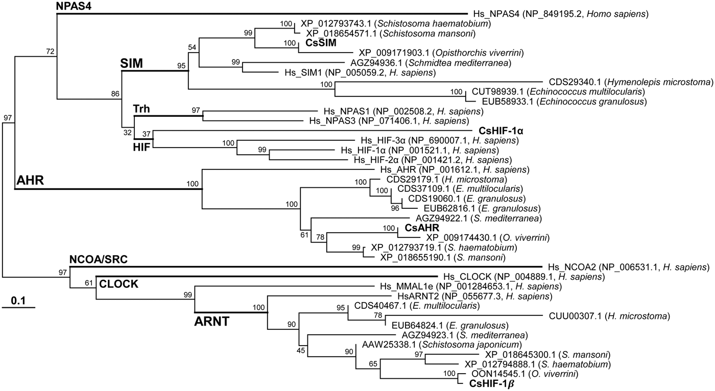
Each of the C. sinensis bHLH–PAS proteins detected unique orthologs during the BLASTp searches of platyhelminth proteomic databases in GenBank. By examining these protein sequences using the SMART program, the amino acid sequences of the bHLH and PAS-A/B domains (183 amino acid positions) were extracted to be applied in a phylogenetic analysis along with the human bHLH–PAS proteins (examples are shown in Supplementary Fig. S2). In the neighbour-joining tree shown in Fig. 1, most platyhelminth proteins including those of C. sinensis were separately positioned in the SIM, ARNT/HIF-1β and AHR subfamily clades. Interestingly, the CsHIF-1α protein was found to be more closely related to the members of HIF subfamily, although the branch that connects them was poorly supported by a statistical test of 1000 replicates (bootstrap value 37).
Phylogenetic analysis of bHLH–PAS proteins identified in platyhelminths, including C. sinensis (labelled in bold letters) and human. The un-rooted neighbour-joining tree was constructed using the concatenated alignment of their amino acid sequences corresponding to the bHLH, PAS and PAS-associated C-terminal domains. In addition to the scientific names of respective donor organisms, the identities of examined proteins are distinguished by their GenBank accession numbers. Numerals at branching nodes indicate their percentages of occurrence in 1000 bootstrap replicates. Branching node that connects the members of each bHLH–PAS subfamily is marked using the subfamily name. AHR, aryl hydrocarbon receptor; ARNT, aryl hydrocarbon receptor nuclear translocator; CLOCK, circadian locomotor output cycles kaput; HIF, hypoxia-inducible factor; NCOA/SRC, nuclear receptor coactivator/steroid receptor coactivator; NPAS4, neuronal PAS domain protein 4; SIM, single-minded; Trh, trachealess.

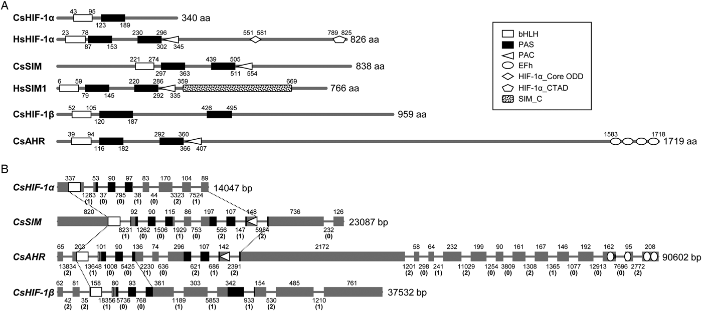
The SMART program predicted several functional domains such as bHLH, PAS-A, PAS-B, PAS-associated C-terminal (PAC), core oxygen-dependent degradation (core ODD) and C-terminal transactivation (CTAD) domains in human HIF-1α. However, neither the SMART nor InterProScan programs detected domain sequences corresponding to PAS-B, PAC and CTAD in CsHIF-1α (Fig. 2A). Moreover, the sequence motif of core ODD (LXXLAP; Huang et al., Reference Huang, Zhao, Mooney and Lee2002) essential for prolyl hydroxylation was not found in CsHIF-1α. The other C. sinensis proteins conserved sequences in most of the bHLH-PAS family-specific domains ranging from bHLH to PAC, while the PAC-related sequence appeared to be highly divergent or deleted in CsHIF-1β. It was also apparent that CsAHR has acquired four EF-hand motifs which were tandemly arrayed in the C-terminus of the protein (Fig. 2A).
Structural characterization of C. sinensis bHLH–PAS proteins and genes. (A) The functional domains of the C. sinensis proteins were predicted using the SMART program. Locations of these domains in the respective proteins are marked with the positions of the initial and terminal amino acids. The domain legend is shown in the right panel. The architectures of human HIF-1α (HsHIF-1α) and SIM (HsSIM) were included for comparison. bHLH, basic helix-loop-helix; PAS, per-arnt-sim; PAC, PAS-associated C-terminal; EFh, EF-hand, calcium binding motif; ODD, oxygen dependent degradation; CTAD, C-terminal transactivation domain. (B) The exon–intron organizations of the C. sinensis bHLH–PAS genes were determined by comparing the genomic and transcribed sequences of the respective genes. The CDSs are presented with grey squares in proportion to their relative sizes and the intervening introns are indicated by solid lines with a fixed length. The phase of each intron, as well as the length of exons and introns in base pairs, are indicated in parentheses. The dotted lines define gene segments that are shared by these C. sinensis genes. The positions of codons in the functional protein domains shown in panel A are marked in the corresponding CDS regions.

The genomic structures of the C. sinensis bHLH–PAS genes were determined by comparing nucleotide sequences between the transcribed and genomic versions of each gene (Fig. 2B). The coding sequences of these genes were variably interrupted by introns with different lengths and insertional phases relative to the reading frames. However, the exon–intron architectures of gene segments encoding the common bHLH–PAS family domains were well conserved among these homologous genes (shown in regions bound by two dotted lines), although the structure might be remodelled in the PAS-B segment of CsHIF-1β and the exon sequence in the downstream region of PAS-A was highly divergent in CsHIF-1α (Fig. 2B).
Prediction of tertiary structures
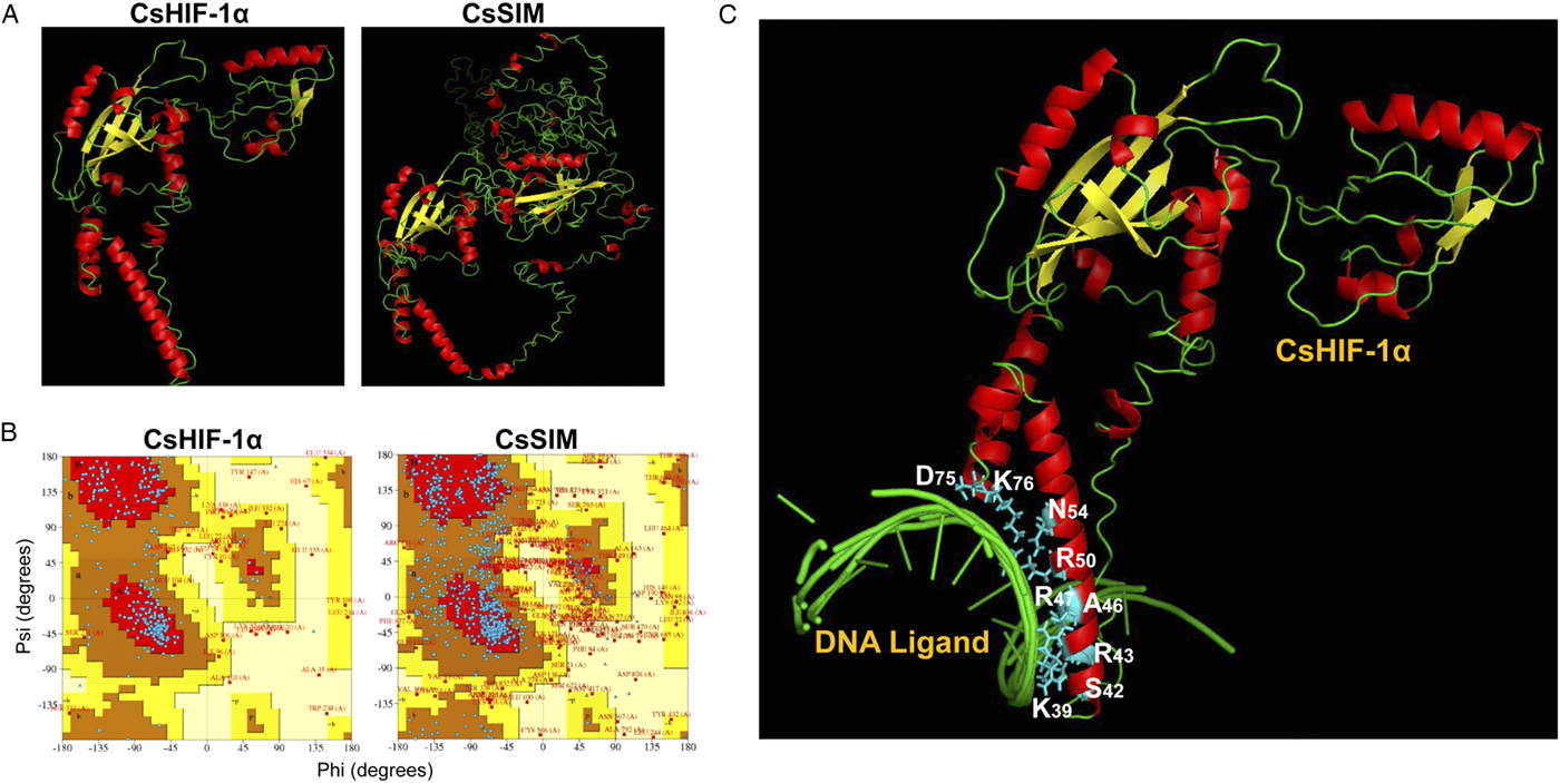
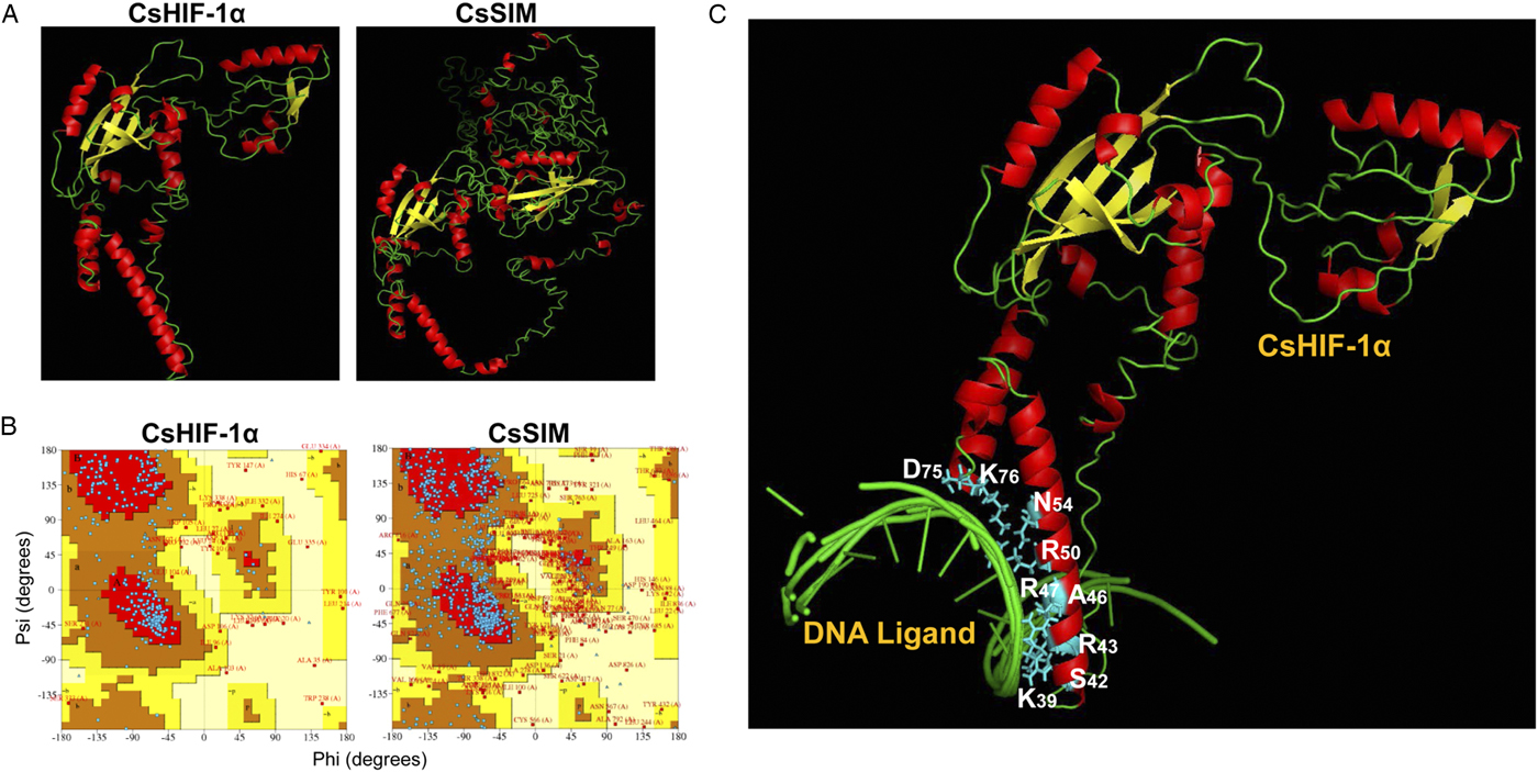
The tertiary structures of CsHIF-1α and CsSIM were simulated using I-TASSER (Fig. 3A). This program suggested five final models for CsHIF-1α using multiple threading templates including the heterodimeric structures of CLOCK:BMAL1 (PDB no. 4F3L) and HIF-2α:ARNT (4ZP4) complexes. Of these models, model 1 exhibited the highest confidence score (C-score; −1.58). The TM-score and RMSD of this model were 0.52 ± 0.15 and 10.1 ± 4.6 Å, respectively. The CsSIM structure was similarly simulated using threading reference templates such as the crystal structures of CLOCK:BMAL1 and NPAS1-ARNT (5SY5) complexes. The C-score, TM-score and RMSD were −3.53, 0.33 ± 0.11 and 17.7 ± 2.5 Å, respectively. As shown in Fig. 3B, the amino acids residing in the most favoured and additional allowed regions were 90.8 and 85.6% in model 1 of CsHIF-1α and CsSIM, respectively. Further, the I-TASSER program indicated a probable tertiary structure of CsHIF-1α:DNA complex based on the HIF-2α:ARNT:HRE DNA complex (4ZPK), along with the amino acids involved in the protein–DNA interaction (Fig. 3C).
Tertiary structures of the C. sinensis HIF-1α and SIM proteins. (A) The three-dimensional structures of CsHIF-1α and CsSIM were simulated using I-TASSER and the ribbon models were represented using PyMol. Red, α-helix; yellow, β-strand. (B) Ramachandran plots were generated to evaluate the stereochemical quality of the predicted CsHIF-1α and CsSIM tertiary structures. The most favoured regions (A, B and L) are in red, while additional allowed (a, b, l and p), generously allowed (−a, −b, −l and −p) and disallowed regions are in brown, yellow and pale yellow fields, respectively. Blue squares and triangles represent non-glycine and glycine residues in the most favoured and additional allowed regions, respectively. Red squares mark non-glycine residues in generously allowed and disallowed regions. (C) Amino acids constituting the DNA-binding pocket of CsHIF-1α were predicted using the I-TASSER program.
Transcript levels of bHLH–PAS genes after hypoxia treatment
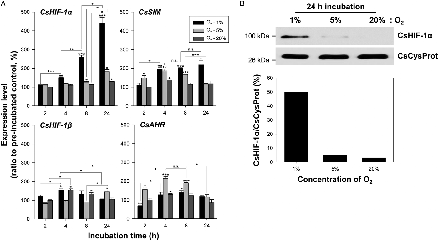
The induction profiles of the bHLH–PAS genes were examined by performing qPCR in live C. sinensis adult worms that were incubated under the 1, 5 or 20%-oxygen conditions. As shown in Fig. 4A, the transcript level of CsHIF-1α was slightly increased under the 20% oxygen condition during the incubation compared to that in pre-incubated worms of the same age (~1.3-fold at 24 h, P = 0.019). However, the transcriptional activity was significantly induced proportional to the incubation period during the hypoxic exposure [1.8-fold (P = 0.011) and 4.4-fold (P = 0.005) at 24 h under the 5 and 1%-oxygen conditions, respectively]. The induction pattern of CsSIM against the hypoxic stimulus was similar to, but less extensive than that of CsHIF-1α, except that CsSIM transcript level reached the maximum level at 4 h (1.9-fold, P = 0.009) and gradually reverted to the normal level under the 5%-oxygen condition (1.1-fold at 24 h, P = 0.105). The highest level of CsAHR expression was observed at 4 or 8 h under all the experimental conditions; however, it was most strongly induced under the 5%-oxygen condition. CsHIF-1β did not respond by exhibiting a recognizable induction pattern, to the hypoxic stimulus. The hypoxic induction of CsHIF-1α expression was confirmed in the C. sinensis worms incubated for 24 h through Western-blot analysis at the protein level (Fig. 4B). The CsSIM-specific antibody did not exhibit any detectable signal, probably owing to the low expression level of CsSIM (data not shown).
Effect of hypoxic stimulus on the expression of the C. sinensis bHLH–PAS genes. (A) The transcription levels of the bHLH–PAS genes were determined by performing qPCR (n = 3, mean ± s.d.) in worms incubated for 2, 4, 8 and 24 h under the 1, 5 and 20%-oxygen conditions. *P < 0.05; **P < 0.01; ***P < 0.001; n.s., not significant. (B) Proteins were extracted from the whole worm bodies of 24 h-incubation groups and examined with CsHIF-1α-specific antibody in Western-blot analysis. The anti-CsCysProt antibody was employed as an internal control to validate equal loading of the protein samples.

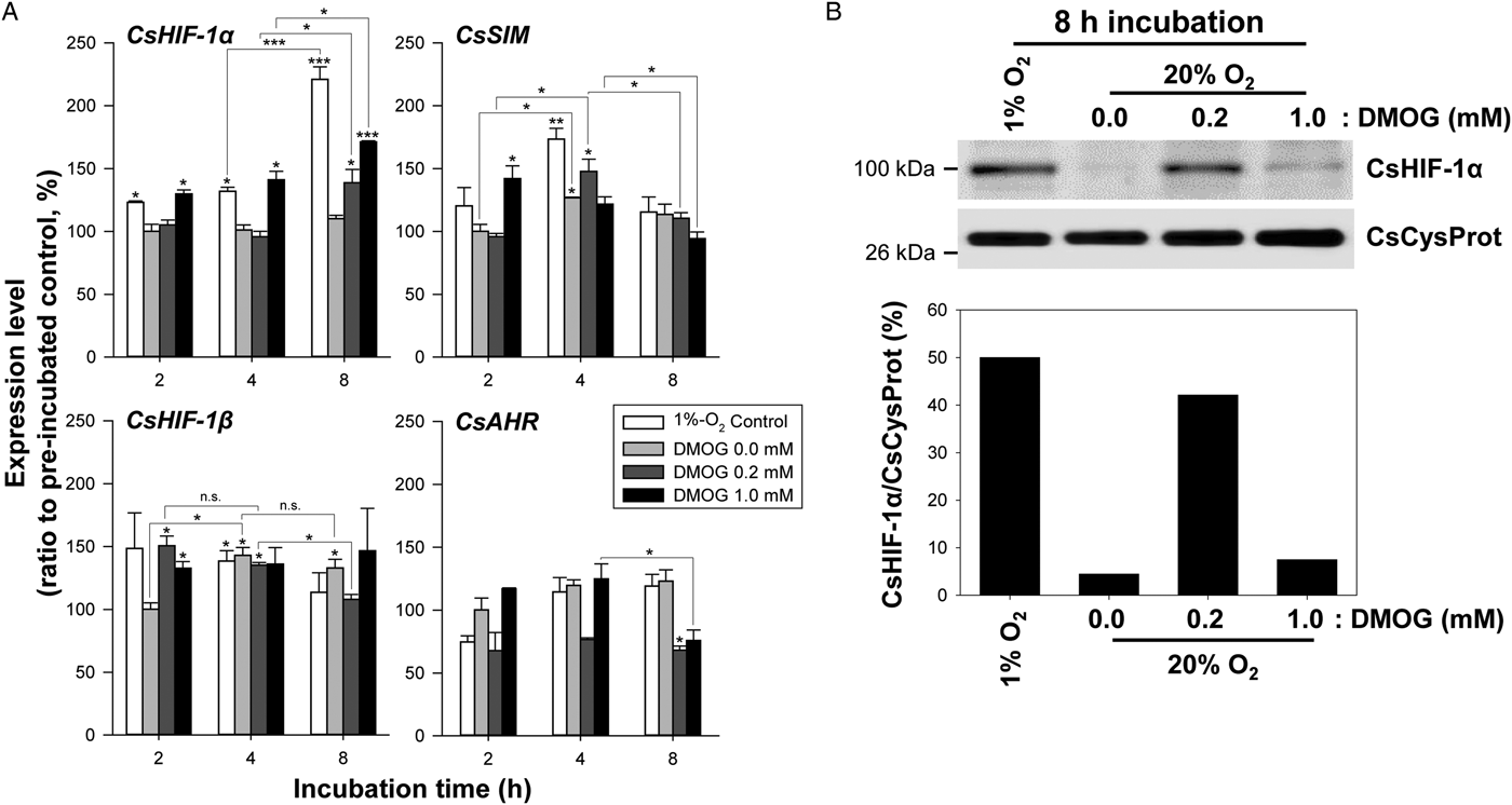
The effect of DMOG on the bHLH–PAS gene expressions was assessed under the 20%-oxygen condition (Fig. 5A). In the group treated with 0.2 mm DMOG, CsHIF-1α was significantly induced at 8 h compared to that in the pre-incubated group (1.4-fold, P = 0.045). However, this gene expression was increased from 2 h in worms treated with 1 mm DMOG. The fold change values in the 1 mm-DMOG group were comparable to those in the 1%-oxygen control group at 2 h [1.3-fold (P = 0.022) vs 1.2-fold (P = 0.029)] and 4 h [1.4-fold (P = 0.022) vs 1.3-fold (P = 0.020)], although the value at 8 h was much lower than that in the control group (1.7-fold vs 2.2-fold, P = 0.020). Despite a certain degree of fluctuation, DMOG did not cause any directional change in the expression of the other bHLH–PAS genes under these experimental conditions (Fig. 5A). At the protein level, the relative amount of CsHIF-1α (% HIF-1α/CsCysProt, 42.1%) was increased by 84.1% compared to that in the 1%-oxygen group (50.0%) after the treatment of 0.2 mm DMOG. However, the CsHIF-1α level was prominently reduced in the 1 mm-DMOG group (% HIF-1α/CsCysProt, 7.46%; Fig. 5B).
Stabilization of the C. sinensis CsHIF-1α protein using DMOG. (A) The expression levels of CsHIF-1α and other bHLH–PAS genes were determined by performing qPCR (n = 3, mean ± s.d.) in worms treated with 0.0, 0.2 and 1 mm DMOG for 2, 4 and 8 h under the 20%-oxygen condition. Worms incubated under the 1%-oxygen condition were used as controls. *P < 0.05; **P < 0.01; ***P < 0.001; n.s., not significant. (B) Proteins were extracted from the whole worm bodies of 8 h-incubation groups and assessed using the CsHIF-1α-specific antibody by performing Western-blot analysis. The anti-CsCysProt antibody was employed as an internal control to validate equal loading of the protein samples.

Effect of nitrogen compounds on the expressions of C. sinensis bHLH–PAS genes
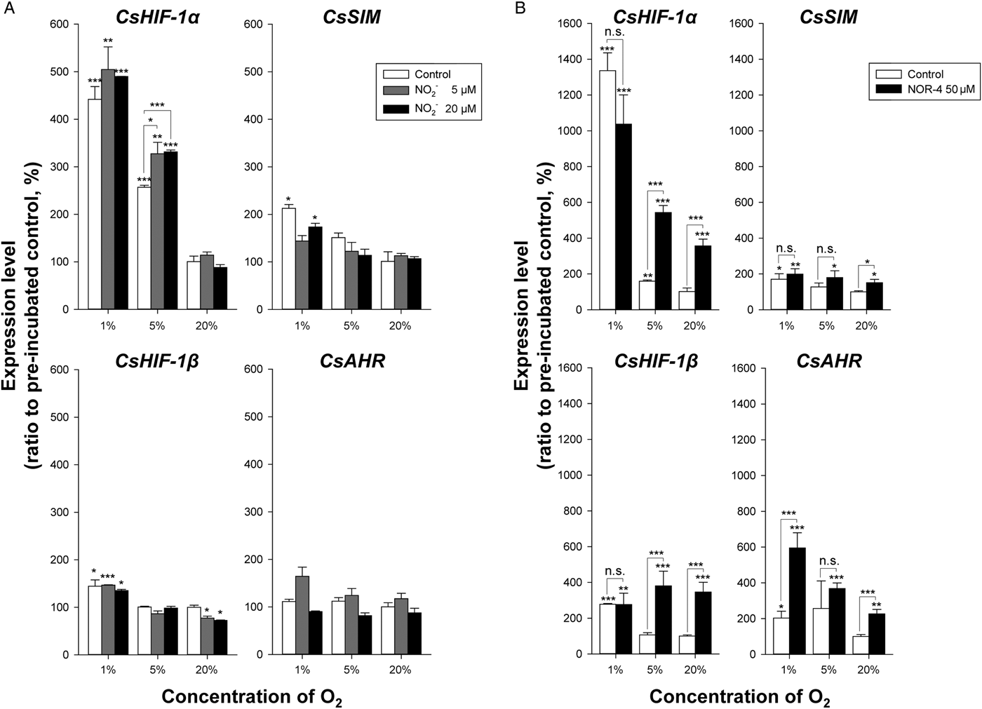
Adult worms were incubated in RPMI-1640 medium supplemented with nitrite (5 and 20 µ m) under the 1, 5 and 20%-oxygen conditions. After 24 h incubation, the transcript levels of the bHLH–PAS genes were evaluated by performing qPCR. As shown in Fig. 6A, nitrite did not considerably affect the expression of these genes under the experimental conditions, except that of CsHIF-1α under the 5%-oxygen condition. The CsHIF-1α expression was increased ~1.2-fold (P < 0.05) by adding nitrite compared to that in the 5%-oxygen control group. However, the levels of the CsHIF-1α and CsHIF-1β transcripts were significantly increased in the liver flukes that were incubated in a NOR-4-containig medium under the 5% (3.4- and 3.6-fold, respectively) and 20%-oxygen (3.5- and 3.4-fold, respectively) conditions (P < 0.001). The NO-induced expression levels of these genes were not significant in worms incubated under the 1%-oxygen condition (Fig. 6B). The transcriptional activity of CsAHR was also up-regulated with the NOR-4 treatment under the experimental conditions, whereas that of CsSIM was marginally influenced by the chemical.
Effect of nitrite and NO on the expression of the C. sinensis bHLH–PAS genes. (A) Adult worms were incubated for 24 h in medium supplemented with various concentrations of nitrite (indicated in the graph legend), under the 1, 5 and 20%-oxygen conditions. The amounts of the CsHIF-1α and other bHLH–PAS gene transcripts were determined by performing qPCR (n = 3, mean ± s.d.). *P < 0.05; **P < 0.01; ***P < 0.001; n.s., not significant. (B) Similar experiments were conducted on the worms incubated for 24 h in a medium containing 50 µ m NOR-4 (NO donor).

Co-incubation of C. sinensis adult worms and human cholangiocytes
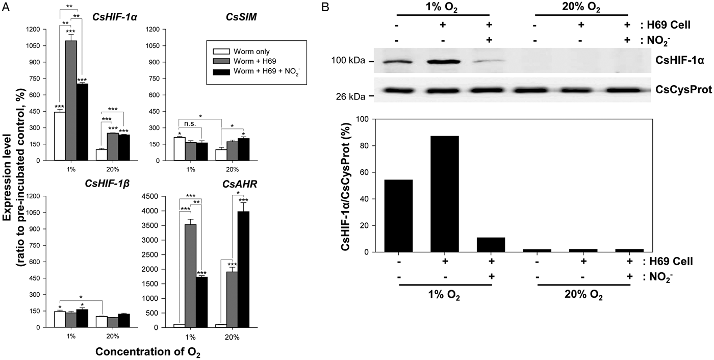
The induction patterns of the bHLH–PAS gene expressions were investigated in worms co-incubated with H69 cells. During co-incubation under the 1%-oxygen condition, the relative level of CsHIF-1α transcripts increased by 10.9-fold (P = 0.001) and 2.5-fold (P = 0.004) compared to that in the pre-incubated and worm-only control groups, respectively (Fig. 7A). Interestingly, the fold-induction values were significantly decreased in another co-incubation group, in which the medium was supplemented with nitrite [7-fold (P = 0.0002) and 1.6-fold (P = 0.006)]. The CsHIF-1α expression was also substantially induced by human cells under the 20%-oxygen condition (2.5-fold relative to the pre-incubated group, P = 0.003). However, the expression level was not downregulated by nitrite in the 20%-oxygen environment. Western-blot analysis using the whole worm extracts demonstrated that the relative levels of CsHIF-1α markedly coincided with those of mRNA transcripts in these experimental groups (Fig. 7B). The CsAHR expression similarly responded to the co-incubation, while the responsiveness of this gene was highly elevated after nitrite treatment under the 20%-oxygen condition. Considering the negligible induction of CsAHR in the 1%-oxygen control group (1.1-fold, P = 0.249), factor(s) other than oxygen might be involved in the CsAHR activation (Fig. 7A). The co-incubation of worms with the human epithelial cells with or without provision of nitrite minimally affected the CsSIM and CsHIF-1β expressions (Fig. 7A).
Effect of cholangiocytes on the expression of the C. sinensis bHLH–PAS genes. (A) The amounts of the CsHIF-1α and other bHLH–PAS gene transcripts were determined by performing qPCR (n = 3, mean ± s.d.) in worms co-incubated with H69 cells for 24 h under the 1 and 20%-oxygen conditions. Experiments using a medium containing 10 µ m nitrite were also conducted during the co-incubations. *P < 0.05; **P < 0.01; ***P < 0.001; n.s., not significant. (B) Proteins extracted from the whole worm bodies of each experimental group were allowed to react with the CsHIF-1α-specific antibody in the Western-blot analysis. The anti-CsCysProt antibody was employed as an internal control to validate equal loading of the protein samples.

Discussion
The molecular features of HIF-1 machinery and its implication in hypoxia were comprehensively investigated in various higher animals, including humans. With respect to the high evolutionary conservation of HIF-1-related molecules, the machinery was believed to operate in most metazoans (Rytkönen, Reference Rytkönen, Williams, Renshaw, Primmer and Nikinmaa2011; Mei and Dvornyk, Reference Mei and Dvornyk2014). Accordingly, HIF-1 and relevant molecules were characterized in the lower metazoan phyla such as Placozoa (Loenarz et al., Reference Loenarz, Coleman, Boleininger, Schierwater, Holland, Ratcliffe and Schofield2011) and Cnidaria (Wang et al., Reference Wang, Yu, Zhen, Mi, Shi, Wang, Wang and Sun2014). A protein(s) immunologically homologous to human HIF-1α was detected in a sponge (Demospongiae; Gunda and Janapala, Reference Gunda and Janapala2009). Parasitic helminths of the phyla Nematoda and Platyhelminthes are exposed to natural and/or host environments comprising a range of oxygen concentration from normoxia to anoxia during their life cycles. A physiological transition to cope with the decreased oxygen availability might be prompted by the special transactivation factors such as HIF-1α in these parasites. Till date, HIF-1α and HIF-1β were characterized in a parasitic nematode, Ascaris suum (Goto et al., Reference Goto, Amino, Nakajima, Tsuji, Sakamoto and Kita2013) as well as in a free-living nematode, Caenorhabditis elegans (Powell-Coffman et al., Reference Powell-Coffman, Bradfield and Wood1998; Jiang et al., Reference Jiang, Guo and Powell-Coffman2001). However, to the best of our knowledge, this is the first report that describes the characteristics and expression profiles of HIF-1-related proteins in platyhelminths.
Based on the current genomic/transcriptomic databases of C. sinensis, the coding DNA sequence of CsHIF-1α was determined to comprise of 1023 bp encoding a 340-amino acid polypeptide (deduced molecular weight, 39.7 kDa). The bHLH and PAS-A domains were instantly mapped in the primary structure of CsHIF-1α. However, the other HIF-1α motifs such as PAS-B, PAC, ODD and CTAD were not recognized in the C-terminal region downstream of the PAS-A domain (Fig. 2; Wang et al., Reference Wang, Yu, Zhen, Mi, Shi, Wang, Wang and Sun2014). Furthermore, the native CsHIF-1α protein appeared to have a molecular weight of ~100 kDa (Fig. 4) and the protein was subjected to the PHD-mediated degradation, which indicated the presence of ODD domain in the protein (Fig. 5). Taken together, these data might suggest that the CsHIF-1α sequence determined in this study is partial. This gene is encoded in a genomic scaffold with a size of 218 414 bp (BADR02003149) and its last exon terminates at the 214 682-bp position. BLASTx/tBLASTx searches of the remaining 3′-region sequence (3732 bp) did not retrieve any hit related to HIF-1α from the non-redundant GenBank databases. The screening of the C. sinensis genomic/transcriptomic databases using the C-terminal sequence of human HIF-1α (positions 302–825; Fig. 2A) did also not detect any homologous sequence. Therefore, investigations to determine the nucleotide sequences of missing chromosomal segment and the 3′-region of CsHIF-1α transcript are necessary to complete the CsHIF-1α structure.
Trematode parasites, including C. sinensis, developed a special mechanism called tanning process for their eggshell formation (Cordingley, Reference Cordingley1987; Bae et al., Reference Bae, Cai, Kim, Sohn and Kong2013b). This process mediated by tyrosinase requires a large amount of oxygen. To cope with the high oxygen demand for the reproductive metabolism as well as the systemic aerobic respiration, C. sinensis adult worm massively secretes myoglobin (Bae et al., Reference Bae, Ahn, Lee, Kim, Cai, Kang, Sohn and Kong2013a). Myoglobin facilitates oxygen transport by the diffusion of oxymyoglobin (oxy-Mb) along with a partial pressure of oxygen (PO2) gradient (Wittenberg, Reference Wittenberg1959; Jürgens et al., Reference Jürgens, Papadopoulos, Peters and Gros2000). In addition to its function as an oxygen transporter, the protein exhibits dual enzymatic activities, i.e. nitrite reductase and NO oxygenase according to its chemical forms [deoxymyoglobin (deoxy-Mb) and oxy-Mb, respectively; Cossins and Berenbrink, Reference Cossins and Berenbrink2008]. Therefore, nitrite and NO can alter the ratio of oxy-Mb to deoxy-Mb depending on the surrounding PO2 in a biological niche. In our study, the effect of nitrite on CsHIF-1α expression was not much prominent in worms incubated alone under the experimental conditions (Fig. 6A). However, the transcript and protein levels of CsHIF-1α were significantly influenced by this chemical under the hypoxic condition in worms co-incubated with H69 cells (Fig. 7). Conversely, the presence of NO in incubation medium highly induced the CsHIF-1α expression under 5 and 20%-oxygen concentrations (Fig. 6B). The biliary secretion is one of the major routes to excrete excess inorganic nitrogen compounds such as nitrite and nitrate (Fritsch et al., Reference Fritsch, de Saint Blanquat and Klein1985). It was also demonstrated that host immune cells recruited at the infection sites generate NO by activating inducible NO synthase (Yang et al., Reference Yang, Shen, Xue, Cheng, Jiang, Yang, Chen and Zhou2015). All these data/facts suggest that various factors from the parasitic worm (myoglobin and HIF-1α-related molecules) and host (oxygen, nitrite and NO) are coordinately involved in the maintenance of oxygen homeostasis in C. sinensis. The PO2-dependent multifaceted functions of myoglobin, if not all (Olson and van der Vliet, Reference Olson and van der Vliet2011), might be intimately associated with the CsHIF-1α regulation.
Healthy mammalian cells/tissues mostly reside in a milieu composed of 2–9% oxygen (Bertout et al., Reference Bertout, Patel and Simon2008), therefore, 1, 5 and 20%-oxygen conditions can be considered as hypoxia, normoxia and hyperoxia, respectively, in the physiological context. The actual oxygen level within the C. sinensis-infected biliary ducts of mammalian hosts remains undetermined. According to our observation demonstrating the heavily impaired intrahepatic biliary duct walls at the infection sites, this situation might be further applied to C. sinensis residing in the host tissues. In this study, adult worms were incubated for 4 h under the 20%-oxygen condition prior to their use in the investigations of gene expression profiling. In addition to exposure to air for a particular time period during worm collection from the experimental animals, the aforementioned equilibration procedure causes metabolic adaptation to the oxygen concentration in worms. Consequently, the 1 and 5%-oxygen conditions might be recognized as severe and mild hypoxic stress by the worms for the induction of CsHIF-1α expression (Figs 4A and 6B).
Under normal physiological conditions, animal cells occasionally experience hypoxic stimulus that is attributed either to reduced blood/oxygen supply or increased local oxygen consumption during the developmental tissue growth. The stressful conditions are generated by various pathologies such as anaemia, pneumonia and cancer (Semenza, Reference Semenza2009). Parasitic helminths, including C. sinensis, provoke chronic inflammation in their vertebrate hosts (Sorci and Faivre, Reference Sorci and Faivre2009; Mao et al., Reference Mao, Xie, Wang, Chen, Ren, Shang, Lei, Tian, Li, Liang, Chen, Liang, Xu, Li, Huang and Yu2015). The chronic inflammation also shares an interdependent association with hypoxia. The alteration in metabolic supply and demand ratio for oxygen leads to inflammatory hypoxia, while the hypoxia-elicited disease is reciprocally characterized by secondary inflammatory conditions (Bartels et al., Reference Bartels, Grenz and Eltzschig2013; Biddlestone et al., Reference Biddlestone, Bandarra and Rocha2015). It appeared apparent that C. sinensis adult worms induce hypoxia at their infection sites by competing with biliary epithelial cells and/or proximal hepatocytes to use oxygen (Fig. 7). Activated inflammatory cells recruited by the hypoxic signal or other parasite-related factors (Mao et al., Reference Mao, Xie, Wang, Chen, Ren, Shang, Lei, Tian, Li, Liang, Chen, Liang, Xu, Li, Huang and Yu2015) would accelerate the rate of oxygen depletion. Meanwhile, the expression of ligand-activated transcription factor AHR was drastically induced in worms co-incubated with human cholangiocytes (Fig. 7A). The diverse natural ligands of AHR include indirubin, bilirubin, lipoxins and prostaglandins (Sinal and Bend, Reference Sinal and Bend1997; Adachi et al., Reference Adachi, Mori, Matsui, Takigami, Fujino, Kitagawa, Miller, Kato, Saeki and Matsuda2001; Seidel et al., Reference Seidel, Winters, Rogers, Ziccardi, Li, Keser and Denison2001). Lipoxins and prostaglandins are produced in epithelial cells and other immune cells from arachidonic acid and intimately involved in the regulation of pathogen-induced inflammatory responses (Chandrasekharan and Sharma-Walia, Reference Chandrasekharan and Sharma-Walia2015; Dennis and Norris, Reference Dennis and Norris2015). Currently, investigations on the AHR ligands, which are probably produced by cholangiocytes stimulated with C. sinensis excretory–secretory products, are being undertaken to gain insights into the molecular and cellular mechanisms of C. sinensis-induced inflammation.
The synthetic α-ketoglutarate (α-KG) analogue DMOG activates cellular HIF and AMP-activated protein kinase (AMPK) signalling cascades that are involved in the maintenance of energy and metabolic homeostasis by inhibiting PHD enzymes. Various mitochondrial α-KG-dependent hydroxylases are also susceptible to the DMOG-mediated inhibition (Zhdanov et al., Reference Zhdanov, Okkelman, Collins, Melgar and Papkovsky2015 and references therein). The direct inhibitory effect on these mitochondrial function and cellular respiration-related enzymes may have partly influenced on the induction profiles of bHLH–PAS genes observed in DMOG-treated C. sinensis worms (Fig. 5A). Nevertheless, the significantly increased level of CsHIF-1α in worms treated with DMOG under the 20% oxygen (Fig. 5B) strongly suggested that PHD serves as an oxygen-sensing component in the liver fluke. A protein (GAA37480.2) homologous to PHDs was identified in the GenBank proteomic database of C. sinensis, although this protein was not characterized in the present investigation. C. sinensis worms invading the hepatobiliary tract of vertebrate hosts are confronted with hypoxia generated by the elevated oxygen-consumption rate during sexual reproduction in worms. Host cholangiocytes and proximal hepatocytes that compete with the liver fluke to utilize local oxygen, in addition to immune cells that are recruited at the infection sites, are likely to accelerate the oxygen depletion. Taken together, all these data suggest that the HIF-PHD machinery constitutes a key regulator of physiological responses to hypoxia in the trematode parasite. Studies on the local hypoxia induced by C. sinensis infection and relevant cellular/molecular factors might provide information to understand pathobiological aspects of clonorchiasis such as chronic inflammation and the development of cholangiocarcinoma. Adaptation to host environments with limited oxygen availability is indispensable for the establishment of parasitic life mode. Therefore, the knowledge obtained by these investigations could reveal the adaptive strategies of parasites in their specific hosts.
Supplementary material
The supplementary material for this article can be found at https://doi.org/10.1017/S0031182018001245.
Financial support
This work was supported by the Basic Science Research Programs of the National Research Foundation of Korea (NRF), funded by the Ministry of Science, ICT and Future Planning (grant number: NRF-2013R1A1A2012011) and by the Ministry of Education (grant number: NRF- 2017R1D1A1B03028355).
Conflict of interest
None.
Ethical standards
Not applicable.