Implications
The influence of maternal nutrition on skeletal muscle development has been at the forefront of scientific research for many years. The main implication of this study is meat quality, which is highly variable. Muscle phenotype characteristics have a direct effect on many meat quality parameters. Maximising meat quality would be of major benefit for the consumer and farmer, and may ultimately lead to an increased production economy. Furthermore, this study provides a model for the developmental programming of diseases such as obesity in the human, and is of relevance to the programming of muscle for athletes.
Introduction
The development of skeletal muscle is crucial to its function. Mammalian myogenesis is a biphasic process involving the formation of an early population of primary (P) fibres and a later population of secondary (S) fibres. In the pig, myogenesis encompasses an intense period of proliferation and differentiation from 35 days of gestation (dg) until approximately 90 dg. An initial rapid wave of primary embryonic myoblast fusion occurs from 35 to 55 dg, resulting in the formation of primary myotubes, which subsequently mature into primary muscle fibres. A longer second wave of differentiation occurs between 55 and 90 dg to produce the larger population of later-forming smaller S fibres. As a direct result of the primary and secondary myogenic waves, central clusters of slow-contracting Type I fibres are surrounded by a ring of fast Type II fibres (Lefaucheur et al., Reference Lefaucheur, Edom, Ecolan and Butler-Browne1995; Quiroz-Rothe and Rivero, Reference Quiroz-Rothe and Rivero2004); a type grouping formation described as a rosette pattern or metabolic bundle. Generally, the central fibre of each cluster of Type I fibres originates from a primary myotube and develops as a slow primary muscle fibre. All other fibres originate from secondary muscle fibres (Wigmore and Stickland, Reference Wigmore and Stickland1983), which mature to either Type I or Type II fibres, depending on muscle function. The total number of fibres is determined in utero, and hence is fixed by the time of birth (Wigmore and Stickland, Reference Wigmore and Stickland1983). In the adult pig, four distinct muscle fibre types have been characterised; slow Type I and fast Type IIA, IIX and IIB (Lefaucheur, Reference Lefaucheur2001). Fibre type classification is based upon the dominant myosin heavy chain (MyHC) isoform expressed by the individual fibre; MyHCIβ, MyHCIIa, MyHCIIx or MyHCIIb. These largely define the biochemical and functional phenotype not only of the individual muscle fibre, but also of the whole muscle. Properties such as contractile performance, oxidative and glycolytic capacity, fibre size and lipid content co-vary between fibre types. Type I fibres are slow contracting, fatigue resistant and highly oxidative. These properties decrease through Type IIA to IIX to Type IIB fibres, which are highly glycolytic and fatigue rapidly.
Developmental programming is becoming an increasingly researched topic with respect to its influence on the variation in growth and developmental performance of an adult in later life. During the critical embryonic period, a foetus is highly responsive to abnormal environmental conditions such as a nutritional stress or stimulus. Altered foetal nutritional and endocrine status may result in developmental adaptations that permanently alter the anatomical structure, physiology and metabolism of the offspring, predisposing them to metabolic, cardiovascular and endocrine diseases in adulthood (for a review, see Wu et al., Reference Wu, Bazer, Wallace and Spencer2006). Postnatal variation in body composition and growth performance may be pre-programmed during foetal development, in utero, by both internal (e.g. genetic) and external factors (e.g. maternal nutrition).
Body composition measurements provide insight into the regulation of normal development as well as the interactions among nutritional, physiological and biochemical functions. The pig is considered to be an ideal candidate for such studies that require an animal model in which body composition at birth, and postnatal changes are comparable to those in human infants (Shulman, Reference Shulman1993). Furthermore, the physiological and anatomical similarity between the pig and the human species is above any other laboratory species (Hughes, Reference Hughes1986; Miller and Ullrey, Reference Miller and Ullrey1987). The semitendinosus muscle (Musculus semitendinosus) is used in this study as it is well characterised with a simple fusiform structure. Anatomically, it contains two portions that exhibit striking differences in muscle fibre composition; a deep, more oxidative area and a superficial, less oxidative area. A distinct transition between the deep and superficial areas exists. The M. semitendinosus is commonly used to assess fibre typing and is also an important meat muscle.
Developmental intra-litter variation in utero affects the postnatal growth of skeletal muscle in the pig (Dwyer et al., Reference Dwyer, Stickland and Fletcher1994; Gatford et al., Reference Gatford, Ekert, Blackmore, De Blasio, Boyce, Owens, Campbell and Owens2003). According to requirements, pregnant sows are restrictively fed. Ad libitum feeding of sows during early gestation shows that the voluntary feed intake is two to three times as high as the requirement (Nissen et al., Reference Nissen, Danielsen, Jorgensen and Oksbjerg2003). It is widely accepted that maternal nutrition affects secondary muscle fibre development, hence having an impact on ultimate muscle fibre composition. The lightest littermate is shown to have a reduced muscle fibre number as a result of a decreased secondary to primary (S : P) fibre ratio, compared to its heaviest littermate pair (Wigmore and Stickland, Reference Wigmore and Stickland1983). Pilot studies in our laboratory have shown that the differential expression of MyHCIIa is significantly greater (P ⩽ 0.05) in the M. semitendinosus of the heavyweight littermate, compared with its lightweight littermate pair between 80 and 100 dg (Ashton, Reference Ashton2002); the mechanism(s) by which this occurs is unknown. Many studies report the effects of nutrition on birth weight, total fibre number (TNF) and S : P fibre ratio, but few have outlined differences in the exact fibre type composition or the oxidative capacity of the muscle, both of which are of fundamental importance. Within an agricultural production system, the composition of fibres within a muscle can ultimately affect many meat quality parameters, e.g. tenderness (Maltin et al., Reference Maltin, Balcerzak, Tilley and Delday2003). Furthermore, there is evidence to suggest that restricted nutrition during gestation could have harmful consequences later in life, including reduced glucose tolerance, Type 2 diabetes and a reduced lifespan (Hales and Ozanne, Reference Hales and Ozanne2003; Ozanne et al, Reference Ozanne, Olsen, Hansen, Tingey, Nave, Wang, Hartil, Petry, Buckley and Mosthaf-Seedorf2003). Studies have linked muscle fibre type with obesity (Tanner et al., Reference Tanner, Barakat, Dohm, Pories, MacDonald, Cunningham, Swanson and Houmard2002), Type 2 diabetes and insulin resistance (Oberbach et al., Reference Oberbach, Bossenz, Lehmann, Niebauer, Adams, Paschke, Schon, Bluher and Punkt2006). In obese women, skeletal muscle possesses fewer Type I fibres and more Type IIB fibres than lean women, exhibiting a reduced oxidative and an increased glycolytic capacity (Hickey et al., Reference Hickey, Carey, Azevedo, Houmard, Pories, Israel and Dohm1995; Tanner et al., Reference Tanner, Barakat, Dohm, Pories, MacDonald, Cunningham, Swanson and Houmard2002). Similar observations have been made in Type 2 diabetic patients (Hickey et al., Reference Hickey, Carey, Azevedo, Houmard, Pories, Israel and Dohm1995; Oberbach et al., Reference Oberbach, Bossenz, Lehmann, Niebauer, Adams, Paschke, Schon, Bluher and Punkt2006).
The aim of this study was to investigate the effects of increased nutrition, throughout three different time periods of gestation, on the muscle fibre phenotype of the M. semitendinosus in the offspring at slaughter weight. We hypothesised that increased maternal nutrition has an influence on skeletal muscle phenotype in the offspring at slaughter weight (c. 158 days old), with a specific effect on the S fibre population.
Material and methods
Nutritional treatments
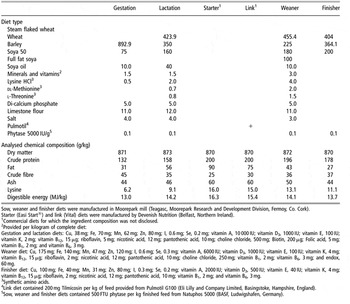
A total of 24 multiparous Landrace × Large White sows were fed ad libitum until service, when they were randomly assigned to one of four gestational nutritional treatments; (T1-control) 30 MJ digestible energy per day (MJ DE/day) throughout gestation (gestation length = 113 days), (T2) same as that for T1 but increased to 60 MJ DE/day from 25 to 50 dg, (T3) same as that for T1 but increased to 60 MJ DE/day from 50 to 80 dg and (T4) same as that for T1 but increased to 60 MJ DE/day from 25 to 80 dg (n = 6 sows/treatment; see Table 1 for details of experimental diets). Between 110 dg and farrowing, all sows were fed 25 MJ DE/day of lactation diet (see Table 1 for diet composition). Once farrowed, sows were liquid-fed the lactation diet by a computerised feeding system (Big Dutchman, Vechta, Germany). A lactation feed curve increasing from 25 MJ DE/kg at farrowing to 95 MJ DE/kg at weaning (28 ± 0.9 days) was used, and sows fed twice daily. Sow feed intake was monitored and recorded during gestation, lactation, and between weaning and oestrus (5 ± 2.1 days after weaning). In the very rare case of a sow refusing to consume her feed, immediate removal from the experiment followed (e.g., this occurred in two cases when sows were sick). However, it must be stressed that it was rare for a sow not to consume the full daily feed allocation.
Ingredient composition and nutrient content of experimental diets (g/kg)

Sow, weaner and finisher diets were manufactured in Moorepark mill (Teagasc, Moorepark Research and Development Division, Fermoy, Co. Cork).
Starter (Easi Start®) and link (Vital) diets were manufactured by Devenish Nutrition (Belfast, Northern Ireland).
1Commercial diets for which the ingredient composition was not disclosed.
2Provided per kilogram of complete diet:
Gestation and lactation diets: Cu, 38 mg; Fe, 70 mg; Mn, 62 mg; Zn, 80 mg; I, 0.6 mg; Se, 0.2 mg; vitamin A, 10 000 IU; vitamin D3, 1000 IU; vitamin E, 100 IU; vitamin K, 2 mg; vitamin B12, 15 μg; riboflavin, 5 mg; nicotinic acid, 12 mg; pantothenic acid, 10 mg; choline chloride, 500 mg; Biotin, 200 μg; Folic acid, 5 mg; vitamin B1, 2 mg; and vitamin B6, 3 mg.
Weaner diet: Cu, 175 mg; Fe, 140 mg; Mn, 47 mg; Zn, 120 mg; I, 0.6 mg; Se, 0.3 mg; vitamin A, 6000 IU; vitamin D3, 1000 IU; vitamin E, 100 IU; vitamin K, 4 mg; vitamin B12, 15 μg; riboflavin, 2 mg; nicotinic acid, 12 mg; pantothenic acid, 10 mg; choline chloride, 250 mg; vitamin B1, 2 mg; vitamin B6, 3 mg; and endox, 60 mg.
Finisher diet: Cu, 100 mg; Fe, 40 mg; Mn, 31 mg; Zn, 80 mg; I, 0.3 mg; Se, 0.2 mg; vitamin A, 2000 IU; vitamin D3, 500 IU; vitamin E, 40 IU; vitamin K, 4 mg; vitamin B12, 15 μg; riboflavin, 2 mg; nicotinic acid, 12 mg; pantothenic acid, 10 mg; vitamin B1, 2 mg; and vitamin B6, 3 mg.
3Synthetic amino acids.
4Link diet contained 200 mg Tilmicosin per kg of feed provided from Pulmotil G100 (Eli Lilly and Company Limited, Basingstoke, Hampshire, England).
5Sow, weaner and finisher diets contained 500 FTU phytase per kg finished feed from Natuphos 5000 (BASF, Ludwigshafen, Germany).
Following weaning at days 26 to 28, the lightest and heaviest littermate pairs of the same sex were selected from each litter based on birth weight (g). Within each treatment, an equal number of males (n = 6) and females (n = 6) were selected. Pigs were housed as individuals at Teagasc, Moorepark Research Centre. Each pig was fed 2 kg Easi Start® (Devenish Nutrition, Belfast, Northern Ireland), 5kg Vital (Devenish Nutrition) and weaner diet to 49 days post-weaning. Finisher diet was then fed as a liquid mix by a computerised wet feed system (Big Dutchman) from 49 days post-weaning up to slaughter at 130 days post-weaning (see Table 1 for diet compositions). Sow feed intake and growth rate (average daily gain) was recorded to slaughter at c.158 days old. Treatment had no effect on sow weight, daily gain or feed conversion efficiency (for details, see Lawlor et al., Reference Lawlor, Lynch, O’Connell, McNamara, Reid and Stickland2007).
Muscle samples
On the occasion a pig was not slaughtered with the rest of the treatment, e.g. due to sickness or being underweight, it was removed from the experiment. Consequently, a standardised n = 10 muscles from five littermate pairs were used for analyses in each treatment. The M. semitendinosus was collected from the left hind-limb of heavy- and light-weight littermate pairs (n = 10/treatment), and immediate weight, length and girth measurements were taken. A standardised complete transverse section (3 cm thick) from the mid-belly region of each muscle, encompassing both the superficial and deep areas, was excised for the purpose of histochemical and immunohistochemical analyses. Samples were quartered, mounted on to a corkboard and snap-frozen in liquid nitrogen. Ten serial transverse sections (12 μm) were obtained from each block with a Bright cryostat (Bright Instruments, Huntingdon, UK) and thaw mounted onto Superfrost slides (Fisher Scientific, Loughborough, UK). Toluidine blue basic general stain (Sigma UK Ltd, Poole, UK) was used as a visual check on the plane of the section and its quality. Sections were air-dried and slides were stored at −80°C until required for histo- and immunohisto-chemical staining.
Histochemical staining
Oxidative differentiation and muscle fibre typing: succinate dehydrogenase staining
Succinate dehydrogenase (SDH) is an enzyme located on the inner mitochondrial membrane. Its activity correlates with the total number of mitochondria, which ultimately determines muscle oxygen demand. SDH staining is stronger for Type I and IIA fibres, and it was thus used as a marker for oxidative capacity. Serial sections were stained for SDH activity according to Nachlas et al. (Reference Nachlas, Tsou, De Souza, Cheng and Seligman1957). Sections were air-dried for 30 min before being incubated in pre-heated succinate substrate solution (15 ml 0.2 M phosphate buffer solution; 15 ml 0.2 M sodium succinate solution and 30 ml tetrazolium salt) at 37°C for 20 min. Following two washes in distilled water, sections were allowed to air-dry for 30 min. They were then placed in a fixative solution (4 ml 10% formalin in 96 ml distilled water) for 10 min before being washed in tap water and mounted in glycerol jelly.
Muscle fibre typing: alkali myosin ATPase staining
Additional sections were stained for myosin ATPase (mATPase) activity according to Guth and Samaha (Reference Guth and Samaha1970). Alkaline pre-incubation was used at pH 10.5 for 15 min at room temperature (RT), and sections were incubated for 1 h at 37°C. Fibre types were classified using this staining method as Type I, Type IIA and Type IIB. However, fibres with ‘IIB’ mATPase profile may contain MyHCIIx, and so could, in fact, be classified as IIX (Lefaucheur et al., Reference Lefaucheur, Ecolan, Plantard and Gueguen2002; Toniolo et al., Reference Toniolo, Patruno, Maccatrozzo, Pellegrino, Canepari, Rossi, D’Antona, Bottinelli, Reggiani and Mascarello2004).
Lipid detection: Oil Red O staining
The number of fibres containing fat was determined using the Oil Red O staining technique (Bancroft and Gamble, Reference Bancroft and Gamble2001). Stock Oil Red O solution was prepared by adding 0.5 g Oil Red O powder to 100 ml 98% isopropanol. Prior to staining, 60 ml of this solution was diluted in 40 ml distilled H2O and left to stand for 24 h. The working solution was then filtered the following day with grade number 42 Whatman filter paper (Whatman, Maidstone, UK) to remove any Oil Red O crystals. Slides were air-dried for 30 min and stained in Oil Red O solution for 10 min. Sections were then de-differentiated in 60% isopropanol (Sigma UK Ltd), washed in distilled water and mounted in glycerol jelly.
Capillary detection: alkaline phosphatase staining
Sections were stained for alkaline phosphatase to reveal the endogenous enzyme in the capillary endothelium (Bancroft and Gamble, Reference Bancroft and Gamble2001). Frozen transverse sections were air-dried at RT for 30 min and then re-hydrated in NTMT buffer (For 50 ml buffer: 1 ml 5 M NaCl; 1.25 ml 1 M MgCl2; 5 ml Tris-HCl pH 9.5; 500 μl 1% Tween 20; 42.25 ml distilled H2O) for 1 min. Muscle samples were stained with 18.8 mg/ml nitro blue tetrazolium chloride and 9.4 mg/ml 5-bromo-4-chloro-3-indolylphosphate toluidine salt (NBT–BCIP) in 67% DMSO (dimethyl sulphoxide) (Sigma UK Ltd, Poole, UK) stain, diluted 20 μl : 1 ml in NTMT buffer, for 8 min at RT. NBT–BCIP is a chromogenic phosphatase substrate, producing a blue-coloured precipitate at the site of enzymatic activity. Sections were washed in NTMT buffer for 1 min and in 1× phosphate buffered saline (PBS) for a further minute, before being mounted in glycerol jelly.
Muscle fibre typing: immunohistochemical labelling of MyHCIIa
Serial sections were stained for Type IIA fibres using SC71 monoclonal antibody (Schiaffino et al., Reference Schiaffino, Gorza, Sartore, Saggin, Ausoni, Vianello, Gundersen and Lomo1989), which has been previously characterised in pig muscle to specifically stain for MyHCIIa (Lefaucheur et al., Reference Lefaucheur, Ecolan, Plantard and Gueguen2002). Slides were air-dried for 30 min. Sections were fixed in 4% paraformaldehyde for 10 min, and then washed three times in 1× PBS (100 mM NaCl, 20 mM phosphate buffer, pH 7.4). Goat serum, at 5%, was used to block the sections. Monoclonal antibody SC71 was used at a concentration of 1 : 200 (previously optimised) and left to incubate overnight at RT. PBS substituting for the primary antibody served as a negative control. The following day, sections were washed three times in PBS containing 0.1% Tween (Sigma UK Ltd). A biotinylated goat anti-mouse IgG secondary antibody, diluted 1 : 500 (Sigma UK Ltd) in 5% goat serum was incubated on sections for 1 h at RT, followed by three further washes with PBS–0.1% Tween. Vectorstain ABC (avidin/biotin) kit (Vector Laboratories Ltd, Peterborough, UK) was applied to the sections for 30 min followed by three PBS washes. The colour reaction was developed using diaminobenzidine tetrahydrochloride, made up as per manufacturers’ instructions (3,3′-Diaminobenzidine (DAB) Peroxidase Substrate tablet kit; Sigma UK Ltd), on the sections for a maximum of 3 min, or until colour reaction developed, and was visualised under a microscope. The reaction was stopped in PBS and dehydrated in a progression of 30% to 100% ethanol. Slides were mounted in Di-n-butylphthalate in Xylene (DPX; Sigma UK Ltd).
Muscle areas
Whole muscle areas were established by drawing around the perimeter of the cut surface onto acetate sheet, and calculating them with a custom-made macro using Kontron Analysis Software (RVC Imaging Suite, London, UK). The transitional line between the superficial (close to the surface of the muscle) and deep (close to the bone) region of each muscle was determined upon analysis of SDH stained sections, allowing the more oxidative deep area to be distinguished from the less oxidative superficial area.
Image analysis
Morphometric analysis of histochemical and immunohistochemical staining was carried out using an interactive image analysis system (Leica Imaging Software, Leica Microsystems Imaging Solutions Ltd, Cambridge, UK). Fibre type percentage was calculated by counting 500 to 1000 fibres from random selected frames in both deep and superficial areas, from which estimations of the TNF were calculated using total muscle area values. Mean fibre cross-sectional area (CSA) was also determined from the same frames for both superficial and deep areas. Fibre shape was determined on the basis of Feret’s ratio (minimum/maximum Feret’s diameter). This is a measure of ellipticity generated by the analysis software, and serves to provide a check that sections were taken on a transverse plane. All fibres were within 10% of being a perfect circle (maximum Feret ratio of 1.0) and therefore measurements generated from them are true transverse representations.
Statistical analysis
Statistical analysis was performed on each treatment (n = 10 littermates), and where applicable on lightweight (n = 5) and heavyweight (n = 5) littermate pairs using GraphPad PRISM® version 5.0. Following initial muscle analysis (see Results for more detail), light- and heavy-weight littermate pairs were not considered as separate groups for all subsequent analyses. Furthermore, no significant gender effects were found; hence males and females were also combined within each treatment.
Data were analysed by one- or two-way ANOVA and, provided a significant F ratio (P ⩽ 0.05) was obtained, post-hoc Bonferroni tests were performed. Comparisons of deep, superficial or whole muscle phenotype × treatment interaction generated numerical values which were presented as means ± standard error of the mean (s.e.) for 10 replicate pigs (combined light and heavy littermate pairs)/treatment.
Results
Growth analysis
Earlier pig growth and performance analysis to slaughter showed no significant effect of treatment on the mean birth weight or weaning weight of pigs (P > 0.05) (Lawlor et al., Reference Lawlor, Lynch, O’Connell, McNamara, Reid and Stickland2007). However, the separation of light- and heavy-weight littermates within each treatment demonstrates a large intra-litter variation, with a significant difference in weight seen at birth and at weaning (light v. heavy littermates) (data not shown). Interestingly, lightweight littermates were heavier at birth in the control treatment than respective littermates in all other treatments; however, by weaning no differences are seen, suggesting these littermates have undergone postnatal catch-up growth. A very similar situation is seen in the heavyweight littermates.
Muscle weight, length and girth
Maternal nutrition had no effect on the intra-litter variation of M. semitendinosus weight, length and girth (data not shown). Similarly, there was no effect of maternal nutrition on muscles from light- or heavy-weight littermates between treatments (data not shown).
Fibre typing
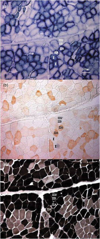
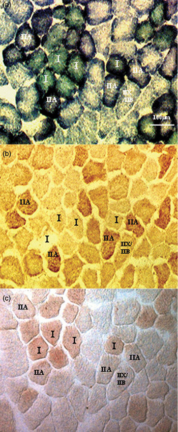
M. semitendinosus samples were stained for SDH activity; MyHCIIa, using SC71 monoclonal antibody; mATPase, following alkaline (pH 10.5) pre-incubation; and lipid, using Oil Red O. SDH and alkaline mATPase methods both produced three levels of staining intensity. Type I fibres are stained a green-blue colour with SDH and negative with mATPase. Type IIA fibres are purple with SDH and mid-brown with mATPase, and Type IIB fibres show very little SDH staining but are black with mATPase (Figures 1a and 2a). Staining with SC71 monoclonal antibody confirmed the identity of IIA fibres (Figures 1b and 2b).
Characterisation of fibre types from histochemical and immunohistochemical staining. Serial sections of postnatal M. semitendinosus samples stained for (a) succinate dehydrogenase (SDH), (b) myosin heavy chain (MyHC)-IIa using SC71 monoclonal antibody and (c) mATPase following alkaline pre-incubation pH 10.5 allow determination of fibre types. Fibres are labelled as Type I, IIA or IIX/IIB. Magnification is ×100 and scale bar is shown in (a).

Identification of oxidative fibre types by histochemical and immunohistochemical staining. Serial sections of postnatal M. semitendinosus samples stained for (a) succinate dehydrogenase (SDH) activity, (b) Myosin heavy chain (MyHC)-IIa using SC71 monoclonal antibody and (c) lipids using Oil Red O indicate that more oxidative fibres have a greater lipid content. Fibres are labelled as Type I, IIA or IIX/IIB. Magnification is ×150 and scale bar is shown in (a).

Primary and secondary fibres were also quantified from mATPase stained sections. P fibres were characterised as those that showed inhibited ATPase activity and were also the largest fibres in the centre of the metabolic cluster. The total number of fibres minus the number of P fibres is the estimate for the number of S fibres, allowing the S : P ratio to be determined.
Comparison between stains allowed the determination of three different fibre types (Figure 1). Serial sections stained for SDH, MyHCIIa and lipids indicate that the more oxidative the muscle fibre type is, the greater is its lipid content (Figure 2). Fibres that stain positively for SDH activity correlate with those that stain positively with Oil Red O. Unfortunately, staining was not consistent enough to analyse more in-depth results on intramuscular lipid content.
Deep, superficial and total muscle area
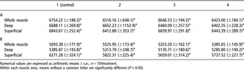
The mean total muscle area from each treatment is outlined in Table 2; increased maternal nutrition had no significant affect on total muscle area. This agrees with the results from gross measurements of the muscle, where no significant differences in muscle weight, length or girth were found (data not shown). Staining for SDH activity was used to identify the transitional line between the deep (oxidative rich) and superficial (less oxidative) regions of the muscle, allowing their percentage areas to be established (Table 2). All treatments significantly increased the deep area of the muscle compared with control, with the greatest increase seen in T2 (P ⩽ 0.001).
The effect of increased maternal nutrition (T2: 60 MJ DE (digestible energy)/day from 25 to 50 dg (days of gestation); T3: 60 MJ DE/day from 50 to 80 dg; T4: 60 MJ DE/day from 25 to 80 dg) from control (T1: 30 MJ DE/day throughout gestation) on the total, % deep, and % superficial M. semitendinosus area

Numerical values are expressed as arithmetic means ± s.e.; n = 10/treatment.
Within each muscle area, means without a common letter are significantly different (P ⩽ 0.05).
Total number of fibres
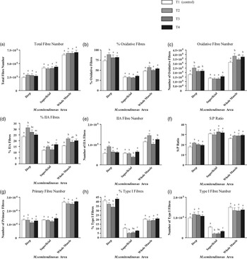
Increased maternal nutrition had no significant effect on the TNF in the deep, superficial and whole M. semitendinosus area, although an increase was evident in all treatments v. control (Figure 3a).
The effect of increased gestational nutrition on the (a) mean total fibre number, (b) proportion of oxidative fibres, (c) absolute number of oxidative fibres, (d) proportion of Type IIA fibres, (e) absolute number of Type IIA fibres, (f) S : P (secondary to primary fibre) ratio, (g) absolute number of primary fibres, (h) proportion of Type I fibres and (i) absolute number of Type I fibres in the deep, superficial, and whole M. semitendinosus area. Serial sections were stained for succinate dehydrogenase (SDH) activity, mATPase activity with alkaline (pH 10.5) pre-incubation, and myosin heavy chain (MyHC)-IIa with SC71 monoclonal antibody. □ = T1 (Control);  = T2;
 = T3; ▪=T4. Numerical values are expressed as arithmetic means ± s.e.; n = 10/treatment. Within each muscle area, means without a common letter are significantly different (P ⩽ 0.05).

Muscle fibre cross-sectional area
The mean CSA of a representative sample of all fibre types present, and specifically of type IIA muscle fibres, were not influenced by increased maternal nutrition in the deep, superficial or whole M. semitendinosus area (Table 3).
The effect of increased maternal nutrition (T2: 60 MJ DE (digestible energy)/day from 25 to 50 dg (days of gestation); T3: 60 MJ DE/day from 50 to 80 dg; T4: 60 MJ DE/day from 25 to 80 dg) from control (T1: 30 MJ DE/day throughout gestation) on muscle fibre (A) and type IIA muscle fibre (B) cross-sectional area (μm2) in the whole, deep and superficial M. semitendinosus area

Numerical values are expressed as arithmetic means ± s.e.; n = 10/treatment.
Within each muscle area, means without a common letter are significantly different (P ⩽ 0.05).
Oxidative capacity
Staining for SDH activity allowed confirmation of oxidative and non-oxidative fibre type proportions (Figure 3b) and numbers (Figure 3c), as well as the identification of the areas of the deep and superficial regions of the muscle (Table 2).
A significant increase in oxidative fibre type proportion was seen in T2 in the whole muscle area (P ⩽ 0.05) and in the deep portion (P ⩽ 0.01). A trend increase is also seen in the whole muscle in T4 (P = 0.08) (Figure 3b). Similarly, muscles from all treatments showed an increase in the number of oxidative fibres in the deep and whole muscle area compared with control, with significance in both T2 (P ⩽ 0.01) and T4 (P ⩽ 0.05). No significant effects were seen within the superficial portion (Figure 3c).
MyHCIIa antibody staining
Immunohistochemical staining with monoclonal antibody SC71 confirmed the histochemical identification of IIA fibres (Figures 1b and 2b). All treatments increased IIA fibre proportion compared with control in the deep, superficial and whole muscle areas. T2 had the greatest effect, increasing IIA fibre proportion by 7% (P ⩽ 0.001) in the deep area and 6% (P ⩽ 0.01) in the whole muscle (Figure 3d). A significant increase was also evident in the deep portion in T3, with an increase of 4% from control, and in the superficial portion and whole muscle in T4, with an increase of 5% and 4.5% IIA fibres respectively, compared to control (P ⩽ 0.01).
Furthermore, T2 had the greatest significant effect on the mean absolute number of IIA fibres. An increase above all other treatments was seen in the deep portion (P ⩽ 0.001 v. control) and in the whole muscle area (P ⩽ 0.01 v. control) (Figure 3e).
S : P ratio and primary fibre number (mATPase staining)
No significant effects on the S : P ratio were seen (Figure 3f); however, an increase is evident in the deep and whole M. semitendinosus area in all treatments, compared with control. Similarly, increased maternal nutrition had no significant effect on the P fibre number (Figure 3g). As a trend increase is seen in the TNF but no change in P fibre numbers, a trend increase in S : P ratio is in accordance.
Type I fibres (mATPase staining)
Type I fibre type proportions are described in Figure 3h, and are consistent with earlier findings that they range from approximately 4% in the superficial portion to 45% in the deep portion (Beermann et al., Reference Beermann, Fishell, Roneker, Boyd, Armbruster and Souza1990). Both the proportion and absolute number of Type I fibres were not affected by increased maternal nutrition in the whole muscle. However, a significant decrease was seen in Type I fibre proportion and absolute number in the superficial portion in all treatments (Figure 3h and i).
Capillary detection (alkaline phosphatase staining)
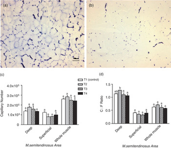
Staining for alkaline phosphatase activity allowed for the detection of capillaries supplying the muscle. Oxidative fibres (Type I and IIA) have a greater capillary density than less oxidative fibres (Type IIX and IIB) (Hudlicka et al., Reference Hudlicka, Brown and Egginton1992). The increased capillary density seen in the deep, more oxidative area compared with the superficial, less oxidative area can clearly be seen in Figure 4a and b, respectively. Increased maternal nutrition had no effect on capillary number or capillary to fibre (C : F) ratio compared with control in any part of the M. semitendinosus (Figure 4c). However, a greater C : F ratio is seen in the deep portion of muscles from T2 compared with T3 and T4 (Figure 4d).
Postnatal M. semitendinosus capillary staining analysis. The deep area (a) is dense in capillaries, compared to the superficial, less oxidative area (b), which has much fewer capillaries. Capillaries stain blue with alkaline phosphatase staining. Magnification is ×100 and scale bar is shown in (a). The effect of increased maternal nutrition on the capillary number (c) and C : F (capillary to fibre) ratio (d) in the deep, superficial and whole M. semitendinosus areas. □ = T1 (Control);  = T2;
 = T3; ▪ = T4. Numerical values are expressed as arithmetic means ± s.e.; n = 10/treatment. Within each muscle area, means without a common letter are significantly different (P ⩽ 0.05).

Discussion
The aim of these studies was to determine the effects of increased maternal nutrition during three different gestational time windows on M. semitendinosus phenotype at slaughter weight. The control treatment delivers a constant level of nutrition (30 MJ DE/day) throughout the entirety of gestation; T2 delivers increased nutrition from 25 to 50 dg, which is immediately before the onset of secondary muscle fibre formation; T3 increases maternal nutrition during secondary muscle fibre formation (50 to 80 dg); and T4 encompasses both developmental time periods (25 to 80 dg). Previously reported data have shown that increased feeding throughout gestation did not affect the birth weight, weaning weight, or intra-litter variation in this study (Lawlor et al., Reference Lawlor, Lynch, O’Connell, McNamara, Reid and Stickland2007). Furthermore, post-weaning weight up until slaughter, actual slaughter weight and the carcass weight were not affected.
A number of studies have investigated the specific effects of maternal nutrition on foetal muscle development (Dwyer et al., Reference Dwyer, Stickland and Fletcher1994; Gatford et al., Reference Gatford, Ekert, Blackmore, De Blasio, Boyce, Owens, Campbell and Owens2003; Nissen et al., Reference Nissen, Danielsen, Jorgensen and Oksbjerg2003; Bee, Reference Bee2004). However, results describing how different maternal feeding strategies affect muscle fibre development in the offspring are not consistent, even where the same time windows during gestation have been examined. This study administers increased maternal feed intake to double that of control (30 MJ DE/day), a level that is approximately 50% of ad libitum. The sows’ requirement for energy during gestation is 15–25 MJ net energy per day (MJ NE/day) (Gatford et al., Reference Gatford, Ekert, Blackmore, De Blasio, Boyce, Owens, Campbell and Owens2003; Nissen et al., Reference Nissen, Danielsen, Jorgensen and Oksbjerg2003). The feeding regime in our study delivers 21.25 MJ NE/day in the control treatment (compliant with requirements), which is increased to 42.5 MJ NE/day in T2, T3 and T4, representing a value that can be argued to correspond to ad libitum levels.
The findings of these studies outline the effect of increased maternal nutrition on skeletal muscle phenotype in the offspring at slaughter weight, with a particular focus on muscle fibre type and oxidative capacity. Increasing the level of maternal intake at all three time periods significantly increased the area comprising the more oxidative deep portion of the M. semitendinosus, with no change in total muscle area. This latter finding is supported by analyses of the gross measurements of the M. semitendinosus, which find that increased maternal nutrition did not influence muscle weight, length or girth. The most significant increase in deep area is seen in the progeny of sows fed increased nutrition from 25 to 50 dg (T2). No significant effects are seen on the TNF. Earlier findings report that the TNF is decreased in the smallest littermate (excluding runts) as a result of in utero or ‘natural’ under-nutrition (Wigmore and Stickland, Reference Wigmore and Stickland1983; Rehfeldt et al., Reference Rehfeldt, Nissen, Kuhn, Vestergaard, Ender and Oksbjerg2004; Gondret et al., Reference Gondret, Lefaucheur, Juin, Louveau and Lebret2006). This often results in a reduced TNF with a larger cross-sectional area. From a meat production perspective, previous studies have shown that pigs which exhibit a greater number of muscle fibres, with a small CSA, correlate with faster and more efficient growth, a leaner carcass and a better meat quality (Dwyer and Stickland, Reference Dwyer and Stickland1991; Oksbjerg et al., Reference Oksbjerg, Peterson, Sorensen, Henckel, Vestergaard, Ertbjerg, Moller, Bejerholm and Stoier2000; Rehfeldt et al., Reference Rehfeldt, Fiedler, Dietl and Ender2000). Our findings show no significant difference in TNF, S : P ratio or fibre CSA due to an increased level of maternal nutrition, and are consistent with earlier studies (Nissen et al., Reference Nissen, Danielsen, Jorgensen and Oksbjerg2003; Bee, Reference Bee2004) that found that increased maternal feed intake had no effect on M. semitendinosus fibre number or area in offspring at slaughter weight.
An increase in the proportion of IIA fibres is seen in all treatments in all areas of the muscle. This is seen to be particularly significant in T2, in which the mean absolute number of IIA fibres is also significantly increased above control in the deep portion and whole muscle area. Pilot studies have shown that at 80 to 100 dg, the heavyweight foetus contains more IIA fibres in both deep and superficial areas (Ashton, Reference Ashton2002). Here we have shown that at 6 months of age, elevated maternal nutrition between 25 and 50 dg significantly increases the proportion of IIA fibres in the M. semitendinosus in the offspring. Similarly, the oxidative fibre proportion and number is significantly increased above the control treatment in the deep portion, and the whole muscle in T2. No treatment effects are seen on the proportion and number of Type I fibres, and therefore the oxidative fibre number, and hence oxidative capacity is increased in the M. semitendinosus due to an increase in Type IIA fibres and not Type I fibres.
The number of Type I fibres was significantly reduced in the superficial portion in all treatments. In conjunction with a reduced percentage area, the superficial portion showed no change in TNF or P fibre number, but a significant increase in IIA fibre proportion. The decreased number of Type I fibres and increase in Type IIA fibres explains why no overall change in oxidative fibre type proportion was seen.
Primary fibre number is believed to be a fixed, genetic effect (Wigmore and Stickland, Reference Wigmore and Stickland1983). The S : P ratio is used to measure the effect of nutrition on S fibres, excluding the genetic component associated with P fibres. The ratio was not affected by increased maternal nutrition in this study. This is inconsistent with earlier findings (Dwyer et al., Reference Dwyer, Stickland and Fletcher1994) where a significant increase in the S : P ratio was found due to increased maternal nutrition from 25 to 50, 50 to 80 and 25 to 80 dg. These findings were explained by an increased number of S fibres, with no effect on P fibre number seen. However, in this study, there was also no significant effect on TNF, muscle weight or muscle and fibre CSA, and hence a significant effect on S : P ratio would not be expected.
The results of this study suggest that as increased maternal nutrition enhances the oxidative capacity of the M. semitendinosus, the lipid content within the fibres is also increased. This is evident in the correlation between fibres that stained positive histochemically for Oil Red O (lipid) and SDH activity (oxidative capacity). The more oxidative fibre types (Type I and IIA) have an increased lipid content compared to less oxidative fibre types (Type IIX and IIB). It has been shown that metabolic phenotype can be altered by stimuli other than increased maternal nutrition. Endurance training (Fluck and Hoppeler, Reference Fluck and Hoppeler2003) and electrical stimulation (Sutherland et al., Reference Sutherland, Salmons, Ramnarine, Capoccia, Walsh and Jarvis2006) both increase the oxidative metabolism of muscle. Fast to slow MyHC isoform transitions in the order MyHC-IIb, -IIx, -IIa, to MyHCIβ occur in parallel with increases in oxidative activity, due to an increase in the number of mitochondria. With endurance training, fat within individual muscle fibres increases in conjunction with the increase in oxidative metabolism as an adaptation of the body to acquire more readily available fuel (Hoppeler, Reference Hoppeler1999). The underlying mechanisms occurring in these studies may also be acting in the model used in this study, in order to achieve an increased oxidative metabolism.
As with lipid content, more oxidative fibres (Type I and IIA) have a greater capillary density than less oxidative fibres (Type IIX and IIB) (Hudlicka et al., Reference Hudlicka, Brown and Egginton1992). Angiogenesis and oxidative capacity are independently regulated (Egginton and Hudlicka, Reference Egginton and Hudlicka2000). Capillary density, and to a lesser extent C : F ratio, are dependent upon fibre CSA (Deveci et al., Reference Deveci, Marshall and Egginton2001). Where an increase in fibre CSA is evident, an increase in capillary number also occurs. Muscle fibre CSA was not significantly affected by increased maternal nutrition. Accordingly, maternal nutrition had no effect on capillary number. If no change in fibre CSA is seen, as in this study, the C : F ratio provides a good indication of angiogenesis. However, the C : F ratio was also unaffected by increased maternal nutrition, but as no change in the TNF or capillary number was seen, a significant effect on the C : F ratio would not be expected. Therefore, although an increase in oxidative metabolism is seen via an increase in the number of Type IIA fibres present and an increased deep area of the muscle, this is not paralleled with an alteration in the capillary supply.
Increasing maternal feed intake from 25 to 80 dg (T4) appears to have no more beneficial effect than increasing it from 25 to 50 dg on the percentage deep area, S fibre number (S : P ratio), and oxidative and IIA fibre proportion or number. In fact, although it encompasses the critical initial time period of 25 to 50 dg, T4 is not as effective at influencing muscle phenotype as T2. The reasons for this are unknown. Although contradictory to the results found in our study, a study by Nissen et al. (Reference Nissen, Danielsen, Jorgensen and Oksbjerg2003) suggests that increased maternal feeding can have disadvantageous effects on muscle phenotype at slaughter weight. Therefore, it can be suggested that in our study, prolonging increased feeding from 50 to 80 dg may decrease the increases seen in T2 back to control, having a balancing or ‘negative’ effect. Increased feeding from 50 to 80 dg (T3) has no significant influence on skeletal muscle phenotype, supporting this idea. However, it must be stressed that this is purely speculation.
From these data, it is concluded that increased maternal nutrition alters skeletal muscle phenotype proportions in the offspring, with a resulting increase in oxidative capacity. The percentage area comprising the deep, more oxidative portion of the M. semitendinosus was significantly increased, as well as the oxidative and IIA fibre type proportion, and S : P ratio. These findings were most significant in T2 and are consistent with other studies which find the time period between 25 to 50 dg as most sensitive to the effects of the plane of nutrition (Dwyer et al., Reference Dwyer, Stickland and Fletcher1994; Gatford et al., Reference Gatford, Ekert, Blackmore, De Blasio, Boyce, Owens, Campbell and Owens2003). This is the most critical time for stimulating the proliferation of presumptive secondary myoblasts, providing the potential for manipulation of overall muscle fibre type composition. Hence, nutrition level in the period immediately before the appearance of secondary myotubes may be more important than that during the actual appearance of muscle fibres, as proposed earlier (Dwyer et al., Reference Dwyer, Stickland and Fletcher1994), and may act to exert an effect on the population of foetal myoblasts available for the development of S fibres.
The mechanisms underlying the altered oxidative metabolism in the M. semitendinosus of slaughter weight offspring due to increased maternal nutrition are as yet unknown. A number of studies have implicated various growth- and transcription-factors in the regulation of oxidative metabolism and the oxidative fibre types (Chin et al., Reference Chin, Olson, Richardson, Yang, Humphries, Shelton, Wu, Zhu, Bassel-Duby and Williams1998; Hughes et al., Reference Hughes, Chi, Lowry and Gundersen1999; Luquet et al., Reference Luquet, Lopez-Soriano, Holst, Fredenrich, Melki, Rassoulzadegan and Grimaldi2003; Gregorevic et al., Reference Gregorevic, Plant and Lynch2004; Khorram et al., Reference Khorram, Khorram, Momeni, Han, Halem, Desai and Ross2007), some of which are shown to be influenced by nutritional status. Altered organ or organ system structure during organogenesis could permanently affect an organism’s metabolism. This developmental ‘programming’ could be explained by the ability of individual cells to generate and respond to external signals within the organism. The precise molecular mechanism(s) by which these effects occur are being pursued.
This study shows changes at the muscle tissue level but not at the whole animal level (Lawlor et al., Reference Lawlor, Lynch, O’Connell, McNamara, Reid and Stickland2007). This suggests that the pigs may have been pushed to their limit in terms of factors such as birth weight and weaning weight, as a result of the high level of genetic selection. With regard to phenotype, the TNF is also unaffected by increased maternal energy. The changes that do occur in the muscle are associated with muscle fibre type composition. This indicates that the fibre type proportions are still malleable, but the TNF and whole animal growth factors may have reached their limit and cannot be increased any further by nutrition. In order to view differences at the whole animal level, it may be necessary to use less selected pigs. Limitations to this study include not controlling for the quantity of milk received by the piglets during the month of nursing, following farrowing and prior to weaning. Differences in intake during this time may have contributed towards the results seen.
Acknowledgements
With thanks to all contributing authors, the team at Teagasc, Moorepark Research Centre where the trial was conducted, and to Aviva Petrie for her statistical advice. This research was funded by the Biotechnology and Biological Sciences Research Council (BBSRC).