Atrial tachyarrhythmia, especially atrial flutter and intra-atrial re-entrant tachycardia is known to occur in the remote phase after percutaneous atrial septal defect closure. Most cases are caused by long-term right atrial volume loading. Atrial tachyarrhythmia is less common in children, because of less atrial enlargement and fibrosis. Reference Chiu, Wu and Tsai1 A large electrical circuit in the atria is necessary for atrial flutter and intra-atrial re-entrant tachycardia to occur. Only a few reports have described intra-atrial re-entrant tachycardia caused by atrial septal defect closure devices. Reference John, George, Joseph and Lokhandwala2,Reference Ravella, Bhoskar, Bachani, Lokhandwala and Dalvi3
Case report
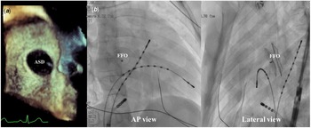
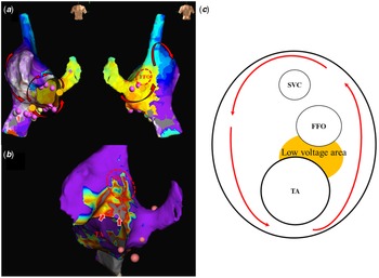
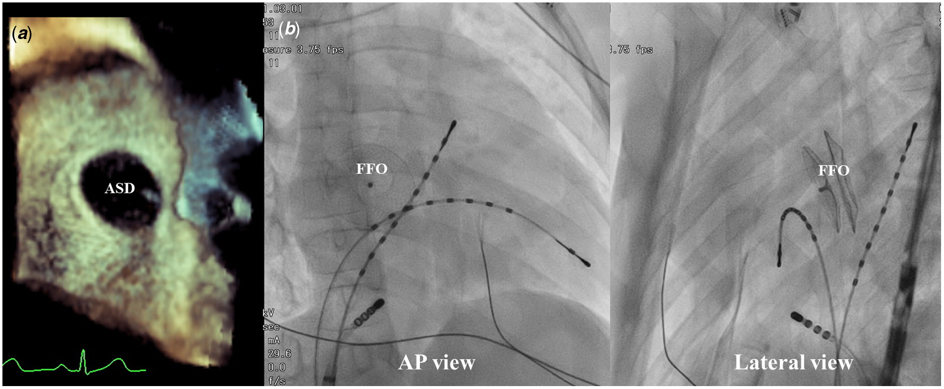
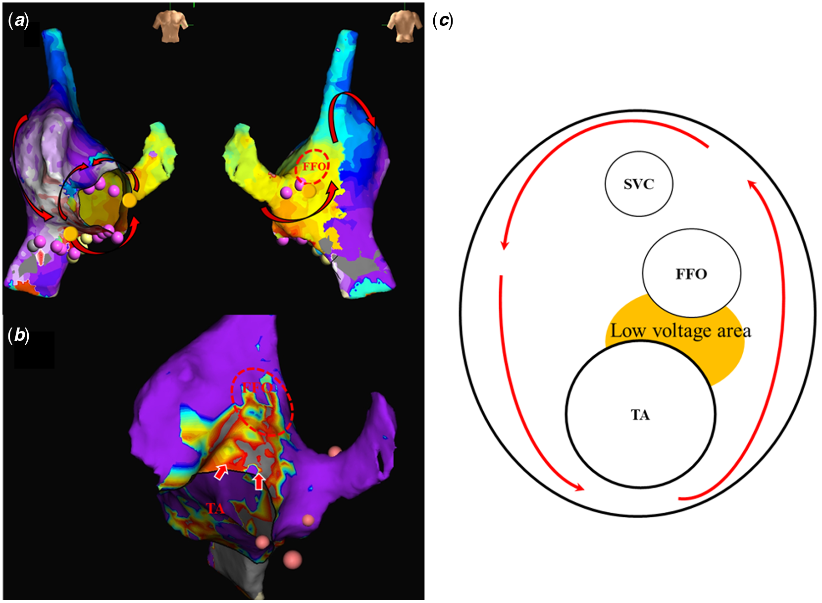
We present a case of a 6-year-old boy who developed intra-atrial re-entrant tachycardia after percutaneous atrial septal defect closure. The patient had been diagnosed prenatally with atrial septal defect, persistent left superior vena cava, and Holt-Oram syndrome, with no previously noted arrhythmias. He underwent percutaneous atrial septal defect closure at the age of 5 years. The transesophageal echocardiography revealed that the defect was 13.0 mm in diameter with adequate surrounding rims for devise closure (Fig 1a). The stretch balloon diameter of defect was 16.7 mm. The distance from the defect to the tricuspid valve was 12.9 mm. A 16.5-mm Figulla Flex II ASD Occluder™ (Occlutech) was deployed (Fig 1b). The patient was discharged without any adverse events. Three weeks later at the follow-up outpatient visit, the patient developed atrial tachycardia without symptoms and required cardioversion. Due to repeated atrial tachycardia, the patient underwent ablation 5 months later. Tachycardia persisted prior to admission to the cardiac catheterisation laboratory, and 12-lead electrocardiography (Supplemental data Fig 1) showed negative p waves in inferior limb leads and an atrioventricular conduction ratio of 2–3:1. Tachycardia cycle length was 245 ms. Using the EnSite Velocity™ Cardiac Mapping System (Abbott), three-dimensional mapping of the right atrium was performed. The results showed apparently common atrial flutter. However, the post-pacing interval at the cavotricuspid isthmus and at the orifice of the coronary sinus, but not at the anterior tricuspid valve leaflet, was consistent with tachycardia. The more anterior the tricuspid valve, the longer the post-pacing interval, so the anterior tricuspid annulus was determined to be outside of the circuit. Three-dimensional mapping of the right atrium was repeated. The activation map showed a large macro-re-entry that ran counterclockwise half way from the lateral lower right atrium behind the tricuspid annulus, ascended atrial septum along the Figulla Flex II ASD Occluder™, and returned to the lateral lower right atrium from the anterolateral wall of the upper right atrium (Fig 2a). The clockwise ascending potential from the lateral right lower atrium to the anterior tricuspid annulus was located at 12 o’clock to the tricuspid annulus and collided with the counterclockwise ascending potential from the tricuspid annulus in the posterior septum (Supplemental data Video 1). The voltage map showed a lower potential in the anterior tricuspid annulus, which was excluded by the Figulla Flex II ASD Occluder™ (Fig 2b). We diagnosed cavotricupid isthmus dependent intra-atrial re-entrant tachycardia and performed linear ablation of the cavotricupid isthmus. Tachycardia terminated during ablation. Thereafter, tachycardia was not inducible. The patient has no recurrence after 1 year.
( a ) ASD image of transesophageal echocardiography. ( b ) FFO Position on the Biplane Fluoroscopic Images. ASD, atrial septal defect; FFO, Figulla Flex & ASD OccluderTM.
( a ) Activation map of intra-atrial re-entrant tachycardia. The clockwise ascending potential from the LLRA to the anterior tricuspid annulus collided with the counterclockwise ascending potential from the tricuspid annulus in the posterior septum at 12 o'clock to the tricuspid annulus. ( b ) The voltage map shows a lower potential in the anterior tricuspid annulus, which was excluded by the FFO. Yellow arrows indicate the low voltage area. ( c ) The tricuspid annulus and FFO fused with the low potential area in front of the tricuspid annulus to form a large circuitLLRA, lateral lower right atrium; FFO, Figulla Flex II ASD Occluder™; SVC, superior vena cava; TA, tricuspid annulus.
Discussion
In young children, atrial flutter and atypical AFL (intra-atrial re-entrant tachycardia) are rare because of a small atrial size and lack of fibrosis. Atrial flutter requires a large circuit to be established in the atrium. To the best of our knowledge, there are two reports of atrial flutter immediately after catheterisation for atrial septal defect. John et al reported a 4-month-old boy who developed intra-atrial re-entrant tachycardia 3 weeks after percutaneous atrial septal defect closure. Reference John, George, Joseph and Lokhandwala2 He was catheter ablated at 2 years of age and the cavotricupid isthmus ablation stopped the atrial flutter, but no clear circuit has been identified. Another report is of a 28-year-old male who developed atrial flutter 4 weeks after percutaneous atrial septal closure, but no ablation was performed. Reference Ravella, Bhoskar, Bachani, Lokhandwala and Dalvi3
In present case, the Figulla Flex II ASD Occluder™ may have inhibited the potential anterior to the tricuspid annulus, resulting in macro-re-entry and a large atrial swirl. This is thought to be due to the fact that the tricuspid annulus and Figulla Flex II ASD Occluder™ fused with the low potential area in front of the tricuspid annulus to form a large circuit, which normally could not be established with a small tricuspid annulus (Fig 2c).
In conclusion, implanting a large ASD closure device in the small heart of an infant can cause changes in the conduction pathways as well as the structure of the heart. If possible, a smaller ASD closure device should be selected.
Supplementary material
To view supplementary material for this article, please visit https://doi.org/10.1017/S1047951122002256
Acknowledgements
We thank Emily Woodhouse, PhD, from Edanz (https://jp.edanz.com/ac) for editing a draft of this manuscript.
Financial support
This research received no specific grant from any funding agency, commercial, or not-for-profit sectors.
Conflicts of interest
None.
Ethical standards
The authors assert that all procedures contributing to this work comply with the ethical standards of the relevant national guidelines on human experimentation and with the Helsinki Declaration of 1975, as revised in 2008. Since this is a case report, no institutional review board approval was necessary.