Non-traumatic intracranial haemorrhage is not common in children; however, it carries significant mortality and morbidity.Reference Al-Jarallah, Al-Rifai, Riela and Roach 1 Several reports suggest that patients with CHD or infective endocarditis are at an increased risk for intracranial haemorrhage or cerebral aneurysm.Reference Lo, Lee, Rusin, Perkins and Roach 2 – Reference García-Cabrera, Fernández-Hidalgo and Almirante 4
Case description
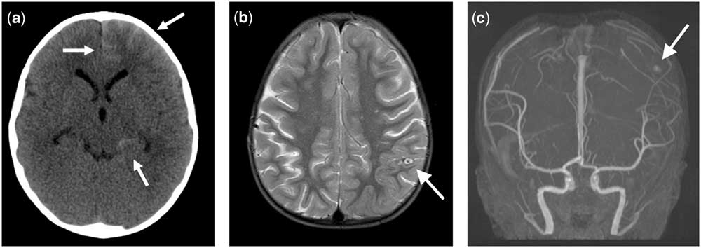
A 2-year-old boy came to the emergency department presenting with headache and vomiting that had lasted for a week. CT of head showed subarachnoid haemorrhage (Fig 1). He had a past medical history of an incomplete atrioventricular canal, status post atrioventricular canal repair at 5 months of age, and status post mitral valve replacement with a mechanical valve at 7 months of age. He had been on Coumadin, and the prothrombin time at the emergency department was 40.8 seconds. He had recently developed subaortic stenosis with a peak pressure gradient of 95 mmHg and was scheduled to undergo subaortic stenosis repair in 2 weeks. An emergency cerebral arteriogram did not show evidence of arteriovenous malformation or cerebral aneurysm. His symptoms improved after he was given fresh frozen plasma and the anticoagulation therapy was discontinued for 5 days. A second head-CT 7 days later showed a resolving subarachnoid haemorrhage with no obvious cerebral aneurysm, and he was discharged on Coumadin. He returned to the emergency department 1 month later because of lethargy and fever; an echocardiogram revealed severely reduced left ventricular function, but no sign of endocarditis or aortic aneurysm. A head CT did not show a recurrence of subarachnoid haemorrhage or cerebral aneurysm. Because of worsening ventricular function and the relatively stable condition of cerebral haemorrhage, the decision was made to urgently perform subaortic stenosis repair.
( a ) Initial head CT showing parietal and occipital subarachnoid haemorrhage (white arrows). ( b ) Postoperative magnetic resonance T2-weighted image showing partially thrombosed cerebral aneurysm in the left central sulcus (white arrow) without recurrence of subarachnoid haemorrhage. ( c ) Postoperative magnetic resonance angiogram showing cerebral aneurysm in the distal M3 branch of the left middle cerebral artery (white arrow).

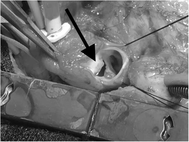
An operation was performed with regular redo-sternotomy and cardiopulmonary bypass. After aortotomy, it was noticed that there was a large tear with a dirty edge on the posterior wall of the ascending aorta (Fig 2) and an undiagnosed sealed rupture of an aortic aneurysm with an abscess between the ascending aorta and the right pulmonary artery. The abscess was adequately debrided and the ascending aortic wall was partially removed. The subaortic stenosis was removed with septal myectomy, and the ascending aorta was reconstructed with end-to-end anastomosis. The patient recovered from the operation well and without neurological sequelae. A blood culture from 1 day before surgery was positive for non-haemolytic Streptococcus, and a microbiology examination from the aortic wall grew Gram-positive cocci. MRI performed 7 days after the operation showed a partially thrombosed cerebral aneurysm without the recurrence of subarachnoid haemorrhage (Fig 1). The patient did well with a 6-week course of antibiotics and had a stable neurological status. An echocardiogram performed 2 months after the operation showed minimal residual subaortic stenosis, improving ventricular function, and a smooth ascending aorta.
Intraoperative photo showing a large tear with a dirty edge on the posterior wall of the ascending aorta (black arrow).

Discussion
Mycotic cerebral aneurysm secondary to infective endocarditis is a well-known cause of intracranial haemorrhage in children, and patients with CHD are at an increased risk for infective endocarditis due to abnormal blood turbulence and the presence of foreign materials used for repair.Reference Awadallah, Kavey and Byrum 5 In this case, no evidence of infective endocarditis or mycotic cerebral aneurysm was found at the time of the initial subarachnoid haemorrhage. However, we suspect that the undiagnosed mycotic ascending aortic aneurysm resulting from subaortic stenosis caused a secondary mycotic cerebral aneurysm. Retrospectively, this patient should have been given antibiotics after the initial subarachnoid haemorrhage because his past medical history was highly associated with endocarditis.
A further challenge with this patient was anticoagulation therapy; it was difficult to balance the need for blood coagulation for the intracranial haemorrhage and the need for anticoagulation therapy for the mechanical heart valve. Moreover, there were no established criteria to determine how long the anticoagulation therapy should have been discontinued or how long the anticipated heart surgery with cardiopulmonary bypass needed to be delayed. In this case, it seemed adequate to delay the cardiopulmonary bypass surgery for 4 weeks to prevent further intracranial haemorrhage.
In conclusion, we report a case of subarachnoid haemorrhage caused by a silent mycotic aortic aneurysm in a child with CHD. For patients with unexplained intracranial haemorrhage and CHD, a detailed survey of the heart and aorta may be necessary.
Acknowledgements
The authors would like to thank Kerry Evans for assistance with manuscript preparation.
Financial Support
This research received no support from any funding agency or commercial or not-for-profit sectors.
Conflicts of Interest
None.