Introduction
Thyroid disorder rates are higher in women, especially during the childbearing age. Thus, thyroid disorder manifestation during pregnancy is common.Reference Glinoer 1 Pregnancy is a physiological condition where a number of modifications may occur to affect thyroid economy. They may occur at any point of gestation, they may be transient or until term. Sometimes these alterations are permanent.Reference Glinoer and De Nayer 2
Foetal thyroid gland produces thyroid hormones (TH) by the 12th week. Therefore, during the first trimester, the foetus is totally dependent on maternal supply. Hence, early detection of maternal thyroid levels and adequate iodine intake during pregnancy are a must to provide sufficient supply of iodine to the growing foetus.
The prevalence of hypothyroidism during pregnancy is estimated to be 0.3–0.5% for overt hypothyroidism (OH) and 2–3% for subclinical hypothyroidism (SCH). The most contributing cause of hypothyroidism is iodine deficiency (ID), affecting 1.2 billion individuals in the world.Reference Ablovich and Glinoer 3 Recent reviews from India suggest that the prevalence of ID (UIE <150 μg/dl) ranges from 30 to 95% among pregnant women.Reference Singh, Fotedar and Lakshminarayana 4 – Reference Srinath 12
Other than dietary iodine insufficiency, ID may occur because of increased glomerular filtration rate (GFR) and its clearance during pregnancy.Reference Glinoer 1 Severe ID may also cause maternal hypothyroxinemiaReference Zimmerman 13 and may lead to irreversible brain damage to the foetus. Mild to moderate ID produces more subtle changes in cognitive functions in the offspring.
Similarly, another essential micronutrient iron also plays a vital physiological role in the growth, development, metabolic reactions, cofactor for number of enzymatic activities and cognitive development of the foetus. Untreated chronic/severe maternal iron deficiency anaemia (IDA) may prove to be detrimental to the developing foetus. The prevalence of anaemia among Indian pregnant women is more than 60%. 14 The WHO estimates that even among the South Asian countries, India has the highest prevalence of anaemia and it also contributes to about 80% of the maternal deaths due to anaemia.Reference Ezzati, Lopus, Dogers, Vander and Murray 15
ID along with IDA may worsen the condition, as iron plays a vital role in the thyroperoxidise enzyme action. Several studies have reported a high prevalence of hypothyroidism in anaemic subjects.Reference Zimmerman 16 – Reference Duntas, Papanastasiou, Mantzou and Koutras 21 Serum ferritin and total iron-binding capacity may be lower in hypothyroidism.Reference Zimmerman 16 This suggests the need of screening for ID in maternity clinics along with the existing system for assessing IDA in India. The screening is important, as 70–80% of women with hypothyroidism remain asymptomatic.
Hence, the study is aimed to determine the prevalence of ID disorders and haemoglobin status among pregnant women during early pregnancy.
Materials and methods
Type of study
Cross-sectional study.
Setting
Jamnabai General Hospital, a Semi-government Hospital, Vadodara.
Duration
Six months (October 2009–March 2010).
Study subjects and sample size
Pregnant women (n = 256) who attended antenatal services during the first trimester (<15 weeks) at the time of screening, without visible signs of ID and healthy singleton pregnancy, were included as study subjects. However, pregnant women with known thyroid dysfunction and twin or triplet pregnancies were excluded at the time of screening.
The pregnant women were enrolled during their first or second antenatal clinic visit. The inclusion criteria were applied and the purpose of the study was explained to the pregnant women. Those who agreed for the participation made the final sample size. Written consent was availed from the concerned hospital authority and from the subjects individually.
Statistical analysis
The quantitative data are expressed as mean ± s.d. To define the normal values, mean, standard deviation and median are considered. Analysis of variance test between normally distributed data and Mann–Whitney test for urinary iodine levels and thyroid hormones were applied. Correlation coefficient was derived to show the interrelation between parameters.
THs
THs including thyrotropin (TSH), free thyroxine (FT4) and total thyroxine (TT4) were assessed using RIA Method at BARC, Mumbai. Five ml of whole blood was collected, centrifuged at 2500 rpm and separated serum was stored at −20°C. The urinary iodine content was estimated using simple microplate method based on Sandell–Kolthoff reaction.Reference Ohashi, Yamaki, Pandav, Karmarkar and Irie 22 Haemoglobin concentration was estimated by acid hematin method (Sahli's Method).
Definitions
As per the guidelines of ‘The Endocrine Society’, 23 maternal thyroid status was defined in various categories. Maternal OH was defined as TSH concentration (>2.3 μIU/ml) and FT4 concentration below normal range (0.65–2.10 ng/dl). In maternal SCH, the patient may not have symptoms, but the concentration of TSH is >2.3 μIU/ml with normal FT4 concentrations. Maternal hypothyroxinemia (HT) was defined as normal TSH concentrations with FT4 concentration below the normal range.
For pregnant women, the haemoglobin concentration of ⩾11 g/dl was considered normal. 24 The haemoglobin concentration between 10.0 and 10.99 g/dl was considered mildly deficient, between 7.0 and 9.9 g/dl was considered moderately deficient and <7 g/dl was considered severe deficient.
ID based on UIC 25 was defined at <150 μg/l as insufficient and ⩾150 μg/l as sufficient levels.
Less than or equal to 4 weeks of pregnancy was classified on the basis of counting days between last menstrual period and the date of pregnancy confirmation. This was observed among subjects with menstruation cycle of <27 days.
Results
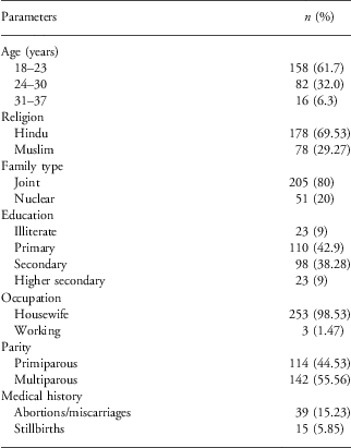
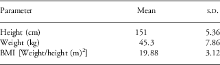
General profile including socio-economic status and anthropometric parameters of the enrolled pregnant women (n = 256) was collected (Tables 1a and 1b).
General profile of the subjects

Anthropometry of the subjects

Of the total population, 39 (15.23%) subjects had history of abortions/miscarriages and 15 (5.85%) had experienced still birth or neonatal mortality during their previous deliveries. Mean body mass index (BMI) was falling into the normal category (18.5–25 kg/m2), whereas 90 (35.15%) subjects had lower BMI (<18.5 kg/m2). A total of 175 (68%) subjects were enrolled during the third month (9 to <15 weeks) from their last menstruation period and 114 (44.53%) subjects were primiparous of all the subjects. Literacy rate in the study population was observed to be 110 (42.9%) with primary and 98 (38.28%) with secondary education. Majority of the subjects were housewives. Average monthly family income was Rs. ⩾3000–5000 per month.
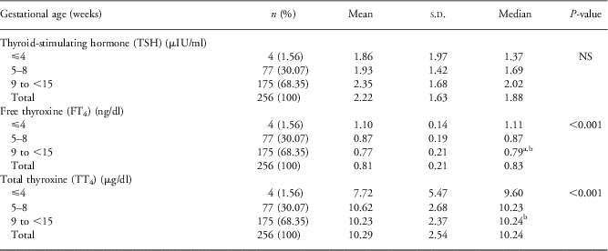
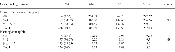
The THs (Table 2), urinary iodine concentration and Hb concentration of the subjects (Table 3 ) are revealed as mean/median and s.d. values.
Thyroid hormone profile of the subjects enrolled at different points of gestation

a P < 0.05 significant difference compared to ⩽4 weeks.
b P < 0.01 significant difference compared to 5–8 weeks.
Iodine and haemoglobin status of the subjects

Our data on TH showed a normal hormonal pattern followed by suppressed TSH level and increased FT4 levels at the beginning of pregnancy, which is also reported by other authors.Reference Springer, Zima and Limanova 26 In our study, majority of the subjects were enrolled towards the end of the second month (5–8 weeks) or the third month (9 to <15 weeks) of gestation. A negative correlation (P < 0.01) between FT4 and TSH levels was observed, which suggests that as TSH levels increase with pregnancy, FT4 decreases. There was also a negative correlation between FT4 and the month of enrolment (P < 0.001), which revealed that as pregnancy progresses there is a reduction in FT4 levels.
TSH was observed to be highest during the third month of gestation ( Table 2 ). However, there was no significant difference between the TSH levels of the subjects enrolled at different weeks. The FT4 levels followed a normal pattern and there was a significant difference (P < 0.001) in the FT4 levels between <4 weeks and the rest two groups. There was also an increase in TT4 values with the progression of gestation within the first trimester. The difference was significant between the groups based on the month of enrolment (P < 0.001). The TSH values ranged (±2 s.d.) between 0.35 and 5.06 μIU/ml, FT4 between 0.46 and 1.13 ng/dl and TT4 between 6.66 and 14.59 μg/dl.
Using spot UIE as an indicator, iodine sufficiency was observed in 213 (78.51%) and 43 (16.79%) showed insufficient levels. Our study population revealed adequate levels of median UIE as 297.14 μg/l, indicating adequate consumption of iodine through diet (Table 3). Our subjects also revealed overall mild iron deficiency with mean Hb concentration as 9.27 ± 1.09 g/dl. On the basis of the WHO-recommended 24 classification for IDA, 235 (91%) subjects had iron deficiency. There were 66.4, 22.7 and 2.73% subjects with mild, moderate and severe deficiency, respectively. There was a non significant difference between all three groups on the basis of the time of enrolment for urinary iodine insufficiency and iron deficiency (P > 0.05). Of the subjects, 16.40% had both iodine and iron deficiencies at the time of registration. Our data also revealed 23% prevalence of SCH, 11% OH and 12% HT using standard cut-offs.
Discussion
During pregnancy, the dietary intake of macronutrients and micronutrients is recommended to be higher than the normal condition; however, in general population, priority is given to the lactation period rather than pregnancy. Hence, it is necessary to assess early gestational nutrition status and generate awareness regarding the importance of adequate dietary intake and implications of nutritional deficiencies during pregnancy. In our study population, one-third of the subjects were observed to be underweight and most of them were primiparous. Majority of our subjects had lower haemoglobin levels, which suggests the importance and need for iron folic-acid supplementation during pregnancy. On the other hand, majority subjects had sufficient and more than sufficient UIE, which suggests either an increased GFR or availability of adequately iodized salt in the community.
Our data also revealed a significant association between BMI and Hb concentrations (P < 0.01). Thus, there is a need to address and develop guidelines that will create awareness among these subjects. There were 15% of the subjects with the history of abortion, who were also the focused population towards our efforts to meet complete nutrition in a feasible cost.
Reference values for TSH followed were using international guidelines 23 , Reference Panesar, Li and Rogers 27 with >2.3 μIU/ml and for FT4 reference range of the commercial kit for RIA was used (0.65–2.10 ng/dl), as no specific reference range for FT4 was available, especially for the first trimester. Considering these reference ranges, remarkable per cent subjects were positive for SCH, OH and HT, which in turn are suggestive of very high prevalence compared with other studies reviewed.Reference Zimmerman 13 , Reference Panesar, Li and Rogers 27 This also depicts mild to moderate ID prevailing in the region after 5 years of reimplementation of the ban on the sale of uniodized salt. This indicates that either there is autoimmunity or environmental factors leading towards ID, despite the availability of iodized salt at household level. Hence, there is a need for thyroid screening in all the maternity clinics.
Pregnancy, a physiological condition due to metabolic changes, leads to ID. Therefore, WHO has recommended daily iodine intake during pregnancy to 250–300 mcg/day that would correspond to 135–155 μg/l, considering 92% absorbed iodine from the diet.Reference Zimmerman 13 In our study, majority of the subjects had iodine sufficiency by UIE may be because of increased GFR, but still few struggled to reach 150 μg/l, which suggests a need for an appropriate supply of iodine and close monitoring for iodine intake, despite the availability of optimally iodized salt.
For healthy pregnant women who reside in areas with a restricted iodine intake or with relative HT, pregnancy may lead to adjustments in the thyroid economy. This also indicates a need for systematic screening at the first prenatal visit as pregnancy has a major impact on thyroid regulation. These effects need to be recognized, assessed, clearly interpreted and correctly managed.
The determination of THs should be investigated thoroughly, for which the reference ranges should be established to have absolute detection of the stage of dysfunction. OH occurs in 2–3% pregnancies, but SCH is more prevalent and frequently remains undiagnosed. However, in our data, the prevalence of thyroid dysfunctions classified as SCH, OH and HT was observed to be comparatively high. This again suggests the need for the evaluation of thyroid function test during pregnancy and particularly in the first trimester, as it is of great importance because of the extra requirement of thyroxin for the growing foetus to prevent the irreversible consequences and damages on pregnancy outcome in the early stage of foetal development.Reference Mansourian 28 Hyperthyroidism and hypothyroidism during pregnancy should be evaluated carefully and assessed properly to avoid the irreversible adverse effects on the growing foetus and pregnant mothers.
Conclusion
In the absence of recommendations for universal screening, aggressive target case finding in high-risk pregnant women is recommended at the first prenatal visit;Reference Ablovich and Glinoer 3 however, targeted thyroid function testing may miss one-third of pregnant women with SCH and OHReference Ablovich and Glinoer 3 as they are asymptomatic. A similar trend was observed in our study. Hence, it is always better to have blanket coverage for ID detection through circulating TH levels at first visit to an antenatal clinic, parallel to the compulsory Hb estimation. This may increase the health-care cost to the government health programme, but the reward is going to be very high as mentally and physically healthy future of the nation's healthy children.
Key points
-
• Maternal ID and thyroid dysfunctions may occur at any stage of gestation. Hence, screening for ID and thyroid dysfunction is very crucial for the early detection of possible adverse effects on the developing foetuses because of maternal iodine insufficiency. Thus, it is recommended to have a mandatory screening programme for maternal ID in the antenatal clinics.
-
• On the basis of the results of the screening, control and treatment measures can be initiated by the government to prevent these micronutrient deficiencies. Attainment of pregnancy requires iodine and iron-sufficient state to provide sufficient supply to the foetuses. Thus, it is recommended that the adolescent girls rather than pregnant women should be supplemented with adequate iodine and iron so that they enter pregnancy with sufficient stores and have healthy outcomes.
-
• However, iodine and iron status of the pregnant women of Vadodara is suboptimal, and thus the recommendation implies strongly to improve the status of the future mothers.
Acknowledgements
We would like to heartily thank all our study subjects for their support and participation to make our study a success. We also extend our thanks to Jamnabai General Hospital authorities for permitting us to carry out our research at their institution, the PPTCT counsellors and laboratory technicians. We thank Prof. M.G. Karmarkar and Dr C.S. Pandav (ICCIDD, Delhi) for providing lab facility for UIE estimations. We thank our Head, Department of Foods and Nutrition, The Maharaja Sayajirao University of Baroda for providing us an institutional platform to conduct this research study.
Financial Support
This research received no specific grant from any funding agency, commercial or not-for-profit.
Conflicts of Interest
None.
Ethical Standards
The study was approved by the ethical committee of the home institution ethical board in compliance with the guidelines issued by Indian Council of Medical Research (No. F. C. Sc FN ME70).