I. INTRODUCTION
One-dimensional (e.g., nanorods, nanowires, etc.) nanostructured materials represent a class of quasi-1D materials in which carrier motion is restricted in two directions so that they usually exhibit significant photochemical, photophysical, and electron transport properties, which is different from that of bulk or nanoparticle materials. When the size of semiconductor nanocrystalline materials becomes smaller and smaller, the surface effect will be even more pronounced, the size and the morphology of nanocrystalline materials will make big effects on the physical property, called quantum size effect.
Bismuth trisulfide (Bi2S3) is a ⅤA-ⅥB compound semiconductor material that crystallizes in the Pbnm orthorhombic space group, having complex band or layered lattice structure. Besides, Bi2S3 nanocrystallines also have prominent quantum size effect, therefore their photophysical and chemical properties have become one of the most active research areas rapidly, among which the ultrafast optical nonlinear response and photoluminescence properties and other characteristics have attracted even more attention.
By reason that Bi2S3 is a highly anisotropic semiconductor material, having layered structure, the growth direction of layered structures is found to form parallelly to the c-axis of the band structure of infinite space by sulfur atoms and the force between molecules linked together. Thus, two ultimate priority factors can decide orthogonal crystal growth direction of Bi2S3: (i) layered structure and (ii) high anisotropy. The Bi2S3 nanocrystallines not only lead to absorption and emission wavelength blue shift, but also produce the nonlinear optical effect, enhancing the redox ability of nanoparticles with superior optical activity. Due to its tunable direct band gap from 1.3 to 1.7 eV, Bi2S3 has unique properties suitable for numerous applications.
For example, Bi2S3 can be applied to photodiode array,Reference Wu, Yu and Chen1 photoelectric converter,Reference Han, Fan, Li and Hu2 electrochemical hydrogen storage,Reference Li, Sun, Huang, Qin, Zhao, Gao, Li, Zhou and Qi3 infrared spectroscopy,Reference Zhao, Xu, Li and Xu4 and economical electron cooling device based on the Peltier effect.Reference Boudjouk and Remington5 Besides, the Bi2S3 nanowire devices that were prepared using the focused ion beam microscopy (FIB) technique have shown better sensitivity and faster responding time than the macroscopic palladium (Pd) wire hydrogen sensor,Reference Yao, Gong, Hu, Liang, Chen and Peng6 so Bi2S3 nanowire with different nanostructures has a good gas and photosensitivity on hydrogen, visible lightReference Peng, Manna, Yang, Wickham, Scher, Kadavanich and Alivisatos7,Reference Aizpurua, Hanarp, Sutherland, Kall, Bryant and Garcia de Abajo8 and laser,Reference Yu and Cao9 especially photosensitivity and response indicate their potential to be used in photodetector and optoelectronic nanodevice applications.Reference Tahir, Ehsan, Mazhar, Upul Wijayantha, Zeller and Hunter10
As Bi2S3 layered network structure can be formed after the crystallization, the possibility of the formation of cylindrical shape and the inherent anisotropy of the quantum transmission have attracted more attention from researchers. Currently, there are some reports about the preparation of Bi2S3. Changhui Ye et al. first synthesized the Bi2S3 nanotubes by gas-phase method without the introduction of reducing gases of dihydrogen (H2) or hydrogen sulfide (H2S),Reference Ye, Meng, Jiang, Wang, Wang and Zhang11 they performed the calcination study under argon (Ar) atmosphere using as-prepared Bi2S3 nanoparticles as precursor and elemental sulfur as raw materials. Sambhaji SReference Sambhaji, Warule, Kashid, Shinde, Chaudhari and Kale12 synthesized and fabricated elegant Bi2S3 nanoflowers in large scale with highly oriented (001) surfaces via a facile hydrothermal/solvothermal route. Xie et al.Reference Xie, Qiao, Zeng, Chen and Gao13 synthesized Bi2S3 and Sb2S3 nanorods with a diameter in the range of 50–400 nm and a length in the range of 1–7 μm from single-source precursors M(S2CNEt2)3 (M = Bi, Sb) via a hydrothermal treatment. Wu et al.Reference Wu, Zhou, Zhang and Zhong14 prepared uniform colloidal Bi2S3 nanodots with an orthorhombic structure at different sizes via a hot injection method. Sigman and KorgelReference Sigman and Korgel15 prepared Bi2S3 nanorods and nanowires by the solventless thermolysis of bismuth alkylthiolate precursors in the presence of a capping ligand species. Using L-cysteine and glutathione as an assembling agent and a sulfur source, Lu and Zhang et al.Reference Lu, Gao and Komarneni16,Reference Zhang, Ye, Hou, Zhao and Xie17 proposed a simple biomolecule-assisted approach for the synthesis of highly ordered Bi2S3 crystals with snowflakelike and flowerlike pattern respectively.
J.D. Desai prepared microcrystalline Sb2S3 thin films by a chemical method using thioacetamide (CH3CSNH2, abbreviated as TAA) as a sulfur source in the presence of a complexing agent,Reference Desai and Lokhande18 CH3CSNH2 was found to be more suitable as an activator for formation of the microcrystalline Sb2S3 than other compounds containing sulfur. So as detailed in this article, single-crystalline Bi2S3 nanowires have been successfully synthesized using BiCl3 as raw materials, and TAA as a sulfur source with 1,2-propanediol as solvent medium and PEG800 as surfactant by a simple reflux method. The preparative parameters such as effect of solvent medium, sulfur source, surfactant type, and molecular weight of the surfactant are optimized. And the influences of such preparative parameters on the morphology are discussed. This synthetic route is simple, effective, easily reproducible/repeatable and provides some reference value for realizing industrial production.
II. EXPERIMENTAL
A. Reagent
Bismuth trichloride (BiCl3), bismuth nitrate (Bi(NO3)3⋅5H2O), thioacetamide (TAA), thiourea (H2NCSNH2), thiosemicarbazide (NH2CONHSNH2), propanediol (HOCH2CH (OH)CH3), polyethyleneglycol (abbreviated as PEG), ethylene glycol ((CH2OH)2), glycerol (HOCH2CH(OH)CH2OH), polyvinylpyrrolidone (relative molecular weight M = 38,400) (PVP), cetyltrimethyl ammonium bromide (CTAB) were all analytically pure, purchased from Shanghai Chemical Reagents Co. and used without further purification.
B. Synthesis
The typical experimental procedure is: 1.26 g of BiCl3 and 0.75 g of TAA were dissolved in 50 mL of propanediol under magnetic stirring, finally form a homogeneous solution. The mixed solution together with 0.2 g PEG800 was then transferred to a three-necked flask (250 mL capacity). After being refluxed at 187 °C for 6 h, the resulting nacarat solid product was centrifuged and alternately washed with acetone, distilled water, and absolute alcohol for several times. Finally, the resultant powder was dried under vacuum at 60 °C for 3 h and characterized.
To study the effect of different reaction conditions on growth of Bi2S3 nanorods, the four experimental parameters including solvent medium, sulfur source, kinds of surfactant, and molecular weight of surfactant must be considered. The experiments were performed by manually adjusting one parameter, but the other three of experimental parameters were kept fixed in each situation in the typical experimental procedure.
C. Characterization
The phase purity of the as-synthesized products was examined by x-ray diffraction (XRD) using D/max 2550 VB/PC x-ray diffractometer (Rigaku, Tokyo, Japan) equipped with Ni-filtered Cu Kα radiation (40 mA, 40 kV, 1° (2θ)⋅min−1). The surface morphology of Bi2S3 products was observed by field emission scanning electron microscopy (FESEM; Hitachi model s4800, Hitachi High-Technologies Corporation, Nishi-Shinbashi, Japan) with accelerating voltages of 15.0 kV. The high-resolution transmission electron microscope (HRTEM) image and the corresponding selected area electron diffraction (SAED) pattern were taken with a JEOL JEM-2100F type transmission electron microscope (TEM) with an accelerating voltage of 200 kV. X-ray photoelectron spectroscopy spectra were recorded on an XPS; Kratos model AXIS ULTRA DLD x-ray photoelectron spectrometer (Shimadzu, Kyoto, Japan) using an Al (Mono) x-ray as the excitation source. The proton nuclear magnetic resonance (1H NMR) and 13C NMR spectrum of the resultant were performed by BRUKER ARX 600 nuclear magnetic resonance spectroscopy.
The diffuse reflectance spectrum was measured on a Shimadzu UV-240 recording spectrophotometer and energy-dispersive analytical x-ray analysis was performed on a Falcon scanning electron microscope operated at 20 kV.
III. RESULT AND DISCUSSION
A. The influence of different surfactants on the Bi2S3 morphology and structure
Surfactant has both lipophilic and hydrophilic groups, on that very account surfactant can not only bridge the interface between hydrophobic and hydrophilic phases for enhancing the compatibility in two-phase system, but also assemble into the nanometer-sized micelles, which may be used as “nanoreactor”. Therefore, the surfactants that are aligned in a certain way in solution are adsorbed on the liquid surface, or interface of two immiscible solutions, and the formed orientation arrangement can be used as template for assembling the ideal nanostructural materials consequently.
According to Murphy and Jana,Reference Murphy and Jana19 the aspect ratio of a shape is defined as the length of the major axis divided by the width of the minor axis. Thus, spheres have an aspect ratio of 1. Murphy pointed out nanorods as materials that have a width of ∼1 ± 100 nm and aspect ratios greater than 1 but less than 20; and he considered nanowires analogous materials that have aspect ratios greater than 20.
It is generally accepted that anisotropic growth of crystals induced by different surface energies using surfactants as “soft” templates is the reason for the formation of most of the long nanowires.Reference Wang, Cai and Zhang20 The surfactant molecules can selectively adsorb and bind onto certain surfaces of the nanocrystal seeds and thus reduce the growth of these surfaces, this selective capping effect induces the nanocrystal elongation along a specific direction to form nanowires.
According to Gangulia et al.,Reference Gangulia, Vaidyab and Ganguly21 some cationic type surfactants are helpful to the formation of rodlike micelles, the shape of the rodlike micelle promotes the formation of rodlike nanomaterials from ionic precursors; in the case of surfactants, medium-strength binding of a surfactant to a growing crystal face helps direct nanorod growth. So the aspect ratio of the resulting nanorods can be controlled by the shape and size of the micellar/microemulsion template, and by the relative concentrations of precursors, salts, and surfactants.Reference Murphy and Jana19
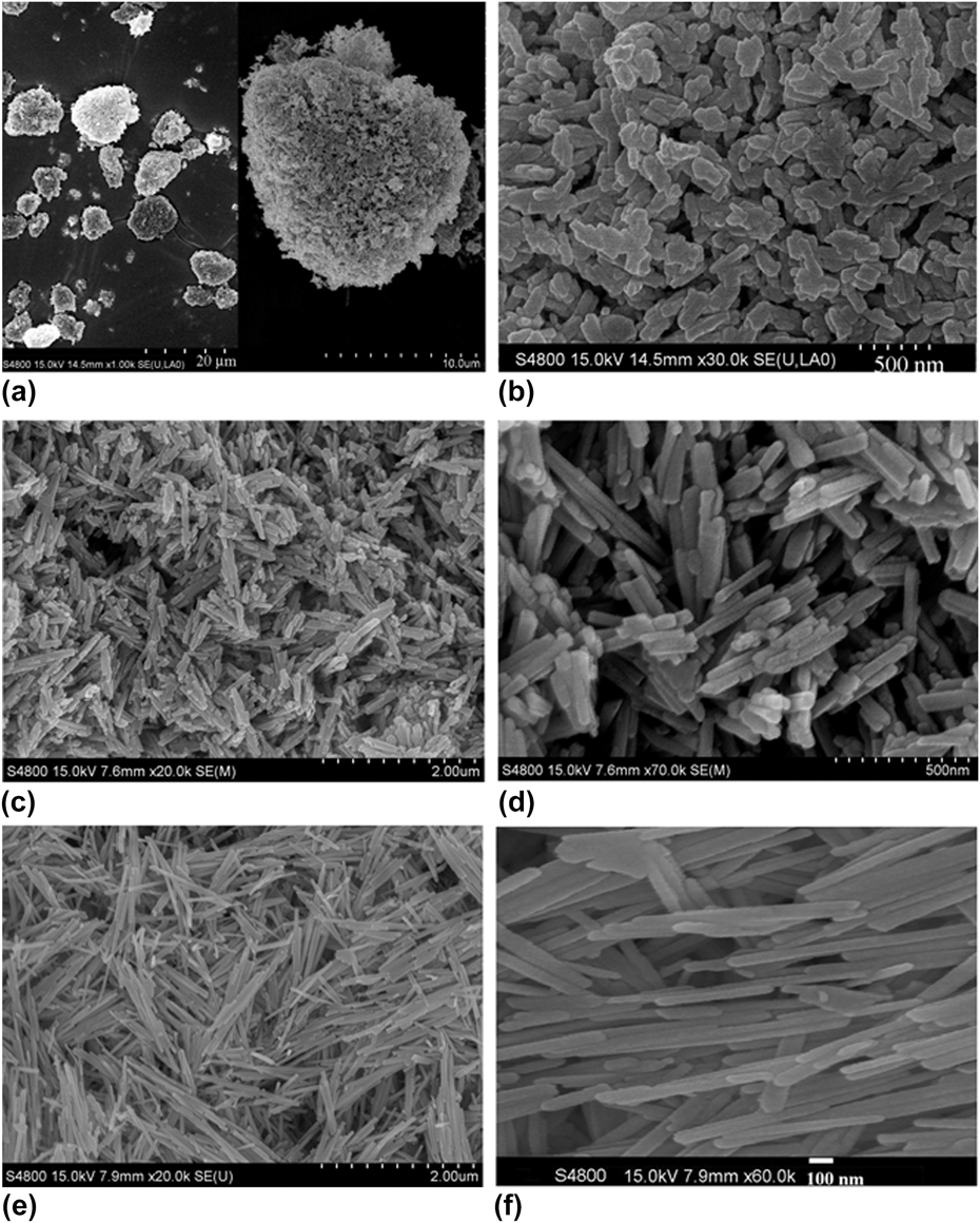
High-resolution scanning electron microscopic (HRSEM) images of the product obtained in propanediol with the assistance of PVP, CTAB, and PEG800 at 187 °C for 6 h are shown in Fig. 1 respectively. The left side of Fig. 1(a) shows that the Bi2S3 particles obtained with the assistance of PVP are composed of nearly circular and elliptical microspheres, the images observed are circular and elliptical shaped, with a diameter of approximately 8.5–15 μm. The image recorded in the right side of Fig. 1(a) is a typical elliptical pattern, which is composed of a number of nonuniform distributed nanorods in clusters (approximately 14–17 μm in diameter).
Field-emission scanning electron microscopy (FESEM) images of the Bi2S3 products synthesized by means of different surfactants (a, b) polyvinylpyrrolidone (PVP), (c, d) cetyl trimethyl ammonium bromide (CTAB), (e, f) polyethylene glycol (PEG)-800, respectively.

However the image recorded in Fig. 1(b) completely displays the fine structure of elliptical microsphere, in which nonuniform rectangular-shaped structure (approximately 300–400 nm long and 125–145 nm wide) can be observed, the average aspect ratio can be estimated as 2.4–2.8. It is clearly observed that these individual rectangular-shaped particles are neither uniformly piled up, nor homoepitaxial growth of one dimensional structures from the center, they clump together to form elliptical clusters under the action of PVP.
The explanation is that PVP with an effective molecular weight of 38,400, have a more rigid structure in comparison to other surfactants used in this article, when the PVP adsorb and bound onto regular surface of growing crystals, the PVP is likely to have more segments in contact with the surface, compared with a flexible polymer.Reference Robb and Smith22 On the other hand, PVP can accept protons either through the O or the N atoms of the pyrrole ring, forming hydrogen bond with other polar compounds, and providing coordination sites for positive ions. In addition, as a result of PVP containing a pyrrole ring with a more rigid structure, the rigid surfactant layer is formed due to restricted orientation of pyrrole ring,Reference Gangulia, Vaidyab and Ganguly21 more segments binding to the surface of growing crystals may hinder bismuth sulfide nuclei growth along the c-axis direction for the one-dimensional structure, this leads to the formation of rectangular-shaped particles with low aspect ratio.
Therefore with the assistance of PVP, only circular and elliptical shaped clusters can form. Due to the presence of van der Waals attraction between the small grains, the grains and small particles clump together to form large grains. The large grain which has higher surface energy is prone to reunite, but the presence of PVP can not effectively reduce surface effects in the nanoparticles, so that the nanoparticles are easily reunited to form the circular and elliptical shaped microspheres.
HRSEM images of the product obtained in propanediol with the assistance of CTAB are shown in Figs. 1(c) and 1(d), the Fig. 1(c) shows that all of the Bi2S3 particles are nanorods. With increasing magnification, the rodlike Bi2S3 nanocrystals (Bi2S3 nanorods) are clearly observed in Fig. 1(d). The average lengths of the long and short axes of nanorods are 310–600 nm and 25–65 nm respectively, with the average aspect ratio being 4.5–12.9. The formation of Bi2S3 nanorods is attributed to the result of the particles being well capped by the cationic surfactants (CTAB).
CTAB is a typical quaternary ammonium surfactant, containing both positively charged head group and long-chain hydrophobic group, which are easily adsorbed on negatively charged solid surface. In aqueous phase, adsorption of CTAB takes place through an ion-pair structure, hence, the cationic surfactant ion can absorb on an oppositely charged solid surface site that is unoccupied by counterions. Due to positively charged head group, the CTAB helps in formation of an assembly of surfactant molecules on the surface of the growing nanorods (bearing negative charge).
Chen et al.Reference Chen, So, Che and Sun23 think that CTAB is easily adsorbed on crystal surface, the CTAB on the surface plays an essential role on distribution in reaction, with its long linear carbon chain preventing the occurrence of reunion. However, due to steric hindrance and electrostatic interactions, the CTAB may form membrane on (001) surface of bismuth sulfide nuclei, this is unfavorable for the growth of bismuth sulfide nanorods with higher aspect ratio along the c-axis direction, leading to formation of shorter nanorods instead. Moreover, rodlike micelles can be formed from the cationic CTAB systems, and the shape of the rodlike micelle promotes the formation of Bi2S3 nanorods from ionic precursors in the cationic CTAB systems.
Unlike the particles obtained in propanediol with the assistance of PVP and CTAB, the Bi2S3 particles obtained in propanediol with the assistance of PEG800 all have longer rodlike morphologies with a uniform distribution, as shown in Fig. 1(e). The observed average lengths of the long and short axes of nanorods in Fig. 1(f) are 1.4–2.1 μm and 41–42 nm respectively, with the average aspect ratio being 34.0–51.5. According to Murphy and Jana,Reference Murphy and Jana19 the longer rodlike particles obtained with the assistance of PEG800 should be considered as nanowires.
In the PEG800 system, it is certain that the TAA plays a critical role in it. TAA, as a complexing agent, has a sulfur functional group, it can coordinate with the propanediol-solvated Bi3+ ions, resulting in formation of bismuth (III) sulfide-complexed clusters, according to Zhang et al. report.Reference Zhang, Ji, Ma, Xu and Yang24 So the crystalline nuclei of Bi2S3 can be formed through pyrolysis of bismuth (III) sulfide-complexed clusters in the early stage of reaction.
It is believed that the Bi2S3 nuclei are microcrystal, therefore, the bonding between the Bi2S3 nuclei and TAA is anisotropic. Accordingly, it is plausible that the dissociation of CH3CSNH2 occurs at local regions of the bismuth (III) sulfide-complexed clusters, where the Bi3+ ions are exposed to the S2− existing in the propanediol solution. Therefore, during the reflux process, the formation of Bi2S3 proceeded along specific directions. In a way, the TAA acted as a “soft template”, leading to the growth of Bi2S3 nanowires.
Due to the hydrogen bonding between the hydroxyl groups, capping agent PEG molecules can form long-chain amphiphilic molecules produced by linking repeating units of ethylene oxide, thus PEG may be self-assembled into rodlike micelles which can act as “soft-template”.Reference Hu, Deng and Huang25 So in the long chain consisting of PEG molecules, Bi2S3 nuclei will be arranged along the direction of chain, much like a number of small particles adsorbed on the chain. With an extension of the reaction time, these Bi2S3 nuclei grow up fast along the direction of chain until they connect to the long strips in the pattern. Simultaneously with the atomic interdiffusion, crystallization proceeds more completely, gradually changing into the Bi2S3 nanowires with a higher aspect ratio.
To verify the formation of bismuth (III) sulfide-complexed clusters before the formation of crystalline nuclei of Bi2S3, the compound of bismuth trichloride with TAA was analog synthesized by solid phase synthesis at room temperature with acetonitrile added as initiator. The actual experiments were performed as follows: 0.9395 g (9 mmol) TAA and 0.9395 g (3 mmol) bismuth trichloride were mixed well together. The mixture was carefully ground in an agate mortar for 30 min and several drops of acetonitrile were added as initiator. At first, the mixture became slightly viscous, indicating that the reaction did happen. Soon after that, the mixture became powdery. Grinding lasted for 8 h, the product was dried in the vacuum at 80 °C for 4 h and then washed with anhydrous methanol until no chloride ion was found in the washing solution. The obtained powder was next dried in the vacuum at 80 °C and finally a complex of TAA with bismuth trichloride was obtained.
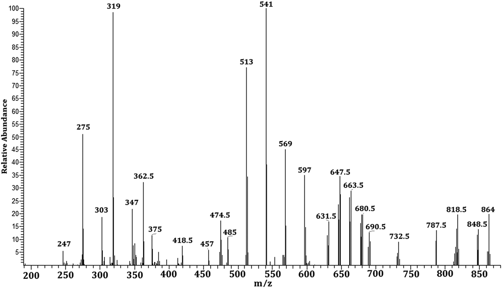
The structural details of the resultant were elucidated by the electrospray ionization mass spectrometry (ESI-MS), as shown in Fig. 2. As concluded from the main peak at m/z = 541 and 319, the species present are assigned to {(S)Bi(μ-S)2Bi(S)-5H+} and {(SC)(S)Bi(μ-S) + 2H+} respectively. Among the major species present, the peaks at m/z = 597, 569, 513, and 275 are assigned to {(S)2Bi(μ-CS)2Bi(S)-5H+}, {(S)Bi(μ-CS)2Bi(S)-H+}, {Bi(μ-S)2Bi(S)-H+}, and {(S)2Bi + 2H+} respectively, the peak at m/z = 864 is assigned to [SC(CH3)=NH]2Bi{μ-(SC(CH3)=NH)}2Bi[SC(CH3)=NH]2+, and is consistent with the composition [SC(CH3)=NH]2Bi{μ-(SC(CH3)=NH)}2Bi[SC-(CH3)=NH]2 (abbreviated as M) of the resultant. Besides, the peaks at m/z = 848.5, 818.5, 663.5, and 647.5 are assigned to [(M-CH3) + H+], [(M-3CH3) + H+], [(M-2CH3-4C(CH3)=NH)-H+], and [(M-2CH3-NH-4C(CH3)=NH)-2H+] respectively.
Electrospray mass spectrum of the resultant.
The finding further demonstrates that TAA can coordinate with bismuth (III) to form [SC(CH3)=NH]2Bi{μ-(SC(CH3)=NH)}2Bi[SC-(CH3)=NH]2 dimeric complexes.
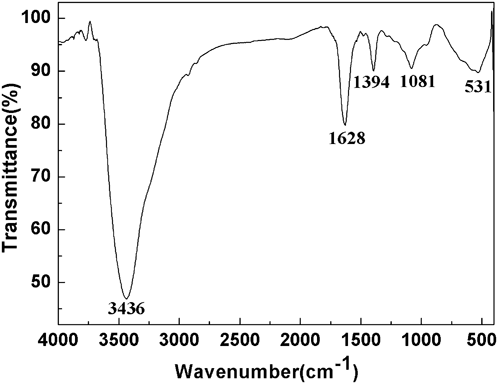
To study coordination properties of the formed dimeric complexes, the IR experiments were performed. As shown in Fig. 3, the IR spectrum of the dimeric complexes displays a medium strong stretching vibration of C=N bond at 1628 cm−1, and a stretching vibration of N–H bond at 3436 cm−1. Besides, the peak around 1394 cm−1 is assigned as a C–H bending vibration in dimeric complexes. It is also found that the peak at 531 cm−1 may be due to the stretching vibration of distinct Bi–S–Bi bridging bond, demonstrating that the TAA coordinate with bismuth (III) to form the dimeric complexes.
The infrared spectrum of the dimeric complexes.
The 1H NMR spectrum of the bismuth (III) sulfide complex can also give another evidence for the interaction between bismuth (III) and TAA. Because TAA is not soluble in CDCl3 and the complex is partially soluble in CDCl3, the 1H NMR spectrum in Fig. 4(a) must be from the H nucleuses in the complex. 1H NMR of the complex in the CDCl3 solution has a strong resonance peak at 7.261 ppm, this is because all the NH groups in ligand SC(CH3)=NH* are equivalent. Moreover, the 1H NMR spectrum of the complex shows several weak peaks within the range of 1.5–1.65 ppm, to observe the these weak peaks, scanning signal accumulation technique was applied to the bismuth (III) sulfide complex, the results are shown in Fig. 4(b).
The proton nuclear of 1H atom magnetic resonance (1H NMR) spectrum of the bismuth (III) sulfide complex.
It can be seen from Fig. 4(b), the 1H NMR spectrum of the bismuth (III) sulfide complex has a resonance peak around 1.58 with the integrated area about 48, probably due to small amount of cluster. The two resonance peaks at 1.253 ppm and 0.814 ppm with the integrated area about 3 may be from the H nucleuses in the CH3 group, however another two resonance peaks at 2.042 ppm and 2.106 with the integrated area about 3 may be also from the H nucleuses in the CH3 group, indicating two CH3 group with different steric configuration. The findings show that probable structure of the cluster is [SC(CH3a)=NH][SC(CH3b)=NH]{Biμ-(SC(CH3)=NH)}14Bi[SC(CH3)=NH]2, in which, CH3a and CH3b are assigned as two CH3 group with different steric configuration.
So when the TAA coordinated with bismuth (III) in the typical experimental procedure, a dimmer of bismuth (III) sulfide complexes should be formed first, the probable structure of the complex for bismuth (III) sulfide can be seen in Scheme 1.
Probable structure of the bismuth (III) sulfide-complexed clusters.
Combined analysis of structure of bismuth (III) sulfide-complexed clusters and morphology of the product obtained in propanediol with the assistance of PEG800, the possible reaction mechanism can be proposed as the following.
i) The formation of dimmer of bismuth (III) sulfide complexes by the coordination reaction between the sulfur sources and the BiIII-propanediol complexes formed at the beginning of the solvothermal process;
ii) Pyrolysis of bismuth (III) sulfide complexes dimmer, and the successive nucleation of Bi2S3 nanocrystals to form crystalline nuclei of Bi2S3 nanowires;
iii) Formation of one-dimensional Bi2S3 nanowires originated from continuous growth of crystalline nuclei along rod-shaped micelles by solid–liquid–solid and Ostwald ripening.
It is well known that the surfactant molecules spontaneously organize into rod-shaped micelles (or inverse micelles) when their concentrations is 10 times more than the critical micelle concentration of the surfactant. These anisotropic structures can be used as soft templates to promote the formation of 1D nanostructured materials when coupled with an appropriate chemical reaction. There are a few functional groups such as S=O, –O–, or C=O on molecules of the surfactant, which are hydrophilic, and they can provide coordination sites for positive Bi3+ ions in propanediol solution, these sites on the molecules of the surfactant provide the necessary heterogeneous nucleation sites. On the other hand, TAA, as a sulfur sources, has a sulfur functional group, which can coordinate with the propanediol-solvated Bi3+ ions, resulting in formation of bismuth (III) sulfide complexes dimmer.
Herein, when temperature is about 120 °C, pyrolysis to Bi2S3 nanocrystals occurs via [SC(CH3)=NH]2Bi{μ-(SC(CH3)=NH)}2Bi[SC(CH3)=NH]2 and by elimination or sequential elimination of C(CH3)=NH and followed by elimination of μ-(C(CH3)=NH). These sulfide complexes dimmer, thus provide a source for Bi2S3 crystalline nuclei.
These crystalline nuclei then continuously grow along rod-shaped micelles by solid–liquid–solid and Ostwald ripening, and finally form one-dimensional nanowires of monocrystalline structure. If surfactant is absent in the reaction system, the small amount of short Bi2S3 nanorods should be obtained. It can be concluded that the anisotropy of Bi2S3 crystal structure plays a key role in the formation of nanowires, the surfactant PEG800 however not only plays a supporting role, but also a stabilizing role in the reaction system.
B. The influence of the different molecular weight PEG on the Bi2S3 morphology and structure
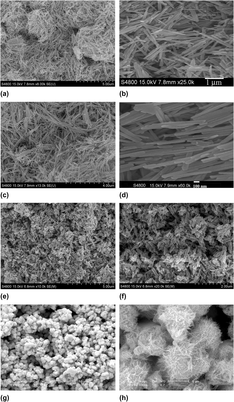
As can be seen from Fig. 5, the morphologies of the product obtained in propanediol in the presence of PEG with different molecular weights are significantly different. PEG as a macromolecular dispersant (general formula HO(CH2CH2O)n-H), which is mainly composed of ethylene oxide repeating units, originates from the chain polymerization of ethylene oxide. Just because of this, the PEG has a good solubility in water. The polymer molecules bear only one hydrophilic “head” group hydroxyl (OH) in the entire molecular chain, the number of functional groups significantly decreases with an increase of molecular weight, and the ability in reducing surface tension of solvent decrease accordingly. Moreover, with an increase of molecular weight, the degree of a chain crosslinking increases, and then hard to soft segment ratio increases accordingly, the effect of the hard part in the polymer becomes stronger and more distinct,Reference Dung26 this leads to the polymer molecules becoming more rigid. So with an increase of molecular weight, more rigid segments binding to the surface of growing crystals may hinder bismuth sulfide nuclei growth along the c-axis direction, leading to the formation of nanorods with low aspect ratio.
FESEM images of the products using (a, b) PEG400, (c, d) PEG800, (e, f) PEG4000, (g, h) PEG20000 as surfactants.
HRSEM images of the product obtained with the assistance of PEG400 are shown in Figs. 5(a) and 5(b), the Bi2S3 particles all have wirelike morphologies with a uniform distribution, the observed average lengths of the long and short axes of nanowires in Fig. 5(b) are 2–3 μm and 40–41 nm respectively, with the average aspect ratio being 50–75.
In contrast to Figs. 5(a) and 5(b), the images recorded in Figs. 5(c) and 5(d) show that the Bi2S3 particles obtained with the assistance of PEG800 all have wire-like morphologies, but the average aspect ratio changes from 50–75 into 34.0–51.5 with the PEG400 being replaced by PEG800. Because PEG400 is “softer” than the PEG800, leading to the anisotropic growth of crystals more easily.
When the PEG4000 was used in place of PEG800 in the typical experimental procedure, Figs. 5(e) and 5(f) show that all of the Bi2S3 particles are nanorods with a nonuniform distribution, the observed average lengths of the long and short axes of nanowires in Fig. 5(f) are 500 nm and 80 nm respectively, with the average aspect ratio being 6.25. Because PEG4000 is more “rigid” than the PEG800, leading to the formation of nanorods with low aspect ratio.
The growth of Bi2S3 crystal includes the special solid–liquid–solid phase transformations and Ostwald ripening process, in which the surfactant plays a key role.Reference An, Wang and Liu27 The PEG polymer, as a surfactant, can also play a critical role in capping agent.Reference Veliotti28 As a result, PEG is able to kinetically control the growth rates of Bi2S3 nanorods or nanowires by interacting with the surface of one-dimensional high-energetic Bi2S3 nanorods through adsorption and desorption. In comparison with PEG800, PEG4000 has a longer chain, it is able to absorb and cap Bi2S3 crystal rods with a larger size, leading to Bi2S3 nanorods growing faster than that with PEG800 as surfactant, and causing nonuniform size distribution of nanorods.
When the PEG20000 was used in place of PEG800 in the typical experimental procedure, the Bi2S3 products are made up of spherical budlike microspheres with chainlike arrangement [Fig. 5(g)]. With increasing magnification, it is observed that the morphologies of spherical budlike microspheres display sproutslike pattern, and they are composed of a number of uniform distributed nanorods in clusters (approximately 7.3–10 μm in diameter), as shown in Fig. 5(h). The explanation is that PEG20000 with an effective molecular weight of 20,000 has a more rigid structure in comparison to PEG800, and PEG4000 surfactants. The rigid chain of PEG20000 binding to the surface of growing crystals may hinder bismuth sulfide nuclei growth along the c-axis direction for the one-dimensional structure, leading to formation of spherical budlike microspheres.
Besides, PEG20000 with long-chain structure, has different surfactant-active from PEG800, and become tangled and reunited easily, also leading to formation of spherical budlike microspheres with the diameter about 10 μm.
C. The influence of solvent medium on the Bi2S3 morphology and structure
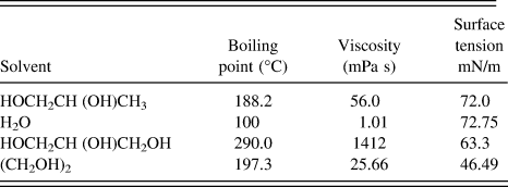
It is believed that the solvent should have the proper boiling point, viscosity, and surface tension, as can be seen in Table I, such physical and chemical properties can influence the solubility, reactivity, and diffusion behavior of the regents and the intermediate.Reference Liu, Liang, Li, Peng and Qian29 In our typical experimental system, to study the influence of solvent on the growth process of Bi2S3, the solvent propanediol was changed with distilled water, glycerol, and ethylene glycol respectively with all the other parameters of the typical reaction unchanged. The experimental results show that the boiling point and viscosity of the solvent used play an important role on the formation of uniform Bi2S3 nanowires.
The physical parameters of the solvent medium.
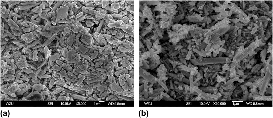
From Table I, it can be seen that the water has the lowest boiling point and viscosity, while the glycerol has the highest boiling point and viscosity among the solvents used in this study described by this article; however the propanediol and the water all have an almost identical surface tension, the largest value among the four surfactants used in experiment, and ethylene glycol has the least surface tension. So in our typical experimental system, when the water was used as solvent instead of propanediol, pure bismuth sulfide phase cannot be acquired. With glycerol as solvent, although the pure phase of orthorhombic type of Bi2S3 can be obtained, the HRSEM images exhibit a majority of uneven-sized blocks with a small amount of nanorods, as shown in Fig. 6(a). Zhu et al.Reference Zhu, Bian, Wang, Li and Chen30 think that the solvent viscosity and surface tension may be an important factor to affect morphologies of the product. Glycerol is one of the most viscous organic solvents, its viscosity is much larger than that of ethylene glycol and water (shown in Table I), which is not beneficial to the dispersion of Bi2S3 nanorods, therefore, well-dispersed uniform Bi2S3 nanorods cannot be formed in the presence of glycerol solvent. The ethylene glycol has an appropriate viscosity with the least surface tension, which is beneficial to formation of well-dispersed uniform Bi2S3 nanorods, but it has a higher boiling point, leading to the reaction system requiring too high temperature.
FESEM images of the products obtained by TAA-assisted reflux at 187 °C in different solvents: (a) Glycerol and (b) Ethylene glycol.
So in our typical experimental system, when the ethylene glycol was used as solvent instead of propanediol, the HRSEM images recorded in Fig. 6(b) exhibit a majority of nonuniform sized nanorods with a small amount of irregular agglomeration of rod-shaped particles. The solvent propanediol employed in our typical experimental system has a moderate viscosity and large surface tension, since its boiling point is lower than that of ethylene glycol, it can not only provide an appropriate temperature to form Bi2S3 crystal with good crystallinity, but also a well-dispersed liquid solvent medium for formation of well-dispersed uniform Bi2S3 nanowires [shown in Fig. 1(f)].
D. The influence of sulfur source on the Bi2S3 morphology and structure
To study the influence of sulfur source on the Bi2S3 morphology and structure, the Bi2S3 particles were first prepared in the typical experimental procedure, and second the Bi2S3 particles were prepared by changing TAA with thiourea and thiosemicarbazide respectively in our typical experimental system. The crystal structure and phase composition of the products were characterized by XRD, energy dispersive spectrometer (EDS), and XPS.
1. Characterization of the product prepared in the typical experimental procedure
X-ray diffraction, EDS, and XPS were adopted to analyze the crystal structure and phase composition of the products.
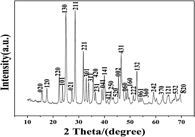
The XRD patterns of the product prepared at 187 °C for 6 h are recorded and shown in Fig. 7. The XRD patterns all display sharp peak with strong diffraction intensity, and low background. It is found that the positions and relative intensities of the reflection peak of Bi2S3 nanowires agree well with standard diffraction card i.e., Joint Committee on Powder Diffraction Standards (JCPDS) 17-320 (Bi2S3), which demonstrates that pure Bi2S3 nanowires can be obtained under the current conditions.
XRD patterns of Bi2S3 nanowires prepared at 187 °C for 6 h by reflux method.
The results of indexing the powder XRD pattern show that all the diffraction peaks in the pattern can be readily indexed by one set of lattice parameters. The relative deviation between the calculated D and experimental D 0 is less than 0.086‰, which indicates that the synthesized products are of a single phase with orthorhombic structure. The crystal structure of synthesized product belongs to the orthorhombic system.
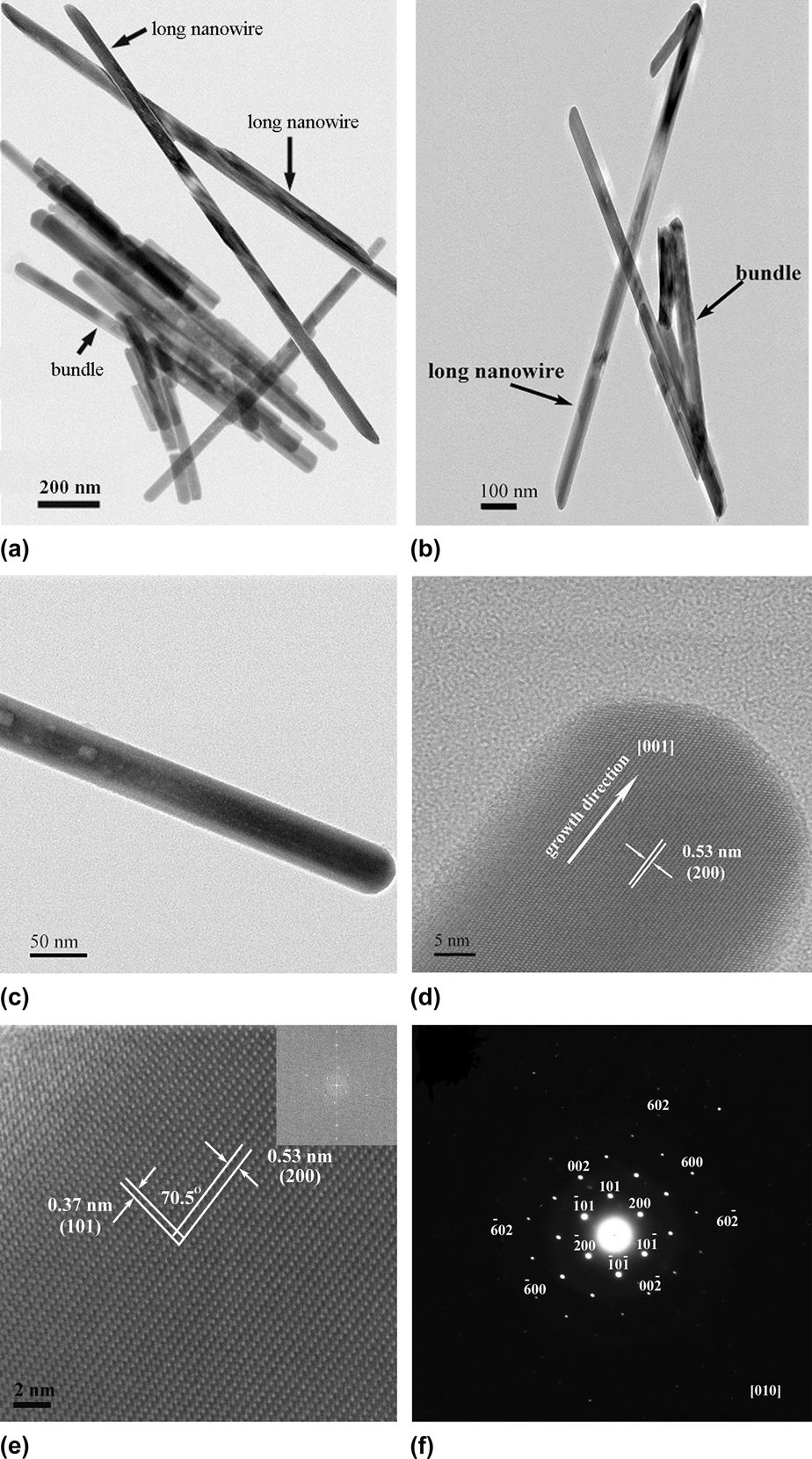
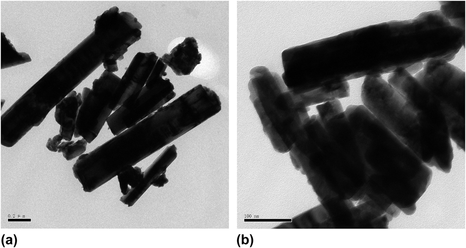
Using BiCl3 and TAA as the raw materials, the representative HRTEM images of the samples obtained after being refluxed at 187 °C for 6 h are shown in Fig. 8. Obviously, long Bi2S3 nanowires with lengths ranging from 1.58 to 1.75 μm and with uniform width of about 40 nm can be seen in Figs. 8(a) and 8(b), some bundles of Bi2S3 nanowires are also observed in Figs. 8(a) and 8(b), closely consistent with those depicted in Fig. 1(f). To achieve further insight into these nanowires, HRTEM images were taken from an individual Bi2S3 nanowire, as recorded in Figs. 8(c) and 8(d). We can see that it crystallizes in a single-crystal orthorhombic structure, showing good crystallinity and clear lattice fringes. As we know, in the orthorhombic system, the parameters a, b, c, crystal indices (h, k, l) and interplanar spacing d, interfacial angle ф have the following two equations,31
 $${1 \mathord{\left/ {\vphantom {1 {{d^2}}}} \right. \kern-\nulldelimiterspace} {{d^2}}} = {{{h^2}} \mathord{\left/ {\vphantom {{{h^2}} {{a^2}}}} \right. \kern-\nulldelimiterspace} {{a^2}}} + {{{k^2}} \mathord{\left/ {\vphantom {{{k^2}} {{b^2}}}} \right. \kern-\nulldelimiterspace} {{b^2}}} + {{{l^2}} \mathord{\left/ {\vphantom {{{l^2}} {{c^2}}}} \right. \kern-\nulldelimiterspace} {{c^2}}}\quad ,$$
and
 $$\cos \;\Phi = {d_1}{d_2}\left( {{{{h_1}{h_2}} \mathord{\left/ {\vphantom {{{h_1}{h_2}} {{a^2}}}} \right. \kern-\nulldelimiterspace} {{a^2}}} + {{{k_1}{k_2}} \mathord{\left/ {\vphantom {{{k_1}{k_2}} {{b^2}}}} \right. \kern-\nulldelimiterspace} {{b^2}}} + {{{l_1}{l_2}} \mathord{\left/ {\vphantom {{{l_1}{l_2}} {{c^2}}}} \right. \kern-\nulldelimiterspace} {{c^2}}}} \right)\quad .$$
(a–c) TEM images, (d, e) HRTEM images, and (f) SAED pattern of Bi2S3.
The results of indexing to the powder XRD pattern give the parameters a = 1.0813 nm, b = 1.1258 nm, c = 0.3940 nm. HRTEM image recorded in Fig. 8(d) shows 200 lattice fringe images on the top of the single Bi2S3 nanowires, the calculated value of the two adjacent spacing is 0.55 nm, which is consistent with the measured value 0.53 nm of orthorhombic Bi2S3 (200) crystal face reported in the literature.Reference Zhang and Chen32
Figure 8(e) is the Fast Fourier Transform photo of Fig. 8(d), the observed fringes of 0.77 nm correspond to the distance between the (101) planes of Bi2S3 nanowires, consistent with the calculated values. The interfacial angle ф between the (200) and (101) plane was measured about 70.5°.
The SAED pattern [Fig. 8(f)], taken from the same Bi2S3 nanowires, shows its single crystal nature and reveals that the corresponding crystal zone axis is [010], the result indicates that Bi2S3 nanorods have [001] directional preferred growth, in agreement with XRD analysis presented above.
Crystal growth behavior is mainly determined by the internal structure of crystals, and complies with the law of the crystal growth under ideal conditions of no external interference. According to the literature,Reference Yu, Jin, Wang, Chen and Peng33 Bi2S3 have a lamellar structure with linked Bi2S3 units forming infinite chains parallel to the c-axis ([001] direction), because the inherent lamellar structure of Bi2S3 determined its preferential growth direction along the [001] zone axis (c-axis) direction. This indicates that strong directional covalent bonds acting perpendicular to the c-axis, can induce Bi2S3 grow along the c-axis at higher speeds.Reference Yu, Jin, Wang, Chen and Peng33 Moreover, it is generally know, Bi2S3 has the strongest chemical interactions among the intrachain S–Bi chemical bonds, but the relatively weak intrachain interaction in each lamella, which leads to the weakest interlayer (van der Waals) interactions between lamellas of layered Bi2S3. Thus it is possible to think that the formation of one-dimensional Bi2S3 nanowires may have originated from the cleavage of lamellar Bi2S3 at the van der Waals planes.
The purity and composition of the as-prepared sample are reflected by EDS analysis. No obvious impurities were detected in the EDS spectrum of Bi2S3 [Fig. 9(a)]. Quantification of the EDS peaks shows the molar ratio of Bi:S from the peak areas is 59.54:40.46 (1.47:1), which is consistent with the stoichiometric composition of Bi2S3. The formation of Bi2S3 is further proved by XPS analysis [Figs. 9(b) and 9(c)], the atomic percentage of Bi and S elements in the sample were determined. Each of components was calculated by integration, the integral image of characteristic spectrum of each Bi element in two components Bi–S and Bi–O can be seen in Fig. 9. The two strong peaks at 158.5 eV and 163.7 eV can be attributed to Bi 4f7/2 and Bi 4f5/2 respectively. The peak at 225.3 eV, appearing in the inset of Fig. 9(b), can be assigned to the S 2s transition. The obtained binding energy values are consistent with those reports,Reference Lu, Han, Yang, Lu and Wang34 indicating the as-prepared sample is composed of one kind of component Bi (III). Then taking into account the atomic sensitivity factors of S and Bi, the molar ratio of Bi-to-S is approximately 2:3 according to quantification of the peak areas of Bi 4f and S 2p, which indicates the component of the as-prepared sample is Bi2S3. It is also found that no peaks of other elements are observed in the wide XPS spectrum except Bi, S, C, N, and O, indicating the high purity of the Bi2S3 product.
(a) EDS analysis of Bi2S3 nanocrystals, (b) XPS survey spectrum of Bi2S3, (c) High-resolution XPS core spectrum for Bi 4f.
2. The influence of sulfur source on the Bi2S3 morphology
Figure 10 shows TEM images Bi2S3 prepared using different sulfur source, when the sulfur source TAA was changed with thiourea and thiosemicarbazide respectively in our typical experimental system, though Bi2S3 nanorods can be obtained, uneven particle size distribution was formed and large-sized microparticles were produced. This indicates that sulfur source in our experimental system has an important influence on the morphology and structure of the Bi2S3 products. When thiourea, thiosemicarbazide and TAA were selected as a sulfur source respectively, crystal structure of the Bi2S3 nanorod may undergo a different formation mechanism, and thus different sized microparticles were produced.
TEM images Bi2S3 prepared using different sulfur source: (a) Thiourea, (b) Thiosemicarbazide.
The former two undergo thermal decomposition through the intermediate formation of the Bithiourea complexed clusters and Bithiosemicarbazide complexed clusters at reflux. While the letter was considered to proceed via a two-step pathway, first, the pyrolysis occurs through the cleavage of the carbon–sulfur bond in Bi-TAA complexed clusters at reflux; second, the formed Bi2S3 crystalline nuclei continue to grow along rod-shaped micelles by solid–liquid–solid and Ostwald ripening to form one-dimensional Bi2S3 nanorods. Besides, our experimental system does proceed through refluxing the solvated Bi3+ ions and sulfur resources in the nonaqueous solvent, during the S2− generation reaction, three kinds of characteristic sulfur sources all undergo thermal decomposition or pyrolysis.
When thiourea and thiosemicarbazide are heated to a temperature above their melting point at 180–182 °C in a solvent, decomposition of thiourea and thiosemicarbazide molecules is accompanied by S2− generation reaction respectively. In comparison to the thiourea and thiosemicarbazide, TAA has a lower pyrolysis temperatures, the pyrolysis of TAA to generate S2− occurs at approximately 120 °C, therefore selection of precursor with different precursor structure often leads to ligands decomposition in different temperature regimes, and thus a different crystal growth rate to form Bi2S3. The uniformity in growth morphology of Bi2S3 products has something to do with the generation rate and pathway of S2− ion from sulfur resources, and the temperature for formation of Bi2S3 products, as a result, an appropriate generation rate and spatial orientation, as well as low thermal decomposition temperature are beneficial to formation of Bi2S3 nanorods.
When the Bi3+ ions complexes based on thiourea or thiosemicarbazide ligands were taken as precursor, S2− generation reaction requires relatively high thermal decomposition temperature; on the other hand, in the sulfur-alkane structure of thiourea and thiosemicarbazide ligands, S atom has a lone pair electron, which can coordinate with Bi3+ directly to form the complexes with low stability. This leads to generation rate of S2− becoming large, thus the larger the thermal generation rate of S2−, the larger speed the crystalline nuclei of Bi2S3 should form by, and finally the large-sized Bi2S3 nanorods with the less uniform distribution are obtained [shown in Figs. 10(a) and 10(b)].
Because of the sulfur-alkane structure of TAA easily changing into the resonance structure of allyl mercaptan with relatively strong nucleophilicity through the resonance, the TAA coordinates to Bi3+ ions to form bismuth (III) sulfide complexes dimmer with relative stability in the form of allyl mercaptan structure (shown in Scheme 2), resulting in low generation rate of S2−. On the other hand, the bismuth (III) sulfide complexes dimmer also has a certain spatial orientation, these two factors are beneficial to formation of uniform long Bi2S3 nanowires [shown in Figs. 8(a) and 8(b)].
The formation of dimmer of bismuth (III) sulfide complexes.
Pyrolysis of bismuth (III) sulfide complexes dimmer.
E. UV–visible absorption spectroscopy and fluorescence spectroscopy
1. UV–visible absorption spectroscopy analysis
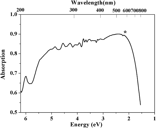
The optical properties of the as-prepared Bi2S3 nanowires are determined by the UV–visible absorption spectra (Fig. 11), which provide a simple and effective method for explaining some features concerning the band structure of nanomaterials.Reference Chen and Sun35 The corresponding band gap values are obtained by the intersection point of the tangent of the absorption edge with extended line of the absorption at lower wavelengths. There is absorption band edge one shoulder peak of 2.17 eV (signed with “*”) that is due to the nanowires, which is 0.87 eV higher than the reported value in the literature bulk Bi2S3 (1.3 eV) material.Reference Variano, Hwang, Sandroff, Wiltzius, Jing and Ong36
UV–vis spectra of the Bi2S3 nanowires synthesized using TAA as sulfur source.
Our Bi2S3 nanowires are mainly about 40 nm × 1.58–1.75 μm in size (according to the analysis of HRTEM image), and we do not expect the corresponding E g to be very different from that of bulk materials. The wider optical band gap of Bi2S3 nanowires may be caused by the quantum size effect, because Bi2S3 nanowires can display a wide range of optical features in the UV–visible region, the experimentally determined E g values of Bi2S3 nanorods are near to the optimum for photovoltaic conversion, suggesting that they may be very promising for applications in solar energy, fluorescent chemical sensors, and photoelectronics.
2. Fluorescence spectroscopy analysis
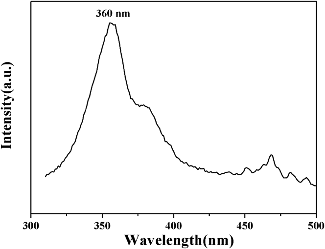
Figure 12 is the PL spectra of Bi2S3 nanowires prepared at 187 °C for 6 h by reflux method, upon excitation with 310 nm light, the Bi2S3 nanowires have an absorption edge of approximately 360 nm (3.44 eV). Comparing with that of bulk material, it is found that there is a blue shift in the absorption peak. Therefore, there is an increased energy gap as compared with the bulk material, the increase in the optical band gap with decreasing the particle size, can be explained by the quantum size effect qualitatively. And the problem would arise from their larger specific surface, higher surface activity and quantum confinement effect.Reference Yu, Cao and Zhu37
PL spectra of Bi2S3 nanowires.
IV. CONCLUSION
In summary, pure single-crystalline Bi2S3 nanowires with lengths ranging from 1.58 to 1.75 μm and with uniform width of about 40 nm were successfully prepared a simple surfactant-assisted reflux method in the presence of the surfactant PEG800 at 187 °C for 6 h. The results of indexing the XRD show that the product is in the orthogonal Bi2S3 phase, the cell parameters are a = 1.0813 nm, b = 1.1258 nm, c = 0.3940 nm. The TAA can not only act as the sulfur source for Bi2S3 nanowires, but also as a “soft template”, directing the formation of anisotropic Bi2S3 crystalline nuclei, which can continuously grow into uniform 1D Bi2S3 nanowires along rod-shaped micelles using PEG-800 templating. When molecular weight of PEG increases from 800 to more than 4000, the degree of a chain crosslinking increases, leading to the morphologies of the product changing from nanowires into nanorods and spherical budlike microspheres. The different sulfur resources have a significant influence on the generation rate and pathway of S2− ion, and thus lead to formation of Bi2S3 nanorods with different uniformity. UV–vis, PL spectra of Bi2S3 show 1D Bi2S3 nanowires have a wider optical band gap, and a blue shift in the absorption peak, indicating Bi2S3 nanowires have excellent optical properties.
ACKNOWLEDGMENTS
We acknowledge the financial supports of National Natural Science Foundation (Grant Nos. 50772075 and 20971043), the Open Project Program of State Key Laboratory of Inorganic Synthesis and Preparative Chemistry, Jilin University.