Introduction
Photodynamic therapy (PDT) is a promising noninvasive localized treatment modality for a diverse range of diseases [Reference Abrahmse and Hamblin1, Reference Avci, Erdem and Hamblin2, Reference Calixto, Bernegossi, Freitas, Fontana and Chorilli3]. A nontoxic dye, called a “photosensitizer” (PS), capable of transferring light energy to other molecules, is administered to the patients with subsequent exposure to a light source of a specific wavelength that leads to the death of the target cell via oxidative damage [Reference Mroz, Hashmi, Huang, Lange and Hamblin4]. PDT combines a photoactivatable PS, light, reactive oxygen species (ROS), and singlet oxygen (1O2), which leads to the death of cells [Reference Lim, Lee, Zhang and Chu5].
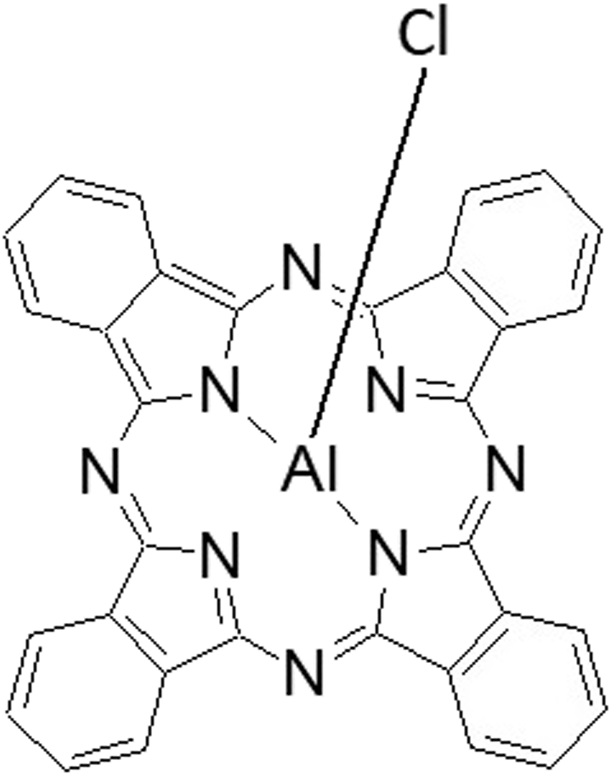
Chloroaluminum phthalocyanine (ClAlPc), which has an aluminum(III) atom coordinated at the central position of the Pc macrocycle (Fig. 1), is a second-generation PS. A remarkable property of ClAlPc is its capability of producing at high quantum yields relatively long-lived singlet and triplet oxygen states. However, ClAlPc is hydrophobic, and thus, it requires advanced technologies to formulate nanostructured delivery systems to promote its dispersion in a physiological environment, improve the specific uptake by targeted tissues, and enhance PDT efficiency [Reference Py-Daniel, Namban, Andrade, Souza, Paterno, Azevedo and Soler6].
Chemical structure of chloroaluminum phthalocyanine.

Due to the hydrophobic nature of phthalocyanine, the drug delivery systems associated with these PSs require complex systems, such as liposomes [Reference Silva, Franchi and Tedesco7, Reference Hinger, Gräfe, Navarro, Spingler, Pandiarajan, Walt, Couffin and Maake8], nanoemulsion [Reference Castilho-Fernandes, Lopes, Primo, Pinto and Tedesco9], nanocapsules [Reference Souza, Ziembowicz, Müller, Lauermann, Kloster, Santos, Lopes, Ourique, Machado and Villetti10], poly(D,L-lactide-co-glycolide) nanoparticles [Reference Simioni, Primo and Tedesco11], and gelatin nanoparticles [Reference Carvalho, Abreu, Ferreira, Gonçalves, Tedesco, Pinto, Ferreira-Strixino, Beltrame-Junior and Simioni12].
Recently, decomposable colloidal particles were introduced as templates for multilayer formation for entrapped bioactive compounds [Reference Pinheiro, Bourbona, Cerqueira, Maricato, Nunes, Coimbra and Vicente13]. First, a tailored polyelectrolyte film is formed on the colloid by electrostatic attraction between oppositely charged species by a technique known as layer-by-layer assembly. Once the film is assembled, a chemical treatment (pH change, oxidation reaction, action chelating agent, etc.) is applied to dissolve the core, leading to hollow spheres [Reference Cuomo, Lopez, Miguel and Lindman14].
Crystallization of manganese carbonate (MnCO3) from supersaturated solution has been the subject of many studies because of applications in catalysis, drug delivery, and controlled release. The porous nature of the MnCO3 microparticles that could be used as decomposable templates to obtain polyelectrolyte spheres offers yet another interesting way to obtain a matrix consisting of a polyelectrolyte complex within the sphere’s interior having a similar chemistry to that of the shell. When the polyelectrolyte multilayer shell is built around the MnCO3 core by the Lbl protocol and core is extracted with acid solution and EDTA, the result is hollow polyelectrolyte spheres (HPSs) [Reference Johnston, Cortez, Angelatos and Caruso15, Reference Liu and Xiaorui16].
The adsorption of biomacromolecules inside the manganese carbonate particles is presumably regulated by electrostatic interactions on the microparticle surface within pores and biomacromolecule–biomacromolecule interaction [Reference Sukhorukov, Volodkin, Günther and Moehwald17].
In a recent study, microsphere design via a manganese carbonate hard template for entrapped silicon(IV) phthalocyanine was reported [Reference Simioni, de Jesus and Tedesco18]. Adsorption of chloroaluminum phthalocyanine (ClAlPc) in MnCO3 microparticles, such as a sacrificial template for fabrication HPS, has not yet been reported in the literature.
The main goal of this study was to prepare and evaluate the potential application of ClAlPc entrapped in HPSs. The HPSs produced were characterized by scanning electron microscopy (SEM). To evaluate whether drug efficacy would be preserved, studies using a steady-state spectroscopic technique were conducted. In sequence, in vitro studies were performed with and without light, as described by the PDT protocols of neoplastic macrophages cells (J774A-1), used as a biological model.
Results and discussion
Fabrication of microcapsules
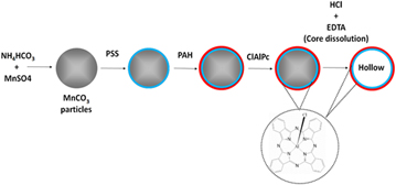
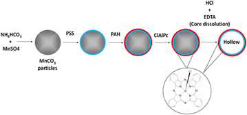
An approach to fabricate matrix-type polyelectrolyte microspheres and a technique of PS encapsulation into the spheres were developed. The scheme illustrating this method is depicted in Fig. 2.
Scheme of HPS fabrication and encapsulation of photosensitizer (ClAlPc) in the spheres.

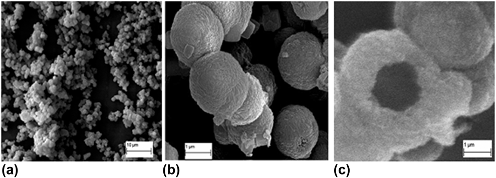
According to the scheme, the porous inorganic MnCO3 microparticles were fabricated by mixing two salt solutions at appropriate conditions. These particles were used as sacrificial templates for sphere fabrication using the Lbl method with PSS and PAH as the polyelectrolytes. The MnCO3 cores with a multilayer coating comprising {PSS/PAH}6 were then dissolved by exposure to HCl and EDTA solution, followed by rinsing with DI water. The coated MnCO3 particles and the spheres following the core dissolution were imaged with SEM (Fig. 3). The method used for fabrication of porous MnCO3 consisted of uniform, homogeneous-size, and nonaggregated microparticles. It is believed that the homogeneous isotropic crystal growth on tiny crystal nuclei yields a spherical shape and uniform distribution of the MnCO3 particles under strong stirring [Reference Jordens, Coker, Gielen, Gerven and Braeken19].
SEM images: (a) MnCO3 microparticles; (b) MnCO3 microparticles in overview, and (c) broken particles (HPS without PS).

As described in the preceding section, the capsule interior has a matrix-type structure, composed of the complex of polyelectrolyte pairs used for the Lbl assembly. When these polyelectrolyte capsules were exposed to a PS, the capture and spontaneous accumulation of the PS occurred within the capsule interior. This phenomenon was demonstrated by Volodkin et al. [Reference Volodkin, Petrov, Prevot and Sukhorukov20] when polyelectrolyte capsules were exposed to a macromolecule (bovine serum albumin). Furthermore, removal of the dissolved cores provides more space for material encapsulation, and the polyelectrolyte coating may function as a protective shell for the encapsulated species [Reference Kim, Choi, Ra, Park, Kim and Lee21].
The prepared inorganic microparticles had an average size of 2.5 µm [Figs. 3(a) and 3(b)]. After coating, MnCO3 template cores were decomposed, as described in section “Results and discussion”, producing HPS–ClAlPc.
Physicochemical characterization of HPS
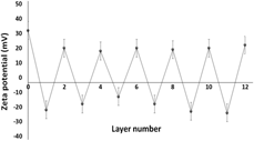
Following the production of MnCO3 particles, the common polyelectrolyte pair PSS/PAH was used to deposit multilayers on the inorganic template. The particles were coated with six bilayers of {PSS/PAH}. The deposition of the layers was controlled by monitoring the zeta-potential after the successive deposition (six layers) of oppositely charged polyelectrolytes. Six bilayers of {PSS/PAH} were deposited on MnCO3 cores, and the surface potential of the microspheres was observed to change regularly from 30 mV for MnCO3 cores and +20 mV for PAH to −22 mV for PSS, indicating the formation of a {PSS/PAH}6 wall architecture (Fig. 4).
Zeta-potential as the function of layer-by-layer deposition of polyelectrolytes.

PSS was chosen as the first layer, despite the slightly negative charge that was present on the particle surface. This procedure was adopted to induce a higher negative charge on the template’s surface, since PSS has surface-active properties and can adsorb onto surfaces with either positive or negative charges [Reference Wu, Xu, Sun, Fu, He and Matyjaszewski22]. The deposition of PSS imparted a higher negative charge to the particles, as expected. The subsequent adsorption of PAH and PSS layers yielded a reversal of the zeta-potential in accordance with the charge of the polyelectrolyte used in each layer. This alternation of zeta-potential is indicative of multilayer growth [Reference Irigoyen, Moya, Iturri, Llarena, Azzaroni and Donath23, Reference Ghostine, Markarian and Schlenof24] with the subsequent deposition of cationic PAH and anionic PSS, suggesting the stepwise formation of alternating PSS and PAH layers on the particles.
UV-vis absorption
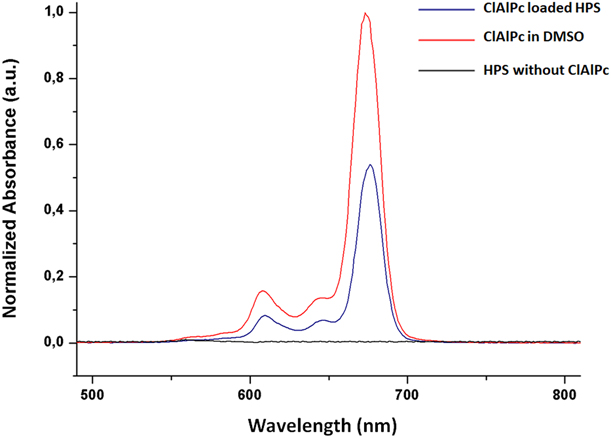
The choice of PS is critical for successful treatment with PDT. The PS should be preferably excited by light of a wavelength in the therapeutic window (600–700 nm), which has greater capacity for tissue penetration. Pure HPSs and ClAlPc in HPS were analyzed by UV-visible absorption spectroscopy. This technique provided physical characterization of the presence of ClAlPc in HPSs as in Fig. 5. Bronze-Uhle et al. [Reference Bronze-Uhle, Costa, Ximenes and Lisboa-Filho25] used UV-visible absorption spectroscopy to show the presence of salicylic acid in albumin nanoparticles.
Absorption spectra of ClAlPc (1 µmol/L) in: DMSO (–), HPS without PS (–), ClAlPc-loaded HPS (–).

The incorporation of the drug into an HPS causes a red shift of the wavelength of maximum absorption at 674 nm (Δλ = 2 nm). A shift in the Q band at I = 674 nm was due to the formation of HPS, but no new Q band was observed. This behavior has frequently been observed and previously described [Reference Sibata, Tedesco and Marchetti26, Reference Silva, Simioni and Tedesco27] for many porphyrin and phthalocyanine dyes incorporated into microheterogeneous media and might also be due to the coating of the drug on the surface of hollow microspheres.
ClAlPc exhibits a strong absorbance in the red region, with a maximum wavelength at 674 nm. The red absorption band allows the drug to be absorbed in the region where there is maximal tissue penetration (600–800 nm, therapeutic window), which should increase the efficiency of cell damage and tumor response to the phototreatment [Reference Hill, Linder, Davies, Sawada, Morgan, Ohulchanskyy and Detty28]. Furthermore, it presents a group of Q bands in the region from 600 to 710 nm and a Soret band at 340 nm. The Q bands represent the vibration levels referring to the first and second electronically excited states. The Soret band is a group of ten electronic transitions with different spins and dipole moment sizes [Reference Stradomska and Knoester29]. The strong absorption regions ranging from 600 to 710 nm (Q band) are associated with the excitation between the HOMO-A1g ground state [(a1u2)(π)] and the degenerate LUMO-[eg(π*)] first excited singlet state, which has Eu symmetry from the (a1u1eg1) configuration. The other band in the UV regions at 340 nm (B band) can be attributed to the excitation between the 1a2u(π) and the 1eg(π*) states. The two characteristic bands mean that the PC ring formed in metal phthalocyanine [Reference Sakamoto and Ohno-Okumura30].
However, the strong π–π interactions between the two aromatic cores of neighboring molecules mean the PS forms dimer and higher-order aggregates both in organic solvents and especially in aqueous media. This aggregate formation leads to significant reduction in ROS formation due to self-quenching of the excited state [Reference Gerhardt, Lewis, Kliger, Zhang and Simonis31]. These properties may also prevent encapsulation into HPSs, and PS encapsulated into HPS may induce aggregation of PS [Reference Gjuroski, Furrer and Vermathen32, Reference Tang, Habimana-Griffin, Lane, Egbulefu and Achilefu33]. PS aggregates in solution exhibit distinct changes in their absorption bands when compared with their monomeric counterparts. From these observed spectral shifts, various aggregation patterns of the dyes in various media have been proposed. The bathochromically shifted J bands and hypochromically shifted H bands of the aggregates have been explained in terms of the molecular exciton coupling theory, i.e., the coupling of transition moments of the constituent dye molecules [Reference Eisfeld and Briggs34]. Thus, if dimeric ClAlPc was present in the medium, changes in the drug’s spectral profile would have been observed, and this did not happen. In this study, there was no band shift in the spectra in the concentration range studied, suggesting that the PS is in a monomeric state.
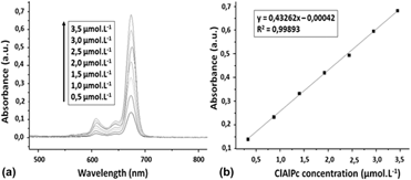
The amount of PS adsorbed on the particles was determined by UV-vis spectroscopy by recording a calibration curve (Fig. 6). The linearity of an analytical method can be defined as the ability to obtain test results that are directly proportional to the concentration of the analyte. Linear regression analyses were carried out by plotting the absorbance intensity versus ClAlPc concentration (µmol/L). Satisfactory linearity was detected for spectrophotometric methods in the 0.5–3.5 µmol/L range (Fig. 6). Least-squares regression for the spectrophotometric method showed an excellent correlation coefficient (r = 0.99893), and the straight-line equation was: absorbance = 0.43262 × [ClAlPc, concentration in µmol/L] − 0.00042. This linear relationship between the absorbance and the concentration suggests the presence of monomeric species.
(a) Absorption spectra of ClAlPc at different concentrations (0.5–3.5 µmol/L). (b) Plot of ClAlPc concentration versus absorbance.

For low absorption intensities (low phthalocyanine concentrations, from 0.5 to 3.5 µmol/L), a linear relationship was observed. However, for more concentrated solutions (absorption maximum, 1.0), a nonlinear relationship was verified, suggesting an aggregation of the particles.
Satisfactory yields and encapsulation efficiency of HPS–ClAlPc were obtained (yield = 68.0% ± 2.5 and EE = 86.0% ± 1.3). The ClAlPc PS was properly encapsulated in the HPS, with an encapsulation efficiency of 86.0%, as expected for hydrophobic drugs [Reference Liu, Ma, Jiao, Xing, Shen and Yan35].
Evaluation of the in vitro drug release performance
There are two distinct ways of releasing the therapeutic cargo once it has reached its intended location: instantly (burst release) or slowly over an extended period (sustained release). Burst release is appropriate when the capsules are to undergo intracellular uptake, whereas sustained release is desirable when the capsules are to remain extracellular, and so high doses of the therapeutic may be dangerous and/or constant levels of the drug are required [Reference Huang and Brazel36].
Sustained release of the therapeutic can be achieved by either slow diffusion of the therapeutic molecules through an intact, but increasingly permeable, capsule wall [Reference Gao, Nie, Zou, Shi and Cheng37] or by gradual release of the therapeutic molecules through a slowly degrading capsule wall [Reference Lee and Yeo38].
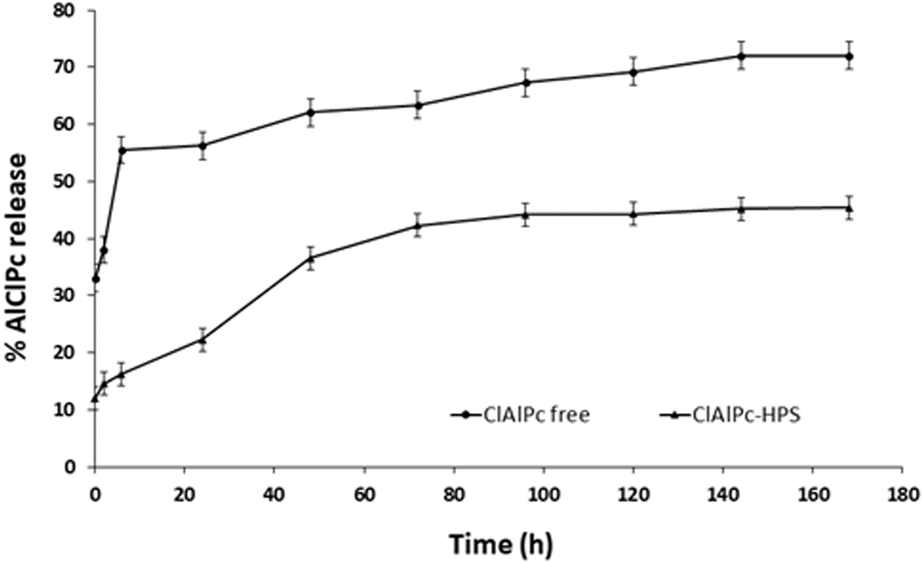
Figure 7 shows the in vitro drug release profiles of free ClAlPc (ClAlPc in DMSO) and ClAlPc-loaded HPS in the first seven days. These in vitro release studies demonstrated that these types of drug carriers allow extended delivery of the drug over more than one week compared with the solubility profile of free ClAlPc.
In vitro drug release profile of free ClAlPc (•) and ClAlPc-loaded HPS (♦).

The analysis of the ClAlPc release profile from HPSs shows approximately 16% released in a burst in the first 6 h, followed by a slow release over seven days that does not exceed 45%. However, when free ClAlPc was incubated with the acceptor media, 60% was rapidly dissolved in the first 120 min. Entrapment of ClAlPc in HPS significantly prolongs its in vitro release (P < 0.05). De Paula et al. [Reference de Paula, Tedesco, Primo, Vilela, Andrade and Mosqueira39] showed the sustained release of ClAlPc loaded in polymeric nanoparticles. The advantages of ClAlPc incorporation were made clear in the release studies. Sustained release systems can maintain a constant drug concentration in the blood plasma for a prolonged period, improving patient compliance (decreasing the number of daily administrations) and therapeutic efficacy [Reference Jethara, Patel and Patel40, Reference Kumar, Bhowmik, Srivastava, Paswan and Dutta41].
Biocompatibility assays
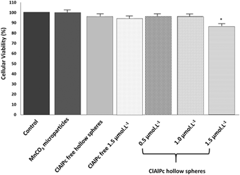
The cytotoxic effects of ClAlPc–HPS on JJ74-A1 cells were found by MTT assay. Figure 8 shows cell viability percentages after the dark toxicity assay. After 3 h incubation in the dark (37 °C, 5% CO2), there was no evidence of a toxic effect of ClAlPc–HPS on J774A-1 cells for any of the tested ClAlPc concentrations. As shown in Fig. 8, there were no significant differences between the viability of cells exposed to HPS and untreated cells (P > 0.05), confirming the HPSs’ biocompatibility.
Cytotoxicity of ClAlPc on J774A-1 cells: cells were incubated varying the ClAlPc-unloaded HPS and ClAlPc-loaded HPS concentrations (0.5, 1.0, and 1.5 μmol/L) for 3 h in the dark (data are presented as mean ± SEM of three independent experiments; *P < 0.05).

Without irradiation, nearly 100% of the cells were viable in the concentration range used.
Schwiertz et al. [Reference Schwiertz, Wiehe, Gräfe and Gitter42] studied calcium phosphate nanoparticles functionalized with different polymers (e.g., PSS/PAH) and porphyrins (m-THPP). Against the macrophage cell line J774-A1, all particles had high dark toxicity and the dark toxicity was independent of the m-THPP concentration. The unloaded CaP/PSS particles showed a comparable dark toxicity, and it was assumed that the calcium phosphate nanoparticles were predominantly taken up by the macrophages.
Photobiological activity
The efficiency of a PS in sensitizing cells by photoinactivation depends on its ability to be taken up by cells, as well as its photochemical properties. Thus, it is important that the PS not only presents good spectroscopy properties, but also is capable of penetrating and selectively accumulating in the target tissues, allowing an efficient phototreatment. This is achievable using a drug delivery system [Reference Mesquita, Dias, Gamelas, Fardilha, Neves and Faustino43, Reference Soboleva, Jans and Rosenkranz44, Reference Fakhar ud, Aman, Ullah, Qureshi, Mustapha, Shafique and Zeb45].
Because each drug presents a range of therapeutic actions, too much being toxic and too little being ineffective, the plasmatic levels depend on the administered dosages. From the clinical point of view, the control of the therapeutic concentration is important.
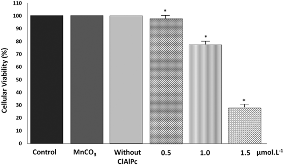
The in vitro experiments showed the system to be not cytotoxic in the dark, yet it exhibits substantial phototoxicity; approximately 1.0 μmol/L of PS and 10.0 J/cm2 of light are sufficient to kill approximately 78% of cells (Fig. 9).
Phototoxicity of ClAlPc on J774A-1 varying the ClAlPc-unloaded HPS and ClAlPc-loaded HPS concentrations (0.5, 1.0, and 1.5 μmol/L) for 3 h and irradiated with laser light at 10 J/cm2 (data are presented as mean ± SEM of three independent experiments; *P < 0.05).

These combinations of PS concentration and the corresponding radiation dose were lethal for macrophage cells. Silva et al. described the preparation of new zinc phthalocyanine conjugated to bovine serum albumin and its photodynamic efficiency in human larynx carcinoma cells (HEp-2 cells). Interestingly, when compared with a classical PS, such as ZnPc, used in PDT, the noncytotoxic concentration is reduced by approximately 20% [Reference Silva, Santos, Gonçalves, Cardoso, Soares and Beltrame-Junior46], but when combined with the light effect, the system has an excellent dose response to the damaged tissue. This factor can be attributed to the maximization of the spectroscopic properties presented by ClAlPc in HPSs that decreases the amount of the drug necessary to reach the photodynamic effect.
The nonaggregation of ClAlPc in biological media also contributes to a higher phototoxicity. The photobiological activity is highly dependent on the PS’s cellular uptake mechanism. In an aqueous medium, ClAlPc is generally absorbed by diffusion across the plasmatic membrane (hydrophobic), leading to a low intracellular concentration. Moreover, a hydrophobic PS, such as ClAlPc, tends to aggregate in aqueous media, which reduce the photodynamic activity [Reference Deda, Uchoa, Caritá, Baptista, Toma and Araki47, Reference Deda and Araki48, Reference Zhao, Yin, Bilski, Chignell, Roberts and He49]. The results showed that the encapsulated ClAlPc in HPSs is an excellent strategy for application in PDT.
Conclusion
This study proved the functionalities of porous manganese carbonate microparticles, besides being merely a decomposable template, by further coating by the Lbl method with suitable polymers for HPSs. A ClAlPc, such as a PS, was adsorbed in MnCO3 to produce a drug delivery system for PDT application. PS photophysical properties were shown to be preserved after HPS encapsulation. The advantages of ClAlPc incorporation were evident in the release studies. The photoactivity of ClAlPc was improved by encapsulation as evidenced by in vitro cell studies. These results reinforce the idea that the use of HPS as PS carriers is useful for PDT of neoplastic tissues.
Experimental
Materials
Sodium poly(styrene sulfonate) (PSS, M w ∼ 70 KDa), poly(allylamine hydrochloride) (PAH, M w ∼ 70 KDa), and chloroaluminum phthalocyanine (AlClPc) were purchased from Sigma Aldrich (St Louis, Missouri). All other chemicals are of analytical grade and were used promptly.
Preparation of MnCO3 microparticles
An equal volume of 0.16 mol/L NH4HCO3 was added to a mixture of 0.016 mol/L MnSO4 and ethanol under vigorous agitation with a final ethanol concentration of 5% (v/v) [Reference Zhu, Stein, Lu, Lvov and McShane50]. The procedure results in highly homogeneous, spherical MnCO3 microparticles with an average diameter of 2.5 µm.
Lbl coating and hollow sphere fabrication
The PSS and PAH solutions used for multilayer assembly were prepared in DI water at 2 mg/mL as described by Zhu et al. [Reference Zhu, Stein, Lu, Lvov and McShane50]. The MnCO3 cores were dissolved by 0.1 mol/L HCl solution, followed by 0.01 mol/L EDTA solution to remove residual manganese ions.
The chloroaluminum phthalocyanine (ClAlPc) (250 mg/mL) was added to the polymer layer after it had attached to the particles. Then, the respective oppositely charged polyelectrolyte species were adsorbed. The amount of PS adsorbed on the particles was determined by UV-visible absorption spectroscopy after recording a calibration curve using the same procedure as previously described by Siqueira-Moura et al. [Reference Siqueira-Moura, Primo, Peti and Tedesco51].
SEM
The MnCO3 microparticles and HPS external morphology were examined by SEM after Au coating using Zeiss EVO MA10 (Carl Zeiss, Oberkochen, Germany) with tungsten filament.
Physicochemical characterization of HPS
Layer-by-layer deposition of polyelectrolytes on the particles was qualitatively monitored by measuring the zeta-potentials of the coated particles with laser light scattering Zetasizer Nano ZS (Malvern, Worcestershire, United Kingdom). An average of three measurements at the stationary level was taken for each data point. The zeta-potential measurements were conducted in deionized water. All experiments were carried out at 25 °C.
UV-vis absorption
Absorption measurements were carried out for the sample in ClAlPc, with a Cary 50 Bio (Varian, Palo Alto, CA) that was background-corrected using matched quartz cuvettes with scanning over the wavelength range from 300 to 800 nm. To establish the linearity of the proposed methods, a calibration curve was constructed at eight concentration levels within the range of 0.50–3.50 μmol/L for the spectrophotometric method. In the spectrophotometric analyses, Beer’s law was obeyed. Least-squares regression analysis was carried out for the data. One-way analysis of variance (ANOVA) and a lack-of-fit test (P = 0.05) were used to determine whether the linear model adequately explains the obtained data.
Process yield (%)
Powdered HPS were obtained after freeze drying. The process yield was calculated by Eq. (1):
 $$Y\left( \% \right) = \left[ {m\left( {{\rm{HPS}}} \right) \div m\left( {{\rm{HPS}} + {\rm{ClAlPc}}} \right)} \right] \times 100\% \quad ,$$
where Y (%) is the process yield, m(HPS) is the mass of HPSs recovered after freeze drying, and m(HPS + ClAlPc) is the mass of HPSs plus mass of ClAlPc in formulation. The encapsulation method was accomplished in triplicate (n = 3).
Determination of the drug encapsulation rate
The total ClAlPc content in HPS was determined using the intended methods. Briefly, 1 mL of ClAlPc and 0.1 mg/mL of HPS entrapped ClAlPc were appropriately dissolved in DMSO (1:1, v/v) and sonicated for 5 min to release all ClAlPc content. After that, the samples were diluted to the final volume (3 mL) and filtered through a 0.45-µm membrane. Besides that, the free ClAlPc content in the formulations was determined by measuring the nonincorporated drug presented that was obtained through separation of the aqueous phase using a centrifuge procedure (Eppendorf centrifuge 5804R, Hamburg, Germany). The association/encapsulation efficiency of ClAlPc into HPS was determined from Eq. (2):
 $${\rm{EE}} = \left( {m1 + m2} \right) \times 100\quad ,$$
where EE is the ClAlPc encapsulation efficiency, m1 is the mass of ClAlPc in HPS, and m2 is the mass of ClAlPc used in the formulation.
The sample solutions were assayed by absorbance measurement at 674 nm, which corresponds to the maximum absorption wavelength (λmax) of ClAlPc. Analyses were performed in triplicate, and the mean results (±SD) are reported. All results obtained were compared using a Tukey’s post-test, and the statistical significance was P < 0.05.
Evaluation of the in vitro drug release performance
To evaluate the in vitro drug release performance of the ClAlPc from HPSs (5.0 mg), they were dispersed into a PBS buffer solution (5 mL, pH = 7.4) and shaken at 37 °C. At different predetermined intervals, the suspension was centrifuged and the supernatant (3 mL) was taken out and replaced by fresh PBS solution (3 mL) to continue the drug release evaluation. The ClAlPc concentration in the supernatant was determined by UV-vis absorbance. The drug release efficiency was calculated by Eq. (3). The data were reported as mean ± SD based on triplicate measurements:
 $${\rm{ClAlPc}}\;{\rm{release}}\left( {\% {{\rm{w}} \over {\rm{w}}}} \right) = {{\left( {{\rm{mass}}\;{\rm{of}}\;{\rm{ClAlPc}}\;{\rm{SPN}}} \right) \times \,\,100} / {\left( {{\rm{mass}}\;{\rm{of}}\;{\rm{ClAlPc}}\;{\rm{FML}}} \right)}}\quad ,$$
where SPN = supernatant and FML = formulation.
Cell culture condition
The cell line used in this study was the mouse macrophage carcinoma line J774 A-1 supplied by American Type Tissue Culture Collection (ATCC, Rockville, Maryland). Monolayer cultures of J774 A-1 were grown in Dulbecco’s Eagle’s minimum essential medium (DMEM) with 10% fetal bovine serum (Gibco Laboratories Inc., Grand Island, NY), 1% L-glutamine, and 1% penicillin-streptomycin (Gibco Laboratories Inc., Grand Island, NY). The cells were used in the logarithmic phase of growth and maintained at 37 °C in humidified 5% CO2 and 95% air.
Biocompatibility assays
For the toxicity test, 96-well microplates were employed. The methodology used to evaluate the toxicity was the classical MTT assay. The tetrazolium salt [3-(4,5-dimethylthiazol-2-yl)-2,5-diphenyltetrazoliumbromide] produced a highly colored formazan dye upon NADH reduction, which reflects a living cellular dehydrogenase [Reference Lunardi, Rotta and Tedesco52]. To investigate the toxicity, a suspension of 5 × 10³ J774 A-1 cells in 200 µL of medium was inoculated into each well in a 96-well microplate. After culturing in a CO2 incubator for 24 hs, monolayer cultures of J774 A-1 were treated with the PS at concentrations of 0.5, 1.0, and 1.5 µmol/L ClAlPc in ethanol and HPS dispersion. After incubation for 3 h, the cells were washed twice, and the volume was completed with the addition of 200 µL DMEM in each well for 24 h. The 1.0 mg/mL MTT solution (50 µL per well) was added to the cells on 96-well microplates, followed by incubation for 4 h, at 37 °C. Next, the crystals formed by the interaction between the mitochondrial dehydrogenases and the MTT reagent were dissolved with isopropyl alcohol. Then, the samples were shaken until complete dissolution of the formed product. After the color reaction was finished, the absorbance was determined by means of a spectrophotometer with a 570-nm filter. The percentage of cell vitality was calculated with respect to the control cells incubated without PS.
Photobiological activity
Suspensions of 5 × 10³ J774 A-1 cells in 200 µL of medium were inoculated into each well in a 96-well microplate. After culturing in a CO2 incubator for 24 h, monolayer cultures of J774 A-1 were treated with the 0.5, 1.0, and 1.5 µmol/L ClAlPc–HPS dispersion. After incubation for 3 h, the cells were washed twice with PBS and fresh DMEM was added before cell irradiation. The results are given as the percentage of the result obtained with the control cultures exposed to PS alone. A dose of light from 10.0 J/cm2 was used. The light source used was one light-emitting diode with an output power of 0.2 W and peak wavelength of 660 nm.
Statistical analysis
All tests were performed in triplicate, and all data were submitted to ANOVA followed by a Tukey’s test. Microsoft Excel was used to create the charts. All the data are expressed as the mean ± SD of three independent experiments. A probability P-value < 0.05 was considered significant in this study.
Acknowledgments
The authors acknowledge the financial support of the Brazilian Agency CNPq (Conselho Nacional de Desenvolvimento Científico e Tecnológico) with the project number 800038/2014-2.