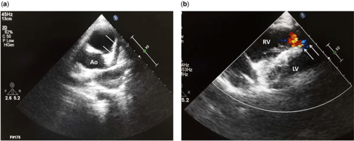
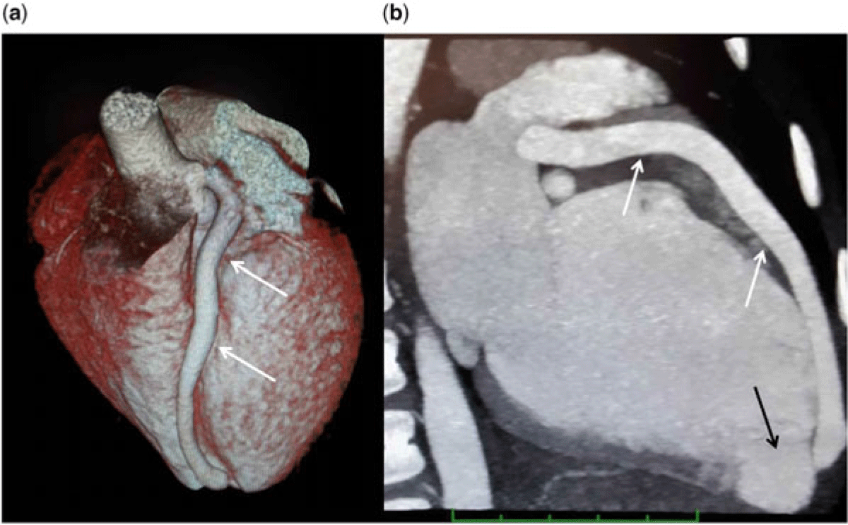
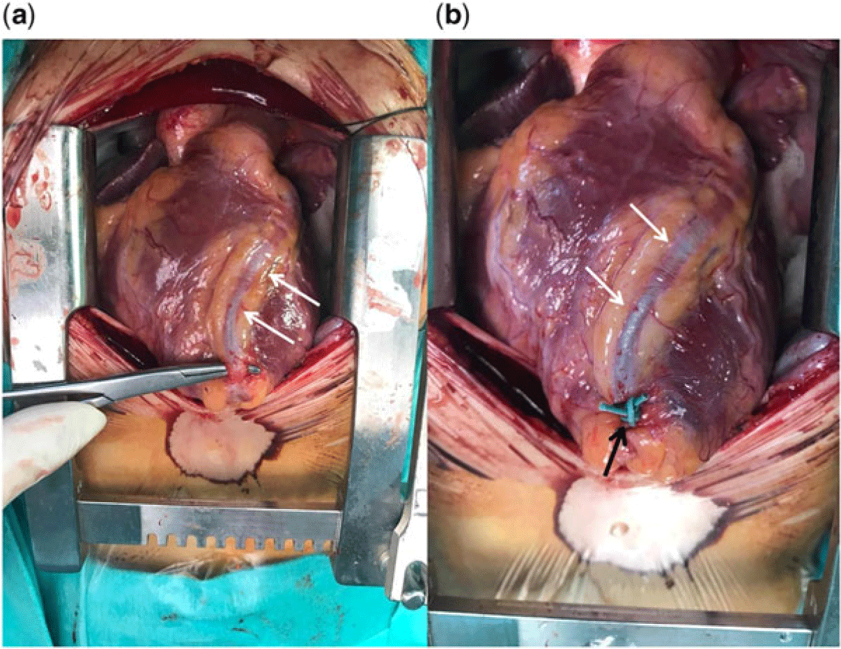
A 7-year-old child was referred to our paediatric cardiology clinic because of heart murmur. She had no history of cardiac or chronic diseases. Initial vital signs were body temperature 36.6 °C, systolic blood pressure 100 mmHg, heart rate 117 beats/min, respiratory rate 21 breaths/min, and 98% oxygen saturation in room air. Echocardiography revealed a larger fistula from the dilated left anterior descending coronary artery with communications to the right ventricle through multiple outlets (Fig 1a and b, Supplementary videos 1 and 2). The cardiac chambers were normal dimensions and not dilated. Coronary computed tomography showed a significant dilation of the left anterior descending artery and a large fistula from the end of the coronary artery emptying into the apical region of the right ventricular chamber with a diverticular dilation (Fig 2a and b). Percutaneous closure was the suggested treatment of choice for coronary artery fistulasReference Thakkar, Patel and Poptani1 and the medical team elected to initially place a closure device into the largest orifice. However, the process was not successful due to the multiple narrow and closely related orifices. So, surgical treatment was found to be optimal treatment option for the case. The closure was performed without cardiopulmonary bypass and a thick suture placed below the left anterior descending artery to close the fistulous trajectories near the apex of right ventricle (Fig 3a and b). The patient was discharged 8 days after surgery. During the follow-up period of 6 months after surgical closure, no residual fistulas were showed by echocardiography.
(a) Transthoracic short-axis view of two-dimensional transthoracic echocardiography shows a dilated left main coronary artery (white arrows, Ao = aorta). (b) Right ventricular view of echocardiogram shows a right ventricle of normal size and colour Doppler shows forward flow into the apical region of right ventricle (white arrows, RV = right ventricle; LV = left ventricle).

(a and b) Coronary computed tomography showed a significant dilation of the left anterior descending artery (white arrows) and a large fistula from the end of the coronary artery emptying into the apical region of the right ventricular chamber with a diverticular dilation (black arrow).

(a) Surgical view of dilated left anterior descending artery (white arrows) and (b) placement of a thick suture (black arrow) below the left anterior descending artery (white arrows) to close the fistulous trajectories near the apex of right ventricle.

Supplementary material
To view supplementary material for this article, please visit https://doi.org/10.1017/S1047951119003305
Acknowledgements
None.
Financial Support
This research received no specific grant from any funding agency, commercial or not-for-profit sectors.
Conflicts of Interest
None.
Ethical Standard
The authors assert that all procedures contributing to this case comply with the ethical standards of the Turkish Council of Medical Research and with the Helsinki Declaration of 1975, as revised in 2008, and has been approved by the institutional committees of Dr. Ali Kemal Belviranlý Obstetrics and Children Hospital, Konya, Turkey.