Introduction
Eardrop delivery via tympanostomy tube insertion followed by tragal pumping (eardrop method) and intratympanic injection are well known and widely used as solution delivery methods into the middle ear. Regarding middle-ear diseases, both methods have been used with corticosteroids for refractory types of otitis media such as eosinophilic otitis media and otitis media with antineutrophil cytoplasmic antibody (ANCA) -associated vasculitis.Reference Masuda, Esu, Iino and Yoshida1, Reference Wang, Han and Yu2
For the eardrop method, otolaryngologists generally perform tympanostomy tube insertion under topical anaesthesia. After eardrop application, tragal pumping must be performed to improve delivery.Reference Hara, Liu, Shah, Saeki, Orita and Sakyo3, Reference Hebert, Vick, King and Bent4 Once the tube is inserted, patients can perform the treatment themselves using a prescribed eardrop with adequate instruction, thereby reducing the need for frequent hospital visits.Reference Chang, Zee, Lee and Tong5 In comparison, patients must visit hospitals frequently for each intratympanic injection as this procedure needs to be performed by otolaryngologists under topical anaesthesia each time.Reference de Cates and Winters6 However, one advantage of intratympanic injection is that otolaryngologists can directly confirm that the ototopical medicine is delivered into the middle ear by visual inspection under a microscope.
While both methods are known to deliver drugs into the middle ear, the volume delivered by the eardrop method compared with intratympanic injection remains unclear. Consequently, treatment selection has largely been based on empirical practice rather than evidence-based criteria. We aimed to compare solution delivery into the middle-ear cavity between the eardrop method and intratympanic injection using a rat model to determine whether the eardrop method can deliver eardrops into the middle ear as effectively as intratympanic injection.
Materials and methods
Experimental design
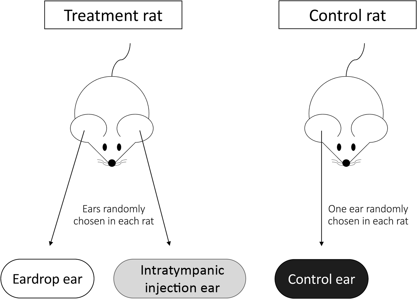
Figure 1 shows the experimental design. To compare the eardrop method and intratympanic injection while avoiding potential biases as much as possible, the eardrop method was performed on one ear (eardrop ear) and intratympanic injection on the other ear (intratympanic injection ear) of the same rat (treatment rat). Ears treated with the eardrop method and intratympanic injection were randomly chosen in each rat. To validate our image analysis method, temporal bones without any treatment were harvested from one ear (control ear) randomly chosen from rats that were not treated with eardrop or intratympanic injection (control rat).
Experimental design. Ears were randomly chosen for the type of treatment (eardrop or ITI). Only one ear in a control rat was randomly chosen as the control ear.

Figure 1 Long description
The diagram illustrates an experimental design involving two rats. The left side shows a 'treatment rat' with two ears labeled as 'Eardrop ear' and 'Intratympanic injection ear', indicating that ears were randomly chosen for each treatment. The right side shows a 'control rat' with one ear labeled as 'Control ear', which was randomly chosen. Arrows point from the rat's ears to the respective labels, emphasizing the random selection process for each ear in the experiment.
Animals
Sprague-Dawley rats were housed in standard polycarbonate rat cages in a temperature- and humidity-controlled room under a 14 hours:10 hours light:dark cycle, with lights on from 07:00 to 21:00 h. Animals were allowed access to water and standard chow ad libitum. Male and female adult breeder rats aged 6–10 months were assigned to this study. Thirty rats were used for the treatment group and another 30 rats for the control group. Adult breeder rats were maintained as male–female pairs, and each pair was sequentially assigned in an alternating manner to either the treatment group or the control group prior to any experimental procedures.
The authors assert that all procedures contributing to this work comply with the ethical standards of the relevant national and institutional guidelines for the care and use of laboratory animals, as approved by the Johns Hopkins University Animal Care and Use Committee (approval number: RA23M358).
Anaesthesia
Rats were placed in a medium-sized induction chamber for induction of anaesthesia with 2–3 per cent isoflurane. Animals were monitored for the depth of anaesthesia by reflexes to toe pinch and respiratory rate. Rats were maintained under anaesthesia via a nose cone with approximately 1.5–2 per cent isoflurane. Eye lubricant was placed in both eyes to prevent corneal desiccation caused by anaesthesia. Rats were placed on a low-heat-setting heating pad to maintain a normal body temperature (37°C).
Eardrop method: tympanostomy tube insertion, eardrop application and tragal pumping
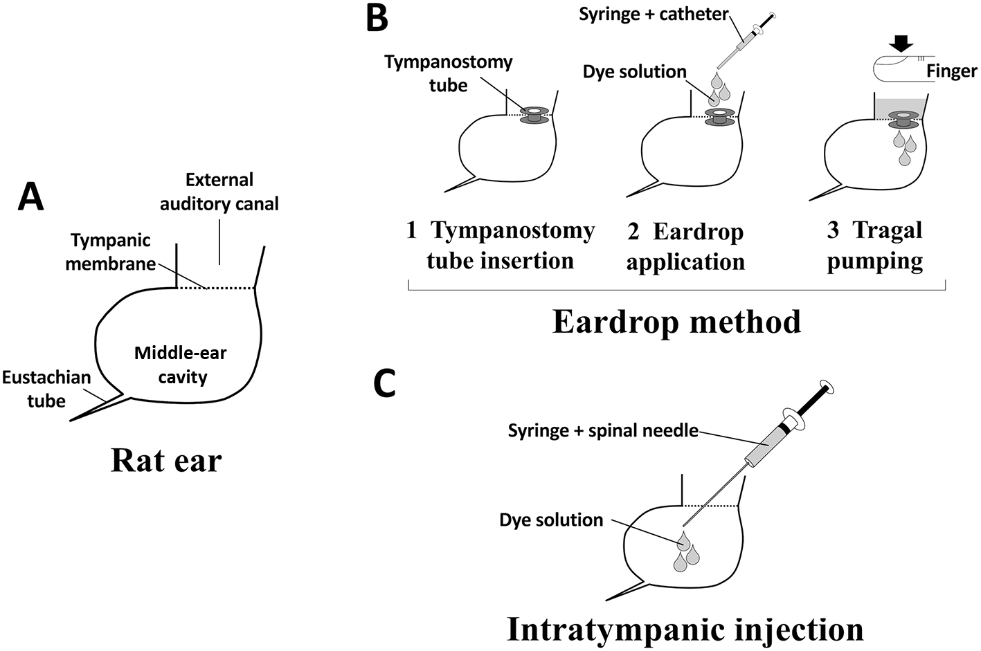
For eardrop ears, tympanostomy tube insertion, eardrop application and tragal pumping were performed according to our previous publication (Figure 2a,b).Reference Yang, Zhao, Chen and Zhao7 Briefly, rats were placed in the lateral position with the eardrop ear upward. The ear was examined to confirm the absence of outer- and middle-ear disease using a microscope and an endoscope. Cerumen in the external auditory canal was removed if necessary. A small incision was made with a myringotomy knife on the posterior half of the pars tensa of the tympanic membrane. A tympanostomy tube (internal diameter, 1.14 mm; Medtronic, Galway, Ireland) was inserted into the tympanic membrane perforation with micro alligator ear forceps. Placement of the tympanostomy tube was confirmed using an endoscope. To detect penetration of the eardrop via the tympanostomy tube, green Davidson® tissue marking dye (1163-1; Bradley Products, Inc., Bloomington, MN, USA) was diluted twofold with distilled water and used as the eardrop solution. A total of 100 µl of eardrop-mimicking dye solution was applied into the external auditory canal using a 22-gauge catheter connected to a 1-ml syringe under microscopic visualisation. Thereafter, the tragus was gently pushed repeatedly for 1 minute. Thirty minutes after eardrop application, the remaining solution in the external auditory canal was removed using the same-size catheter and syringe, and the tympanostomy tube was then removed with ear forceps under a microscope.
A, The model section of a rat ear. B, Description of the eardrop method. C, Description of the intratympanic injection.

Figure 2 Long description
A shows a diagram of a rat ear, highlighting the external auditory canal, tympanic membrane, middle-ear cavity and Eustachian tube. B illustrates the eardrop method in three steps: tympanostomy tube insertion, eardrop application using a syringe and catheter and tragal pumping with a finger. C depicts the intratympanic injection process, showing a syringe with a spinal needle administering dye solution into the ear.
Intratympanic injection
Figure 2c shows a diagram of this procedure. Rats were placed in the lateral position with the intratympanic injection ear upward. Using an endoscope, the integrity of the external auditory canal and tympanic membranes was examined. Cerumen in the external auditory canal was removed if necessary. To perform intratympanic injection precisely and consistently, we used an instrument with an endoscope and a 27-gauge spinal needle (Figure 3). To visualise the tip of the needle during the procedure and insert it accurately into the posterior half of the pars tensa of the tympanic membrane, the tip of the needle was positioned 5 mm ahead of the tip of the endoscope. With needle insertion into the tympanic membrane, 100 µl of dye solution was slowly injected into the middle-ear cavity of the intratympanic injection ears. The overall average volume of the middle-ear gas space in Sprague-Dawley rats has been reported to be 42.0 µl.Reference Kania, Herman, Tran Ba Huy and Ar8 Therefore, an injection volume of 100 µl was determined to be sufficient for middle-ear delivery, consistent with volumes used in prior intratympanic injection studies in Sprague-Dawley rats.Reference Park, Kim, Han, Park and Park9 Thirty minutes after injection, the solution in the external auditory canal was removed using the same method as in the eardrop-ear procedure.
Our instrument for intratympanic injection. A, The instrument was made with a 1-mL syringe, a 27-gauge spinal needle, an endoscope, and an adapter for a camera. B, The tip of the instrument.

Figure 3 Long description
The image A shows an instrument for intratympanic injection held by a gloved hand. It consists of a one milliliter syringe attached to a 27-gauge spinal needle, an endoscope and a camera adapter. The image B shows a close-up of the instrument's tip, highlighting the needle and endoscope alignment.
Histological analysis
Rats were euthanised immediately after completion of all procedures described above and were under general anaesthesia until euthanasia. After euthanasia, both temporal bones were harvested for histological analysis. Histological and image analyses were performed according to our previous publication.Reference Yang, Zhao, Chen and Zhao7 Briefly, after fixation in formaldehyde solution and decalcification, the tissue was processed for embedding. Multiple thin sections were then cut horizontally and stained with haematoxylin and eosin.
To compare solution delivery between the two methods, the area stained by dye in the middle ear on histological sections was analysed using Image J software. Figure 4 shows the representative images of a temporal bone section analysed using Image J software by a scorer blinded to the experimental groups. First, the temporal bone section was cropped to include only the middle-ear cavity (Figure 4a,b). Next, the green-stained area and unstained area were selected and measured using the colour threshold function of Image J software. In this process, the selected area for measurement is shown in red. Figure 4c shows the green-stained area highlighted in red. Residual pale green areas were considered unstained (below the threshold on Image J software) because the green staining is weak. Figure 4d shows the unstained area highlighted in red. The stained area ratio (per cent) was calculated as (stained area)/(stained area + unstained area).
Representative temporal bone section images during the image analysis. A, An original image. B, The cropped image of the middle ear cavity. C, The green-stained area. D, The white unstained area.

Figure 4 Long description
The image A shows a temporal bone section with labeled anatomical features: external auditory canal, tympanic membrane, middle-ear cavity and Eustachian tube. The image B shows the cropped section focusing on the middle-ear cavity. The image C highlights the green-stained area in red, indicating the stained region. The image D highlights the unstained area in red, showing the region below the staining threshold.
Statistical analyses
Descriptive statistics were presented as numbers (per cent) and means ± standard deviations for categorical variables and continuous variables, respectively. The stained area ratio was compared between the eardrop and intratympanic injection ears within the same rat using a paired t-test. Baseline characteristics between the treatment and control cohorts (age and body weight) were compared using independent samples (Student’s) t-tests. Categorical variables (sex and side of the ear) were compared between cohorts using chi-square tests. A p value less than 0.05 was considered statistically significant. All analyses were performed using SPSS Statistics version 28 (IBM, Armonk, NY, USA).
Results and analysis
Animal profiles
Table 1 shows the animal profiles of the eardrop, intratympanic injection and control ears. In the treatment group, 15 males and 15 females were included for the eardrop and intratympanic injection ears. The mean age was 266.5 ± 32.6 days, and the mean body weight was 452.1 ± 89.6 g. The eardrop method was applied to the left ear in 15 rats and to the right ear in the remaining 15 rats. An additional 15 males and 15 females were included in the control group for the control ears. The mean age was 262.4 ± 37.2 days and the mean weight was 453.6 ± 93.7 g. For the control ears, left ears were used in 15 rats and right ears in the remaining 15 rats. There were no significant differences in age (p = 0.649), body weight (p = 0.949) or sex (p = 1.000) between the treatment and control groups.
Profiles of rats in the treatment and control groups

Table 1 Long description
The table compares characteristics of rats in treatment and control groups, each consisting of 30 rats. Both groups have similar mean ages, with treatment group at 266.5 days and control group at 262.4 days, showing no significant difference (p=0.910). Sex distribution is identical, with 50% males in each group (p=1.000). Body weight averages are also similar, with treatment group at 452.1g and control group at 453.6g (p=0.949). The side of the ear treated is evenly distributed, with 50% right ears in both groups (p=1.000). All p values indicate no significant differences between the groups, suggesting comparable profiles.
Treatment group rats were used for eardrop ears and intratympanic injection ears. Control group rats were used for control ears.
* Student’s t-test;
† chi-square test
Histological analysis
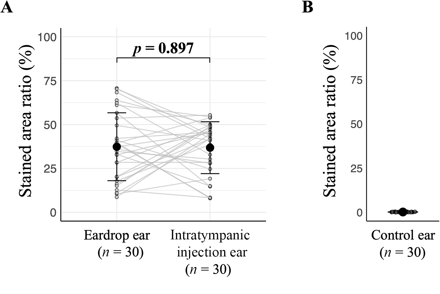
Figure 5 shows representative images of the eardrop, intratympanic injection and control ears. Images of the eardrop and intratympanic injection ears, obtained from the same rat, showed that large regions of the middle-ear cavities were stained (Figure 5d,e). In contrast, no substantial green signal was observed in the control ear, confirming the absence of false-positive signal detection (Figure 5f). Image analysis and data processing revealed that the stained area ratios in the eardrop and intratympanic injection ears were 37.4 ± 19.3 and 36.8 ± 14.8 per cent, respectively. A paired t-test was used to compare the stained area ratio between the eardrop and intratympanic injection ears, as both were derived from the same rat under similar conditions. Figure 6a shows that no significant difference was detected in the stained area ratio of the middle-ear cavity between the eardrop and intratympanic injection ears (p = 0.897; 95 per cent confidence interval, −7.81–8.88), suggesting that the solution delivery efficacy of the eardrop method is comparable to that of intratympanic injection. Figure 6b shows that the stained area ratio in the unstained control ear was 0.15 ± 0.09 per cent, validating the visual inspection.
Representative images of temporal bone sections in eardrop, ITI, and control ears. A, B, C. Original images. D, E, F. The green-stained areas. G, H, I. The white unstained areas.

Figure 5 Long description
The image consists of three sets of temporal bone sections labeled as eardrop ear, intratympanic injection ear and control ear. Each set includes three images: original images, stained areas highlighted in red and unstained areas highlighted in red. The original images show the anatomical structure of the ears. The stained areas highlighted in red indicate regions where staining occurred, with the eardrop and intratympanic injection ears showing substantial staining compared to the control ear. The unstained areas highlighted in red show regions without staining, with the control ear having more unstained areas compared to the other two. Each section is clearly labeled to differentiate between the types of ears and the staining conditions.
The mean stained area ratio (large black dots) with standard deviation error bars for each ear. A, Eardrop and ITI ears. The paired ears of individual rats are connected. B, Control ear.

Figure 6 Long description
The image contains two graphs labeled A and B. Graph A shows the stained area ratio in percentage for eardrop ears and intratympanic injection ears, each with a sample size of 30. The data points are connected and the mean values are indicated with large black dots. The p-value is 0.897, suggesting no significant difference between the two methods. Graph B displays the stained area ratio for control ears, also with a sample size of 30, showing minimal staining with clustered data points near the bottom of the graph.
Discussion
The aim of this study was to provide additional evidence regarding the efficacy of the eardrop method for middle-ear drug delivery. To this end, we compared solution delivery into the middle-ear cavity between the eardrop method and intratympanic injection using a rat model and found no significant difference. The stained area ratio was approximately 0 per cent in the control ears, indicating the accuracy of the image analysis method used in this study. Based on these results, we confirmed that the eardrop method for solution delivery into the middle-ear cavity appears to be as efficient as intratympanic injection in this rat model.
To compare solution delivery by the eardrop method and intratympanic injection precisely, we applied these methods to each ear of the same rat. This study design was used to minimise the effects of anatomical differences in the middle-ear cavity among individual rats, including differences in cavity size. Such individual differences may alter the movement of the dye solution in the middle-ear cavity, thereby interfering with precise comparison of the two methods. The Eustachian tube is also an important factor that should be considered. After drug administration into the middle ear, patients are generally required to remain supine with the treated ear positioned upward and to avoid swallowing, yawning or speaking to prevent leakage into the Eustachian tube.Reference de Cates and Winters6 Although Eustachian tube function is often impaired in patients with refractory otitis media with effusion, it has been reported that Eustachian tube function may be related to solution penetration via a tympanostomy tube.Reference Yang, Zhao, Chen and Zhao7, Reference Mills, Albizzati and Todd10 We therefore believe that comparing the eardrop method and intratympanic injection within the same rat using a paired t-test reduces potential biases, as both ears are likely to be under similar conditions, including the anatomy of the external auditory canal and middle-ear cavity and the influence of Eustachian tube function.
To replicate the experimental conditions used in previous human studies, we used a grommet tympanostomy tube with an internal diameter of 1.14 mm, which has also been used in human studies,Reference Chandrasekhar, Tsai Do, Schwartz, Bontempo, Faucett and Finestone11, Reference Basura, Adams, Monfared, Schwartz, Antonelli and Burkard12 to evaluate the eardrop method. The internal diameter of the tympanostomy tube is an important factor in this study because solutions can pass more easily through tubes with larger diameters than through those with smaller diameters.Reference Mills, Albizzati and Todd10 Different tube shapes have also been shown to result in different levels of penetration.Reference Mills, Albizzati and Todd10 Further studies evaluating the outcomes of the eardrop method using tympanostomy tubes of various sizes and shapes are needed to determine the most appropriate tube for this method.
Tragal pumping following tympanostomy tube insertion and eardrop application is also important for the eardrop method. It has been reported that eardrops do not pass efficiently or consistently (only up to 4–5 per cent) through a tympanostomy tube without tragal pumping.Reference Hara, Liu, Shah, Saeki, Orita and Sakyo3, Reference Hebert, Vick, King and Bent4 Tragal pumping can significantly increase solution penetration from the external auditory canal into the middle-ear cavity via a tympanostomy tube,Reference Hara, Liu, Shah, Saeki, Orita and Sakyo3, Reference Boyd and Gottschall13 therefore tragal pumping following eardrop application should be performed to facilitate eardrop penetration.
Because we used a specially designed instrument equipped with an endoscope and a spinal needle for the intratympanic injection procedure, we were able to visualise the needle tip during the procedure and insert it accurately. Although this procedure can also be performed under a microscope, the use of an endoscope facilitated intratympanic injection by allowing visualisation of the entire tympanic membrane.
Intratympanic injection is also widely used for the treatment of inner-ear diseases such as idiopathic sensorineural hearing lossReference Chandrasekhar, Tsai Do, Schwartz, Bontempo, Faucett and Finestone11 and Ménière’s disease.Reference Basura, Adams, Monfared, Schwartz, Antonelli and Burkard12 Previous reports have also demonstrated the efficacy of the eardrop method using dexamethasone eardrops for idiopathic sensorineural hearing lossReference Chang, Zee, Lee and Tong5 and gentamicin-betamethasone eardrops for Ménière’s disease.Reference Kaplan, Hehar, Bance and Rutka14 In the present study, we did not investigate solution delivery into the inner ear, therefore to evaluate the feasibility of the eardrop method as an alternative to intratympanic injection for patients with idiopathic sensorineural hearing loss or Ménière’s disease, further studies measuring specific drug concentrations in the inner ear following administration by both methods are required.
This study has potential limitations because it was not designed as a non-inferiority study.Reference Ranganathan, Pramesh and Aggarwal15 A substantially larger number of animals would be required to conduct a non-inferiority study to determine whether the eardrop method is superior to intratympanic injection.
• Medications for middle-ear diseases are commonly delivered into the middle-ear cavity either by the eardrop method with a tympanostomy tube followed by tragal pumping or by intratympanic injection
• Both methods have been used in middle-ear diseases, but the actual efficiency of drug delivery between the two approaches has not been fully compared
• In clinical practice, the choice between the eardrop method and intratympanic injection has been determined empirically rather than based on evidence-based criteria
• In a rat model, the eardrop method achieved comparable solution delivery into the middle-ear cavity to intratympanic injection, with no significant difference in the stained area ratio (p = 0.897)
• This work provides experimental evidence supporting the eardrop method as a potentially practical alternative to intratympanic injection for middle-ear drug delivery, which may reduce the burden of frequent hospital visits for patients.
Conclusion
We found that dye solution was delivered into the middle-ear cavity with efficacy comparable to that of intratympanic injection in a rat model. This study provides insight into solution delivery into the middle ear in patients with middle-ear diseases.
Acknowledgements
This work was supported by JSPS KAKENHI Grant Number 24K19757 (to Satoshi Hara). Funding for this research was made possible by Subsidies for Current Expenditures to Private Institutions of Higher Education from the Promotion and Mutual Aid Corporation for Private Schools of Japan through a subaward from the Juntendo University. Its contents are solely the responsibility of the authors and do not necessarily represent the official views of the Promotion and Mutual Aid Corporation for Private Schools of Japan and Juntendo University (as applicable).
Competing interests
T.Y. and H.O. receive research funding through their institution from Renee Medical Corporation.
Ethical Standards
The authors assert that all procedures contributing to this work comply with the ethical standards of the relevant national and institutional guidelines for the care and use of laboratory animals, as approved by the Johns Hopkins University Animal Care and Use Committee (approval number: RA23M358).