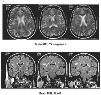
We present herein the case of a 58-year-old Caucasian woman who was admitted to the Neurological Department because of progressive difficulty in eyelid opening over the previous 3 months. Her past medical history was unremarkable except for high blood pressure, treated with angiotensin 2 receptor antagonist, as well as symptoms consistent with Raynaud phenomenon. However, upon questioning she admitted to have been having xerostomia for the last 3 years. Clinical examination revealed bilateral blepharospasm and was otherwise unremarkable. The ophthalmologic assessment defined no pathologic conditions that could contribute to or trigger significant eye irritation. Mild xerophthalmia – Schirmer's test 5 mm, tear breakup time 3–4 s and negative Rose Bengal staining – was insufficient to explain the severity of the symptoms. Routine laboratory tests and immunologic profile were unremarkable, except for a moderately increased Erythrocyte Sedimentation Rate (ESR) of 52 mm/h and a positive rheumatoid factor of 158 IU/ml. A minor salivary gland biopsy was consistent with Sjögren's syndrome (SS) – 2 mononuclear cell foci, of at least 50 cells each, per 4 mm2 of tissue. Brain magnetic resonance imaging (MRI) scan confirmed the involvement of the central nervous system (CNS), showing multiple lesions with increased signal in T2 and FLAIR sequences detected in the basal ganglia, thalamus, the periventricular white matter and brainstem bilaterally (Fig. 1), which did not enhance with gadolinium. The patient was treated with botulinum toxin injections that had limited effect on the blepharospasm. Subsequently, she received 1 g of methylprednisolone intravenous infusion, which resulted in moderate improvement of the symptoms. Unfortunately, she was lost to follow up, so that no further treatment was administered and long-term response to treatment could not be established.
Brain magnetic resonance imaging scan showing multiple lesions with increased signal in T2 (a) and FLAIR (b) sequences at the basal ganglia and in the periventricular white matter bilaterally.

Sjögren's syndrome is an autoimmune process whose spectrum ranges from an exocrinopathy to systemic disease, with the potential of evolving to non-Hodgkin's lymphoma. Neurologic manifestations have been reported in about 20% of patients. Although CNS involvement in SS has been a matter of considerable controversy, several reports have suggested that when it occurs, brain MRI abnormalities are usually disseminated in space, affecting mainly the deep white matter (Reference Alexander, Tatal, Moutsopoulos and Kassan1–Reference Binder, Snaith and Isenberg3). Nevertheless, we are not aware of any published report of bilateral blepharospasm as the presenting symptom of SS and not caused by severe xerophthalmia. A previous report described three patients with SS presenting with blepharospasm because of severe xerophthalmia that resolved with botulinum toxin injections Reference Spiera, Asbell and Simpson(4). In contrast with the previous reported cases, our patient had mild xerophthalmia – according to the ophthalmologic tests applied – and severe blepharospasm that could not be attributed to local eye irritation. In addition, the patient reported as a primary symptom difficulty in opening her eyes and not eye irritation. Thus, it was speculated that the extensive lesions in the basal ganglia, thalamus and brainstem might have been responsible for her symptomatology. Indeed, several studies argue that a variety of CNS lesions involving the rostral brainstem, thalamus and basal ganglia are associated with blepharospasm and other forms of cranial dystonia (Reference Jankovic5–Reference Berardelli, Rothwell, Hallett, Thompson, Manfredi and Marsden7) Finally, SS has been associated with other forms of dystonia related to CNS involvement, and a recent case report has described a patient with orofacial dystonia that dramatically improved with corticosteroid treatment Reference Van Den Berg, Horstink, Van Den Hoogen and Oyen(8,Reference Papageorgiou, Kontaxis, Bonakis and Kalfakis9). We conclude that patients with blepharospasm should also be evaluated in the direction of autoimmune diseases and patients with SS and blepharospasm should be investigated for possible CNS involvement, especially when there is no significant eye irritation.