Introduction
Neurodegenerative disease, characterized by the loss of structure, function or quantity of specific neurons in the human brain, is a generic term covering a range of incurable and debilitating conditions that progressively lead to disorders of our nervous system. A series of pathophysiological symptoms include memory/cognitive impairment and motor dysfunction, caused by diseases such as Alzheimer's disease (AD), Parkinson's disease (PD), Huntington's disease and amyotrophic lateral sclerosis (ALS) (Kumar et al., Reference Kumar, Singh and Ekavali2015; Gitler et al., Reference Gitler, Dhillon and Shorter2017). The onset of the diseases is prone to be prevalent in the middle-aged and elderly at an increasing rate (Jaul and Barron, Reference Jaul and Barron2017). Though many approaches have been introduced with the potential to understand and treat these neurodegenerative diseases, the outcomes of clinical research have yet to be assessed. Therefore, novel experimental approaches and insights are essential to better understand the pathogenesis and to find new therapeutic agents against the diseases.
As loss of protein homeostasis (proteostasis) occurs, the proteins associated with the nervous system fail to sustain their native conformation, start to aggregate and deposit in the brain through two different aggregation routes (Hipp et al., Reference Hipp, Kasturi and Hartl2019). The first route is to form amorphous aggregates, which are usually present in undefined structures. Alternatively, monomeric proteins aggregate into soluble oligomers and then form highly ordered amyloid fibrils (Dobson, Reference Dobson2003; Selkoe, Reference Selkoe2003; Adamcik and Mezzenga, Reference Adamcik and Mezzenga2018). One of the pathological hallmarks of AD is the finding of senile plaques deposited in the brain. The core component of the plaques was demonstrated to be the insoluble amyloid-beta (Aβ) fibrils (Tagliavini et al., Reference Tagliavini, Giaccone, Frangione and Bugiani1988; Fukumoto et al., Reference Fukumoto, Asami-Odaka, Suzuki and Iwatsubo1996). Numerous other neurodegenerative disorders share this common pathology with amyloid proteins, such as Parkinson's α-synuclein (α-syn), Creutzfeldt–Jakob disease's prion protein (PrP), and ALS's superoxide dismutase 1 (Chiti and Dobson, Reference Chiti and Dobson2017). Although most disease-related amyloids are assumed to be non-functional, there are some amyloids, found from microbes to humans that are functional, and even disease-related amyloids can, sometimes, possess functions (Fowler et al., Reference Fowler, Koulov, Balch and Kelly2007; Pham et al., Reference Pham, Kwan and Sunde2014; Brothers et al., Reference Brothers, Gosztyla and Robinson2018). Therefore, amyloid proteins can be both protective and destructive, critically dependent upon the amyloid homeostasis (Chen et al., Reference Chen, Xu, Yan, Zhou, Jiang, Melcher and Xu2017), which involves protein synthesis and folding, conformational maintenance and degradation (Fowler et al., Reference Fowler, Koulov, Balch and Kelly2007; Hipp et al., Reference Hipp, Kasturi and Hartl2019).
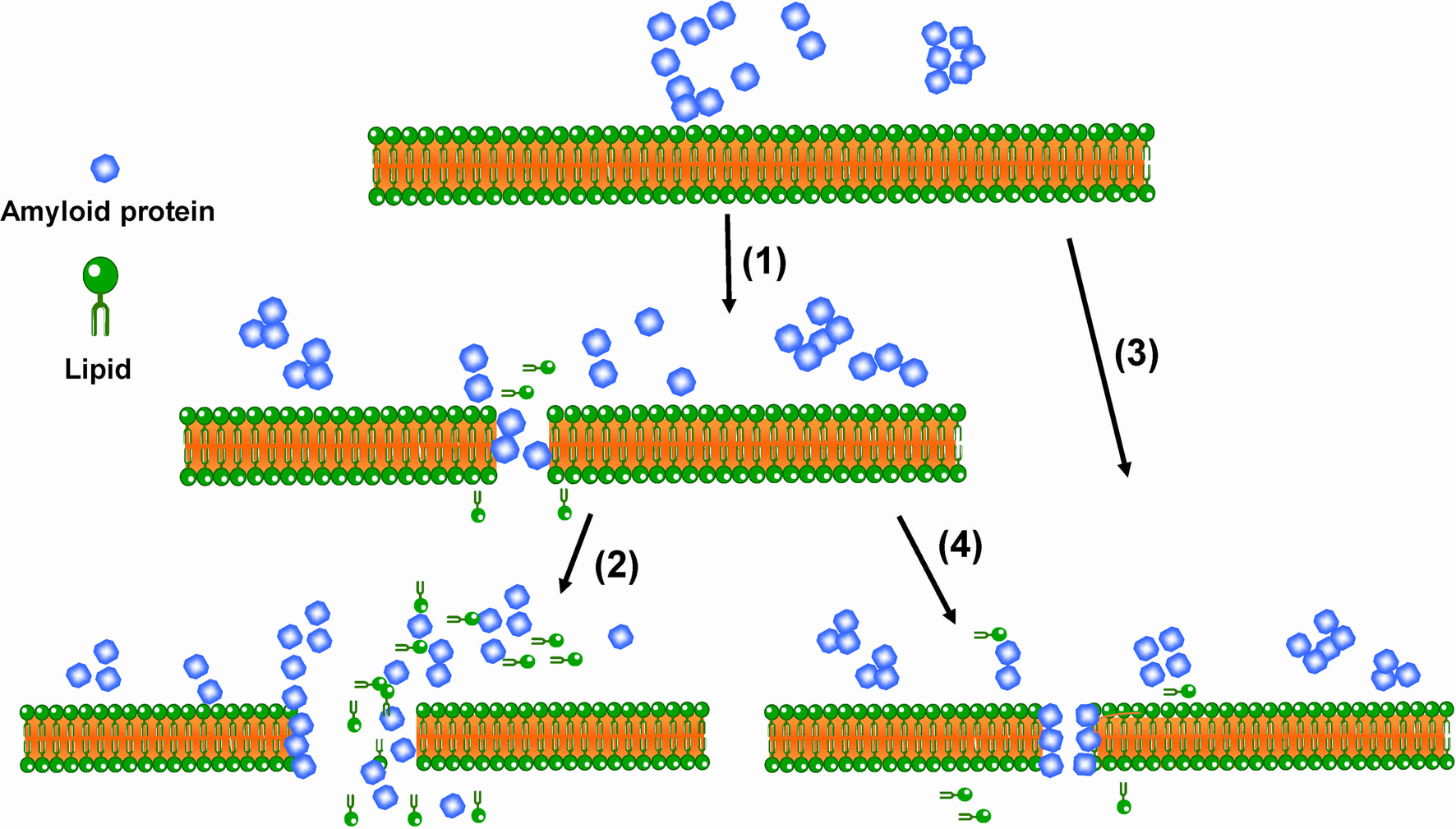
Amyloid fibril formation has been proposed as a ‘nucleation-dependent polymerization’ process (Chiti and Dobson, Reference Chiti and Dobson2006), where the kinetics falls into three phases: the lag phase, the elongation and the final plateau. First, toxic oligomers form in the initial lag period and may act as a template to nucleate amyloid fibril formation in the later phases. Likewise, amyloid fibrils facilitate the formation of new fibrils through a process of secondary nucleation (Jahn and Radford, Reference Jahn and Radford2008; Eichner and Radford, Reference Eichner and Radford2011; Tipping et al., Reference Tipping, Van Oosten-Hawle, Hewitt and Radford2015). A number of studies have indicated that soluble oligomers or protofibrils are the main contributing factor of the cellular toxicity rather than the final-step mature fibrils (Klein et al., Reference Klein, Krafft and Finch2001; Walsh and Selkoe, Reference Walsh and Selkoe2007; Benilova et al., Reference Benilova, Karran and De strooper2012). Although consensus has not been reached on the pathogenesis of amyloids, a prevalently accepted mechanism states that the early aggregates interact with lipid membranes, disrupt the integrity of cell membranes, resulting in uncontrolled extracellular Ca2+ influx and imbalance of other biomolecules (Lashuel and Lansbury, Reference Lashuel and Lansbury2006; Williams and Serpell, Reference Williams and Serpell2011; Lee et al., Reference Lee, Kim, Arce, Gillman, Jang, Kagan, Nussinov, Yang and Lal2017). However, several questions remain to be answered. For example, what sizes of the aggregates are responsible for the damage to cellular membranes? What are the toxic mechanisms by which amyloid oligomers can disrupt cellular membranes? Do these aggregates thin the membranes, form pores or step-wise increase membrane permeability, or a combination?
There are several reasons why these questions remain to be answered. First, it is difficult to detect individual oligomers, because the fibrillation process is a dynamic equilibrium process, giving rise to a highly heterogeneous mixture. The heterogeneity hinders the analysis of the size, conformation and structure of the neurotoxic aggregates. In addition, monomers and small aggregates (from dimers to hexamers) in vivo are likely to interconvert in a dynamic equilibrium (Klein et al., Reference Klein, Krafft and Finch2001; Johnson et al., Reference Johnson, Steel and Gafni2014). Moreover, at physiological conditions, amyloidogenic proteins exist at a low concentration and vary between intracellular regions and brain cell types. For example, concentrations of intercellular α-syn might be likely in the low-nanomolar range (Mollenhauer et al., Reference Mollenhauer, Cullen, Kahn, Krastins, Outeiro, Pepivani, Ng, Schulz-Schaeffer, Kretzschmar, McLean, Trenkwalder, Sarracino, VonSattel, Locascio, El-Agnaf and Schlossmacher2008; Hannestad et al., Reference Hannestad, Rocha, Agnarsson, Zhdanov, Wittung-Stafshede and Höök2020), while the soluble Aβ42 were only found at the picomolar levels in human AD brain entorhinal cortex and superior frontal gyrus regions (Lue et al., Reference Lue, Kuo, Roher, Brachova, Shen, Sue, Beach, Kurth, Rydel and Rogers1999; McLean et al., Reference Mclean, Cherny, Fraser, Fuller, Smith, Beyreuther, Bush and Masters1999). Such low concentrations are below the detection capability of conventional biophysical assays. Using higher (non-physiological) concentration in the assays may result in completely different interactions or pathways that may not representthe in vivo processes. In addition, the conventional biophysical techniques such as fluorescence assay and circular dichroism (CD) (Whitmore and Wallace, Reference Whitmore and Wallace2008; Ruggeri et al., Reference Ruggeri, Adamcik, Jeong, Lashuel, Mezzenga and Dietler2015) represent the averaged data of whole populations, smoothing out the low ‘noise’ signal from an individual aggregate (Lord et al., Reference Lord, Lee and Moerner2010). These ensemble methods are therefore not ideal for the investigation of the energetically metastable, heterogeneous states of amyloid proteins. To overcome some of these limitations, single-molecule (SM) amyloid characterization at the physiological concentration is thus of great importance.
SM techniques (SMTs) have unique advantages of recording the behavior of individual molecules in a heterogeneous sample (Seisenberger et al., Reference Seisenberger, Ried, Endreß, Büning, Hallek and Bräuchle2001; Zhang et al., Reference Zhang, Ha and Marqusee2018). The required concentration for most SMTs is close to the endogenous level (pM to nM). Such conditions assist in the formation of stabilized heterogeneous and transient oligomers at a time that allows studying their structures and dynamics, which are easily hampered or altered at higher concentrations (Mukhopadhyay et al., Reference Mukhopadhyay, Krishnan, Lemke, Lindquist and Deniz2007). Moreover, the experimental parameters of SMTs can reach a nanometer spatial resolution and a microsecond temporal resolution (Greenleaf et al., Reference Greenleaf, Woodside and Block2007). SMTs may give the appropriate and accurate analysis of transient amyloid aggregates at the SM level. SMTs can be classified into three types: SM fluorescence-based imaging (SMFI), SM force spectroscopy (SMFS) and single-nanopore electrical systems (SNSs). SMFI methods are localization-based imaging techniques that can normally track fluorophore-tagged single particles or vesicles without perturbation (Shashkova and Leake, Reference Shashkova and Leake2017; Hannestad et al., Reference Hannestad, Rocha, Agnarsson, Zhdanov, Wittung-Stafshede and Höök2020). SMFS generally applies the tension or torsion to investigate the mechanical responses of biological systems (Neuman and Nagy, Reference Neuman and Nagy2008). SNSs allow analyzing the behavior of both small- and large-sized oligomers in a single nanopore system by recording the dwell time and residual current when they assemble into nanopore (Houghtaling et al., Reference Houghtaling, List and Mayer2018). The principles of these techniques and their applications on amyloid proteins will be discussed in further detail in section ‘SMTs for biophysical studies of amyloid proteins’.
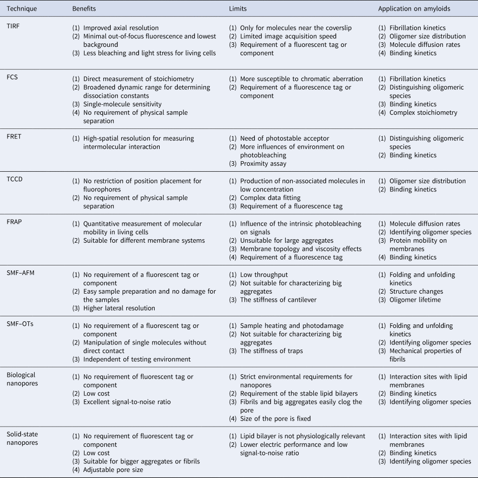
Our paper aims to give an overview of the current understanding of the molecular mechanisms involved in amyloid misfolding, by focusing on SM approaches in contrast to the more commonly used bulk or ensemble methods. In particular, we focus on the application of the three main types of SMTs mentioned above for the characterization of the biophysical properties of amyloid proteins. Here, we will briefly state the working principles, benefits and limitations of each SMT, and combined SM dual measurements will be briefly discussed in this review. We will also summarize the detailed insights they can give for different species of amyloid proteins, the interaction with membranes and their roles in amyloid diseases. In addition, our paper covers the perspectives of each SMT for the diagnosis of amyloidosis.
SMTs for biophysical studies of amyloid proteins
In comparison to the conventional methods studying ensemble systems, SM approaches provide insights into the heterogeneity of amyloid proteins and distinguish contributions of different amyloid oligomers, and therefore hold the potential to gain a deeper understanding of the structure and function of different amyloid morphologies. We subsequently discuss three main types of SMTs and their variants in three separate sections and show how these techniques have been relevant to amyloid protein studies at the molecular level.
SM fluorescence imaging (SMFI)
Fluorescence imaging is based on the emission of light by a fluorophore. After the excitation of an electron to a higher energy state, the fluorophore relaxes back to the ground state and re-emits a new photon. A protein of interest can be directly observed with a non-covalent fluorophore or covalent fluorophore after conjunction with a probe (Jaiswal et al., Reference Jaiswal, Mattoussi, Mauro and Simon2003; Keppler et al., Reference Keppler, Gendreizig, Gronemeyer, Pick, Vogel and Johnsson2003). SMFI techniques visualize the changes of fluorescent properties of labeled proteins such as fluorescence lifetime, quantum yield or polarization to reveal intermolecular interactions, reaction kinetics and conformational dynamics.
Total internal reflection fluorescence (TIRF), fluorescence resonance energy transfer (FRET), two-color coincidence detection (TCCD), fluorescence recovery after photobleaching (FRAP) and fluorescence correlation spectroscopy (FCS) are common fluorescence-based methods for tracking single particles. For an overview of the history and some of the recent discoveries in the life sciences made via SMFI, we refer to the review by Shashkova and Leake (Reference Shashkova and Leake2017). In this review, we will focus on the implementation of these techniques for amyloid protein studies.
TIRF imaging
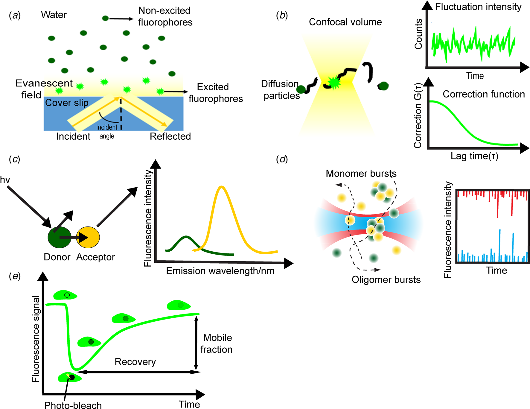
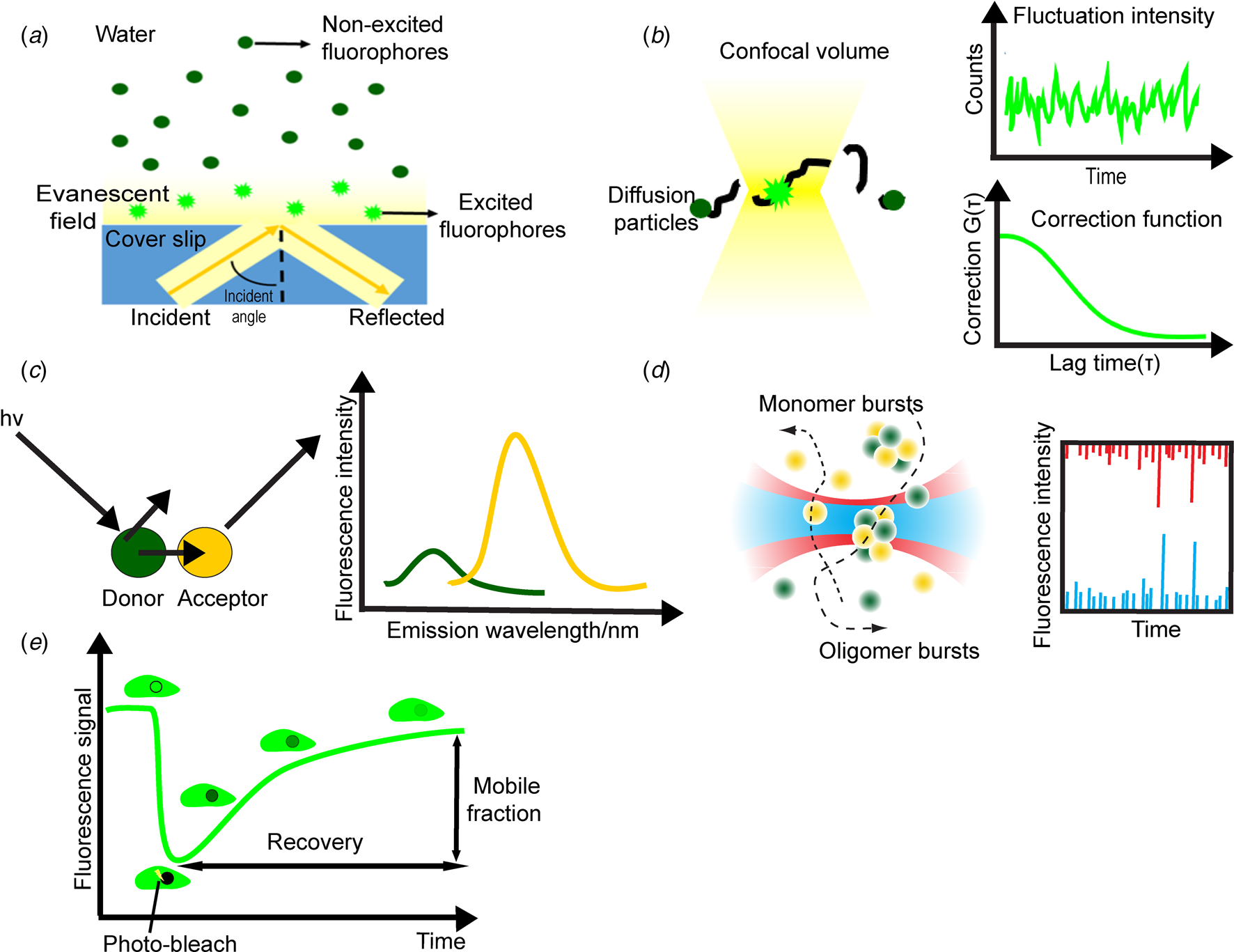
TIRF imaging is now one of the most common fluorescence microscopic imaging to track single particles by selectively illuminating one area to minimize background signals. The innovation of TIRF has greatly enhanced the signal-to-noise ratio by reducing the background fluorescence, resulting in better contrast for detecting single molecules (Schneckenburger, Reference Schneckenburger2005). For TIRF, a solid–liquid interface with different refractive indexes, such as a glass coverslip/slide and a water-based physiological solution, is needed (Fig. 1a) (Shashkova and Leake, Reference Shashkova and Leake2017). The incidence angle with a 90° refraction is called a ‘critical angle’. At larger angles than the critical angle, the excitation light will not transmit through the liquid surface, instead, a total internal reflection of the incident light occurs at the interface and an ‘evanescent field’ is created. The intensity from this field drops off exponentially with the increasing distance from the interface whereas the depth of penetration can actually be adjusted over a wide range. Therefore, TIRF imaging can restrict the excitation to fluorophores that are positioned close to the surface, making this technique particularly suitable for identifying molecules integrated into membranes.
Schematic illustrations of the basic concept of SMFI techniques. (a) TIRF uses total internal reflection at the interface where an ‘evanescent field’ is produced and the fluorophores close to the glass surface are efficiently excited by the evanescent field. (b) In FCS method, fluorescent molecules diffuse through a tiny confocal volume and the fluctuations of fluorescence intensity are detected to determine the number of particles in this area. The autocorrelation function provides information about the concentration and the thermodynamic and kinetic parameters. (c) FRET represents a non-radiative energy transfer between a donor and an acceptor chromophore molecule via the long-range dipole–dipole coupling when these molecules are in close proximity (left). The donor fluorescence is quenching while the emission of the acceptor is increasing (right). (d) TCCD is based on oligomers with two distinct fluorophores but cannot exhibit high FRET give coincident fluorescent bursts after emitting the photons (left). The fluorescence intensity derived from the two-color channels (red and blue) presents the numbers of monomers that are labeled in one oligomer (right). (e) FRAP can be used to quantify the recovery of fluorescence intensity over time after a region of interest in a cell or tissue is photo-bleached with a high-intensity laser and subsequently probed by a low-power laser.
TIRF has been applied for the direct visualization of amyloid aggregates. As using covalently labeling proteins might affect the aggregation process (Zheng et al., Reference Zheng, Xu, Yang, Peng, Wang, Yu, Hua, Zhao, He and Hong2018) and it is challenging to tag samples extracted from in vivo sources, Horrocks et al. (Reference Horrocks, Lee, Gandhi, Magdalinou, Chen, Devine, Tosatto, Kjaergaard, Beckwith, Zetterberg, Iljina, Cremades, Dobson, Wood and Klenerman2016) relied on the addition of a dye (Thioflavin-T (ThT)) that non-covalently binds to oligomers and fibrils with β-sheet structures. Small β-sheet aggregates of α-syn, Aβ and tau in vitro are observed with the sizes <250 nm at the beginning of aggregation and the enriched oligomers contain on average 25 monomers. Amyloid fibrillation kinetics can be tracked as well. In the presence of ThT, Yuji's group (Ban et al., Reference Ban, Hoshino, Takahashi, Hamada, Hasegawa, Naiki and Goto2004) clearly observed the real-time individual Aβ40 fibrils growth with a speed-dependent manner and found the constant rate of fibril growth is similar with the steady-state enzymatic kinetics. Another study showed that glucagon fibrils grow exclusively by forming branches along sides of the parent fibril with an angle of 35°–40° (Andersen et al., Reference Andersen, Yagi, Manno, Martorana, Ban, Christiansen, Otzen, Goto and Rischel2009), which differs from Aβ fibrils that extend themselves from the end of the preformed seeds in a linear elongation. By using TIRF, Patil et al. (Reference Patil, Mehta, Jha and Alexandrescu2011) revealed three different human islet amyloid polypeptide (hIAPP) aggregation patterns that demonstrate the role of secondary nucleation mechanisms where the preformed fibrils serve as a new template for fibril formation. All these studies illustrate that TIRF microscopy is an effective tool to visualize entire aggregates and fibril growth kinetics.
TIRF was also applied to depict the permeation of single vesicles or cells filled with fluorescent dye by measuring the Ca2+ influx induced by oligomers on the membranes. After the penetration of synthetic Aβ oligomers (down to 200 pM) into the live neuronal astrocyte cells (Drews et al., Reference Drews, Flint, Shivji, Jönsson, Wirthensohn, De Genst, Vincke, Muyldermans, Dobson and Klenerman2016; Flagmeier et al., Reference Flagmeier, De, Wirthensohn, Lee, Vincke, Muyldermans, Knowles, Gandhi, Dobson and Klenerman2017), tetramers or larger oligomers were found as the most toxic Aβ oligomer aggregates across the membranes. This suggests that membrane permeabilization is one of the toxic mechanisms induced by oligomers. The extracellular chaperone clusterin, antibody or nanobody that specifically binds to oligomers could prevent Ca2+ influx induced by synthetic Aβ oligomers (Drews et al., Reference Drews, De, Flagmeier, Wirthensohn, Chen, Whiten, Rodrigues, Vincke, Muyldermans, Paterson, Slattery, Fox, Schott, Zetterberg, Dobson, Gandhi and Klenerman2017), whereas these inhibition effects are not obvious under cerebrospinal fluid (CSF) conditions. This implies that these SM assays might be a cost- and time-effective approach before clinical trials. Recently, with TIRF, Tanaka and coworkers showed that Aβ42 oligomers on the surface of live hippocampal neurons impair the ability of learning and memory in the early stage of AD via suppressing the increase of fluorescently labeled (glutamate receptor) GluA1 number and exocytosis frequency in both post-synaptic and extra-synaptic membranes (Tanaka et al., Reference Tanaka, Sakaguchi and Hirano2019). The increase of GluA1 is important for the cellular basis of synaptic plasticity (Jarosz-Griffiths et al., Reference Jarosz-Griffiths, Noble, Rushworth and Hooper2016). Based on this result, a feasible therapy to recover mild memory impairment could be to target the increase of exocytic GluA1 on neuronal cells and TIRF could be an effective imaging tool to validate this potential.
A novel approach, label-free scattering microscopy has been used to image the surface-bound lipids and detect the specific protein-binding to single vesicles without labeling the lipids (Agnarsson et al., Reference Agnarsson, Lundgren, Gunnarsson, Rabe, Kunze, Mapar, Simonsson, Bally, Zhdanov and Höök2015). Scattering microscopy produces the ‘evanescent field’ by optical waveguide instead of using the objective in TIRF. In this method, a more uniform illumination profile is obtained. Recently, Hannestad et al. investigated the kinetics of monomeric α-syn interaction with different lipid vesicles under both fluorescence and light-scattering emission at the single-vesicle level. They found α-syn induces obvious step-wise structural changes and asymmetric membrane deformation of individual phosphatidylglycerol vesicles with a very low α-syn concentration 10 nM (~100 proteins per vesicle) (Hannestad et al., Reference Hannestad, Rocha, Agnarsson, Zhdanov, Wittung-Stafshede and Höök2020). The pore formation mechanism was not concluded to result in membrane leakage when α-syn binds to the vesicles. This finding might enlighten the abnormal behavior of monomeric α-syn in vivo. Scattering microscopy was also used to distinguish misfolded proteins from normal ones in Aβ plaques of an AD mouse brain model (Ji et al., Reference Ji, Arbel, Zhang, Freudiger, Hou, Lin, Yang, Bacskai and Xie2018). Aβ plaques were found to form a typical core–shell structure that is surrounded by a halo-like shell of lipid-rich deposits (Schweikhard et al., Reference Schweikhard, Baral, Krishnamachari, Hay and Fuhrmann2019). This result indicates the ability of scattering microscopy in providing novel perspectives into the biochemical constitution of pathological structures.
FCS method
FCS is a quantitative analysis method with the ultimate SM sensitivity for the detection of fluorescent molecules (Rigler, Reference Rigler2010; Elson, Reference Elson2018). In FCS, the fluctuations of fluorescence intensity, originating from the diffusion of fluorescent molecules (physiological nanomolar to picomolar) through a very tiny confocal volume (~1 μm3) is measured with a high-temporal resolution (nanosecond time scale) (Fig. 1b). The detected fluorescence real-time fluctuations and autocorrelation function provide the thermodynamic or kinetic parameters such as diffusion coefficient (DC) or time, nucleation boundary radius and the free energy barrier for nucleation (Medina and Schwille, Reference Medina and Schwille2002; Garai et al., Reference Garai, Sahoo, Sengupta and Maiti2008; Kitamura and Kinjo, Reference Kitamura and Kinjo2018). Knowing these parameters can assist in developing an exact thermodynamic model of Aβ aggregation. Moreover, understanding the energy landscape of amyloids may estimate the stability of intermediates, which could help the quest for a method to reduce its population (Garai et al., Reference Garai, Sengupta, Sahoo and Maiti2006).
FCS has been used to characterize the amyloids aggregation process and determine the presence of different oligomeric species, both in well-equilibrated systems in vitro and more complicated environments in vivo (Liang et al., Reference Liang, Lynn and Berland2010; Guan et al., Reference Guan, Cao, Cantlon, Elbel, Theodorakis, Walsh, Yang and Shah2015; Tiiman et al., Reference Tiiman, Jarvet, Gräslund and Vukojević2015). Mittag et al. (Reference Mittag, Milani, Walsh, Rädler and Mcmanus2014) fitted FCS data to Gaussian distribution models to clearly distinguish different Aβ oligomeric species in vitro. They estimated the sizes of different amyloid types, including monomer (<1 nm) and low-molecular-weight oligomers (1–5 nm), high-molecular-weight oligomers (5–40 nm), protofibrils (40–4000 nm) and fibrils (4000–20 000 nm). Additionally, Rigler's group observed that Aβ peptides have a highly cooperative polymerization process where the fibril is predominantly built up from monomers or dimers rather than free oligomers (Tjernberg et al., Reference Tjernberg, Pramanik, Björling, Thyberg, Thyberg, Nordstedt, Berndt, Terenius and Rigler1999). The size of small Aβ aggregate particles in vitro is 1.7–2.1 nm and big aggregates have sizes of 100 nm or higher (Garai et al., Reference Garai, Sureka and Maiti2007). Tiiman et al. (Reference Tiiman, Jarvet, Gräslund and Vukojević2015) later suggested that the smallest ThT-visible Aβ aggregates in vitro with a diffuse time ~200 μs, is comprised of 40–70 Aβ monomers. By directly tracking Aβ42 concentration changes, Novo et al. (Reference Novo, Freire and Al-Soufi2018) found that the critical aggregation concentration of Aβ42 to form oligomers is about 90 nM. Based on this, they suggested that the assembly process is spontaneous and cooperative which is similar to the process of surfactants self-assembling and produces the stable micelle-like oligomers formed from 50 monomers (hydrodynamic radius, R h, 7–11 nm). In terms of complex samples, with FCS, Pitschke et al. (Reference Pitschke, Prior, Haupt and Riesner1998) detected the very large multimers of single Aβ aggregates in all CSF samples from 15 AD patients but in none of the 19 age-matched healthy controls. Funke et al. (Reference Funke, Birkmann, Henke, Görtz, Lange-Asschenfeldt, Riesner and Willbold2007) also easily differentiated three CSF AD samples from the control ones by the presence of Aβ aggregates. FCS is also suitable for identifying the inhibitors of the initial formation of amyloid oligomers by analyzing the average size and number of oligomeric particles. D-enantiomeric amino acid peptides were demonstrated to reduce the average size of Aβ aggregates but increase their number (Wiesehan et al., Reference Wiesehan, Stöhr, Nagel-Steger, Van Groen, Riesner and Willbold2008). These results demonstrate the effectiveness of FCS in determining heterogeneous oligomeric species. FCS can also be used to quantitatively investigate the binding of amyloid proteins on membranes. Fluorescently labeled α-syn has been demonstrated to preferentially bind to acidic vesicles (Miraglia et al., Reference Miraglia, Ricci, Rota and Colla2018). Less bulky head groups of anionic lipids enhance this binding possibly due to the anionic lipids being able to pack more closely together, thus producing a high-charge density (Rhoades et al., Reference Rhoades, Ramlall, Webb and Eliezer2006; Middleton and Rhoades, Reference Middleton and Rhoades2010). The requirment of binding a single α-syn molecule is a minimum of 30 lipid molecules or a 60-molecule bilayer patch (Rhoades et al., Reference Rhoades, Ramlall, Webb and Eliezer2006). Additionally, α-syn prefers to bind small vesicles such as synaptic vesicles, over large multi-lamellar vesicles. It was suggested that the ideal match of vesicle curvature with α-syn would be as found in the N-terminal helix (Middleton and Rhoades, Reference Middleton and Rhoades2010). Another study showed that labeled Aβ40 and Aβ42 oligomers exhibit stronger binding to neuronal and somatic cell membranes than monomers at physiological concentrations (250 nM) (Sarkar et al., Reference Sarkar, Das and Maiti2013). The negligible binding to cell membranes by monomers explains why they are non-toxic. Additionally, on mammalian cell membranes, the size distribution of small Aβ40 aggregates (2.3 nm average R h) peaks at both tested concentrations of 150 and 350 nM, while only large assemblies of Aβ40 (≫10 nm average R h) appear at a concentration of 350 nM (Nag et al., Reference Nag, Chen, Irudayaraj and Maiti2010). These results indicate that the formation of large Aβ complexes on living cells relies on Aβ concentration. This discovery might partially explain why the Aβ targeting drugs fail in clinical trials since the much higher physiological concentration of Aβ is used in vitro. Recently, Simpson et al. (Reference Simpson, Szeto, Boukari, Good and Leach2020) identified differences of Aβ aggregation and cytotoxicity between 2D in vitro models (typically culture cells on flat plastic or glass substrates) and 3D environments, such as collagen hydrogels in tissue culture models. Combined with FCS, they found that the size of Aβ aggregates in 3D collagen hydrogel is 25–200 times larger in diameter than 2D culture, suggesting a vastly different aggregation process. It appears that the cytotoxicity of Aβ gets attenuated in 3D compared to 2D environments. The difference between crowding and confinement is the main contribution. This discovery might provide another reason for failed Aβ drug screening models that might miss these two factors.
Fluorescence cross-correlation spectroscopy (FCCS), which is an extension of FCS, was developed by Schwille et al. (Reference Schwille, Meyer-Almes and Rigler1997). In FCCS, there are two differently labeled fluorescent species whose concentration and diffusion characteristics are measured in parallel. In addition, information about whether the two molecules interact can be obtained. The principle is the same with FCS, but the overlapping diffraction-limited confocal volumes exists in FCCS due to two different lasers focusing onto the same spot. FCCS has been used to study the interaction of aggregation inhibitors such as antibodies (Bonito-Oliva et al., Reference Bonito-Oliva, Schedin-Weiss, Younesi, Tiiman, Adura, Paknejad, Brendel, Romin, Parchem, Graff, Vukojević, Tjernberg, Terenius, Winblad, Sakmar and Graham2019) and kinesin-1 (Zheng et al., Reference Zheng, Tian, Peng, Yang, Fu, Jiao, Zhao, He and Hong2016) with Aβ peptide. Two kinesin-1 subunits, KLC-E and E-KLC, were tagged to determine the interaction of kinesin-1 on Aβ peptide at the SM level (Zheng et al., Reference Zheng, Tian, Peng, Yang, Fu, Jiao, Zhao, He and Hong2016). Data revealed both KLC-E and E-KLC have an inhibitory effect on Aβ42 aggregation over a period of time, and KLC-E shows a much higher binding affinity than E-KLC, which might result from the spatial conformation of KLC-E.
FRET, TCCD and FRAP techniques
FRET is one of the most commonly used techniques when studying physical interactions between two molecules within several nanometers (30–70 Å range), a distance sufficiently close for intermolecular interactions to occur (Roy et al., Reference Roy, Hohng and Ha2008; Schuler and Eaton, Reference Schuler and Eaton2008). The principle of FRET is based on the excitation energy transfer from a donor to an acceptor chromophore molecule with the reduced donor fluorescence/lifetime and increased emission of acceptor fluorescence/lifetime. For protein interaction studies, fluorophore-tagged antibodies or fusion proteins are the two common methods to adhere to the fluorophores to the interacting proteins.
Based on the direct observation of coincident bursts of oligomer fluorescence, two distinct structural types of α-syn oligomers were inferred during α-syn aggregation in vitro (Cremades et al., Reference Cremades, Cohen, Deas, Abramov, Chen, Orte, Sandal, Clarke, Dunne, Aprile, Bertoncini, Wood, Knowles, Dobson and Klenerman2012). Oligomers of types A and B, with medium and higher FRET efficiencies, respectively, were found at α-syn aggregation different stages. Type B oligomers with a more compact structure and proteinase-K resistance were monitored in later aggregation stage, indicating type A might undergo a structural reorganization to form type B. Structural conversion has been assumed to be the key step of α-syn aggregation. This result is consistent with the hypothesis of the sequential finding of discrete oligomeric intermediates in the early stage of α-syn aggregation (Conway et al., Reference Conway, Lee, Rochet, Ding, Williamson and Lansbury2000). Additionally, they detected toxic α-syn oligomers originating from disaggregation of mature α-syn fibrils, suggesting that the disaggregation of fibrils can also be a source of toxic species and result in toxicity (Cremades et al., Reference Cremades, Cohen, Deas, Abramov, Chen, Orte, Sandal, Clarke, Dunne, Aprile, Bertoncini, Wood, Knowles, Dobson and Klenerman2012). This implies that attention should also be paid to the disaggregation of fibrils at the final stage beside the toxic forms in the early stage of the aggregation process. Later, Horrocks and co-workers combined fast flow microfluidics with FRET to investigate the oligomerization of α-syn (Horrocks et al., Reference Horrocks, Tosatto, Dear, Garcia, Iljina, Cremades, Dalla Serra, Knowles, Dobson and Klenerman2015). Isolating type A from type B oligomers by dilution, they discovered that type A dissociates but type B stabilizes in the lower ionic strength of the solution. Moreover, type B oligomers from two α-syn mutant forms (A53T and A30P) displayed different FRET efficiencies, suggesting that the variants contribute to the structural diversity of oligomeric ensembles (Tosatto et al., Reference Tosatto, Horrocks, Dear, Knowles, Dalla Serra, Cremades, Dobson and Klenerman2015). By systematic analysis of the FRET efficiency values in a wide range of starting α-syn monomer concentration (from 0.5 to 140 μM), the required number of seeds to affect the initial aggregation was predicted to be in the order of 104 species in a single cell (10 μm)3 (Iljina et al., Reference Iljina, Garcia, Horrocks, Tosatto, Choi, Ganzinger, Abramov, Gandhi, Wood, Cremades, Dobson, Knowles and Klenerman2016b). This demonstrates that FRET is suitable to quantitatively explore the seeding process of amyloid aggregation. The same setup also demonstrated that tau protein has a similar structural trajectory evolving from type A to type B oligomers prior to fibril formation (Shammas et al., Reference Shammas, Garcia, Kumar, Kjaergaard, Horrocks, Shivji, Mandelkow, Knowles, Mandelkow and Klenerman2015). Combining FRET with the FCS method, the fraction of small Aβ oligomer containing <10 Aβ molecules were detectable as low as 2 ± 2% in solution at physiological concentrations (Wennmalm et al., Reference Wennmalm, Chmyrov, Widengren and Tjernberg2015). Additionally, Kaminski Schierle et al. (Reference Kaminski Schierle, Bertoncini, Chan, Van Der Goot, Schwedler, Skepper, Schlachter, Van Ham, Esposito, Kumita, Nollen, Dobson and Kaminski2011) found that labeled α-syn in living worm Caenorhabditis elegans has the slowest decrease in fluorescence lifetime, followed by in vitro samples, then by neuronal cells. It indicates that the aggregation rate of α-syn is environmentally-dependent with the fastest rate of aggregation in cells. This result shows that FRET is an effective tool to track the kinetics of α-syn self-assembly in vitro and in vivo, and the molecular environment should be taken into consideration when revealing the nature of aggregation species.
Recently, FRET has been used to identify the binding of Aβ oligomers to the cholinergic nicotinic ACh receptor α7 (α7nAChR) (Cecon et al., Reference Cecon, Dam, Luka, Gautier, Chollet, Delagrange, Danober and Jockers2019). Loss of activity of α7nAChR is believed to impair cognitive performance in AD (Bertrand et al., Reference Bertrand, Lee, Flood, Marger and Donnelly-Roberts2015). The result showed that the Aβ binding site on α7nAChR overlaps with the orthosteric ligand binding sites comprising loop C with a high-binding affinity (pK D 10.3 ± 0.2). FRET was also used to evaluate different α7nAChR-related compounds of interfering with the binding abilities of Aβ to α7nAChR (Cecon et al., Reference Cecon, Dam, Luka, Gautier, Chollet, Delagrange, Danober and Jockers2019). In addition, FRET was applied for the screening of various compounds against Zn2+–Aβ interaction (Lee et al., Reference Lee, Lee, Kang, Yang, Kim, Ghisaidoobe, Kang, Lee, Lim and Chung2019). Eight natural inhibitors (out of 145 compounds) were demonstrated to have efficient inhibition effects (>80%) and six of them are capable of decreasing the toxicity induced by Zn2+–Aβ on living cells. The two studies indicate that FRET is suitable for measuring the binding activity of peptides/metal ions to amyloid proteins and very effective to assess potential drug candidates against neurodegenerative disorders. Zadorsky's group used FRET to demonstrate that the newly found amyloid protein Toh1, which is localized in the yeast cell wall, could interact with other PrPs Rnq1 and Sup35. The result highlighted that the interactions among different amyloid proteins not only occur between molecules with a similar primary structure but also between proteins that have a close secondary structure and conformational folds (Sergeeva et al., Reference Sergeeva, Sopova, Belashova, Siniukova, Chirinskaite, Galkin and Zadorsky2019).
Besides investigating the interactions of two molecules, FRET is also suitable to explore the interaction of membranes and amyloid proteins. Reynolds et al. found that α-syn aggregation appears to extract lipids from the membranes making them thinner. These extracted lipids could be the initial step to a more intense disruption of membrane integrity. Larger damage to membranes was observed at higher α-syn concentrations, demonstrating the concentration dependency of α-syn aggregates on membrane toxicity (Reynolds et al., Reference Reynolds, Soragni, Rabe, Verdes, Liverani, Handschin, Riek and Seeger2011). Terzi et al. (Reference Terzi, Hölzemann and Seelig1995) reported that Aβ preferentially interacts with anionic membranes and Wong et al. found more net negative charge increases the binding affinity of low-molecular-mass species of Aβ40 (<10-mer) with membranes. The very weak interaction of Aβ40 with the pure dipalmitoylphosphatidylcholine neutral liposomes confirms that Aβ40 membrane insertion is highly dependent on the negative charge of the membrane (Wong et al., Reference Wong, Schauerte, Wisser, Ding, Lee, Steel and Gafni2009). The tight binding of Aβ40 to anionic membranes was further confirmed by TIRF imaging but with a much lower dissociation constant of the peptide from the membrane (<470 pM) (Ding et al., Reference Ding, Schauerte, Steel and Gafni2012), compared to Wong's report (38.8 ± 7.0 μM). The main reason is the very low peptide concentration in the latter case (sub-nanomolar), which leads to a very low peptide/lipid ratio. This result indicates that the ratio of peptide/lipid concentration should be considered when calculating the binding affinities of amyloid proteins with lipid membranes.
In addition to the interaction of membranes with oligomers, the fluorescent lipid membranes were found to directly bind to fibrils and accelerate fibril formation (Gorbenko et al., Reference Gorbenko, Trusova, Girych, Adachi, Mizuguchi, Akaji and Saito2015; Lindberg et al., Reference Lindberg, Wesén, Björkeroth, Rocha and Esbjörner2017). The vesicle lipid–fibril interaction reinforces Aβ42 secondary nucleation at the surface of existing Aβ fibrils (Lindberg et al., Reference Lindberg, Wesén, Björkeroth, Rocha and Esbjörner2017). The charged plain lipid membrane has a similar effect on Aβ fibrillation. Additionally, Widenbrant et al. (Reference Widenbrant, Rajadas, Sutardja and Fuller2006) found the Aβ aggregates bind to the giant vesicle membrane, rapidly cross the vesicle bilayer, then incorporate into the interior small vesicle membrane. Meanwhile, the incorporated membrane was found to possess reduced membrane fluidity, which is indicative of the pathophysiology of disease-affected cells. The reason for membrane penetrations and finally cell death was suggested to be due to induced membrane leakage by the localized aggregates, not due to pore formation mechanisms.
TCCD technique uses two fluorophores that cannot exhibit high FRET and which are illuminated by two lasers compared to one laser for FRET (Orte et al., Reference Orte, Clarke and Klenerman2010). Monomers tagged with a single color are only detected by one of the detecting channels while the oligomers with two distinct fluorophores result in coincident fluorescent bursts after emitting the photons (Fig. 1d, left). The signal of fluorescence intensity derived from the two-color channels presents the numbers of monomers that are labeled in one oligomer (Fig. 1d, right). As different oligomers show different coincident bursts of fluorescence when they diffuse through the confocal volume, TCCD is sensitive and quantitative to provide the information about the concentration and the stoichiometries of associated molecules in a heterogeneous environment. Klenerman's group used TCCD to trace the oligomer ensemble of the SH3 domain from bovine phosphatidylinositol-3-kinase (Orte et al., Reference Orte, Birkett, Clarke, Devlin, Dobson and Klenerman2008). The heterogeneous oligomeric species of SH3 with the most cytotoxic properties have a median size of 38 ± 10 molecules. The size distribution of these oligomers remains constant while their stability is gradually increasing during aggregation. The TCCD method was also applied to determine the interaction of Aβ peptide with neuroserpin, a member of the serine protease inhibitor, which can suppress the toxicity of Aβ peptides (Chiou et al., Reference Chiou, Hägglöf, Orte, Chen, Dunne, Belorgey, Karlsson-LI, Lomas and Klenerman2009). It was found that Aβ increases the rate of neuroserpin's polymerization by forming a weak complex with neuroserpin. As Aβ is later released from the complex and not incorporated into the neuroserpin polymers, it was suggested that Aβ40 acts as a catalyst to enhance polymerization. Iljina et al. (Reference Iljina, Garcia, Dear, Flint, Narayan, Michaels, Dobson, Frenkel, Knowles and Klenerman2016a) quantitatively determined the composition of self- or co-oligomer formation of Aβ40 and Aβ42 under physiological conditions. The main oligomers are Aβ40 at the physiologically relevant ratio of 9:1 of Aβ40–Aβ42. Dimers are the predominant oligomers when the total Aβ concentration is ~1 nM. The oligomerization of α-syn, as well as their co-aggregation with Aβ40, Aβ42 and tau, was also determined by TCCD (Iljina et al., Reference Iljina, Dear, Garcia, De, Tosatto, Flagmeier, Whiten, Michaels, Frenkel, Dobson, Knowles and Klenerman2018). At a very low concentration, close to the physiological conditions, co-oligomerization is more favorable than pure α-syn oligomer formation, but co-oligomers also showed lower lipid membrane disruption than α-syn assembled oligomers.
FRAP is another SMFI technique to measure molecular mobility and turnover processes. The basic principle of FRAP is that a region of interest in a cell or tissue is photo-bleached with a high-intensity laser and subsequently probed by a low-power laser to quantify the recovery of fluorescence intensity over time (Fig. 1e). The recovery curve provides information about the mobile fraction and the DC of the labeled component (Axelrod et al., Reference Axelrod, Koppel, Schlessinger, Elson and Webb1976). Nonetheless, specific interactions, membrane topology and viscosity can also lead to differences in DCs (Reits and Neefjes, Reference Reits and Neefjes2001). Thus, great considerations should be taken when performing FRAP experiments in different cell lines or compartments to compare DCs and mobile fractions.
By counting the number of photobleaching steps, FRAP can directly distinguish the labeled Aβ oligomers with a different number of subunits at a very low concentration (1 nM). Combined with confocal microscopy, Ding et al. differentiated labeled Aβ40 oligomers that were deposited onto a cover glass and found the size varies from dimers to hexamers, but mainly the dimers, trimers and tetramers. This result is in agreement with the peak estimation from gel filtration chromatography (Ding et al., Reference Ding, Wong, Lee, Gafni and Steel2009). Nevertheless, some oligomer species such as pentamers and hexamers are only observed by FRAP but not the gel filtration method, indicating the advantage of SMTs on quantitatively measuring oligomer species. Additionally, the determination of Aβ oligomer species on lipid membranes can also be obtained by forming the lipids on the clean cover glass. It was shown that the sizes of formed oligomers highly depend on the concentration of amyloids on lipid membranes. At close to physiological concentration (2 nM), a large fraction of Aβ species (34%) on supported anionic lipid membranes are trimer/tetramer (Ding et al., Reference Ding, Schauerte, Steel and Gafni2012). Similar smaller pore-forming oligomers were observed directly by high-resolution atomic force microscopy (AFM; Quist et al., Reference Quist, Doudevski, Lin, Azimova, Ng, Frangione, Kagan, Ghiso and Lal2005), indicating these species might be the smallest in the membrane and the source of pre-pore forming oligomers and neurotoxicity. At a relatively high concentration (100 nM), faster oligomer formation and larger size distribution with a very wide range from around pentamers to 60-mer were observed. In this situation, the fast forming oligomers might serve as the nuclei for the direct insertion into a membrane or for the initial formation of fibrils and then lead to membrane permeabilization (Ding et al., Reference Ding, Schauerte, Steel and Gafni2012). Additionally, combined with TIRF, they found the binding to anionic lipid membranes starts with the monomers and the subsequent incubation leads to the formation of oligomers in the membranes. To get a more detailed picture of the toxic mechanism at the SM level, Schauerte et al. (Reference Schauerte, Wong, Wisser, Ding, Steel and Gafni2010) combined membrane conductivity measurements with FRAP to identify the structure of amyloid proteins when causing membrane-permeabilization. The ability of the oligomers to permeabilize the membrane was determined by simultaneously measuring the conductance as well as their size distributions. At a low concentration below 10 nM, labeled Aβ monomers uniformly bind to the membrane while they do not induce any detectable conductance. At intermediate concentrations (from 10 to 100 nM), small oligomeric Aβ species (mainly dimers to 14-mer) are identified and the smallest size of oligomers to show a detectable conductivity are hexamers but not dimers. When the concentration is >100 nM, large Aβ structures and large-conductance are detected, indicating the serious interruption of these aggregates on membranes. This observation supports the pore-formation model where monomers interact with the membrane surface followed by surface diffusion and then assemble into pore structures. By analyzing DC curves, the change of membrane fluidity, which is important for biological functions in cell membranes (Vereb et al., Reference Vereb, Szöllosi, Matkó, Nagy, Farkas, Vigh, Mátyus, Waldmann and Damjanovich2003; Marguet et al., Reference Marguet, Lenne, Rigneault and He2006), can also be determined when membranes interact with the amyloid peptides (Sasahara et al., Reference Sasahara, Morigaki, Okazaki and Hamada2012, Reference Sasahara, Morigaki and Shinya2013; Schreiber et al., Reference Schreiber, Fischer and Lang2012). hIAPP shows a more dramatic change of average DC signal before and after adding it into the lipid bilayer (from 1.07 ± 0.2 to 0.004 ± 0.004 μm2 s−1), compared to Aβ42 peptide (from 0.99 ± 0.1 to 0.07 ± 0.01 μm2 s−1) (Sasahara et al., Reference Sasahara, Morigaki, Okazaki and Hamada2012; Reference Sasahara, Morigaki and Shinya2013). The deposition of hIAPP on the surfaces of bilayers results in the insertion to the bilayers (Sasahara et al., Reference Sasahara, Morigaki and Shinya2014). The membrane mobility decreases considerably when Aβ42 concentration increases from 10 to 20 μM (Meker et al., Reference Meker, Chin, Sut and Cho2018). These results address the involvement of lipids in amyloid aggregation when proteins penetrate the fluid membranes. FRAP has proved also useful for assessing molecular mobility in living cells. α-syn aggregates in the SH-SY5Y human neuroblastoma cells showed a very low fluorescence recovery signal compared with the area without aggregates, which showed complete signal recovery (Roberti et al., Reference Roberti, Jovin and Jares-Erijman2011). The diffusion constant of α-syn is relatively small (0.03–0.04 μm2 s−1) in living cells, suggesting that the α-syn aggregates remain predominantly immobile. These results demonstrate that FRAP is powerful to investigate the effects of amyloid protein on the membrane fluidity and function.
SM force (SMF) microscopy
Force microscopy (widely, though inappropriately, named as force spectroscopy) is a set of techniques used for the study of individual molecule interactions and the binding forces that include AFM, optical tweezers/traps (OTs) and magnetic tweezers (MTs), which have been extensively reviewed before (Neuman and Nagy, Reference Neuman and Nagy2008; Adamcik and Mezzenga, Reference Adamcik and Mezzenga2012). Here we focus on the techniques that have been used and will be of specific relevance in the future to study single amyloid molecules and their interactions.
SMF–AFM
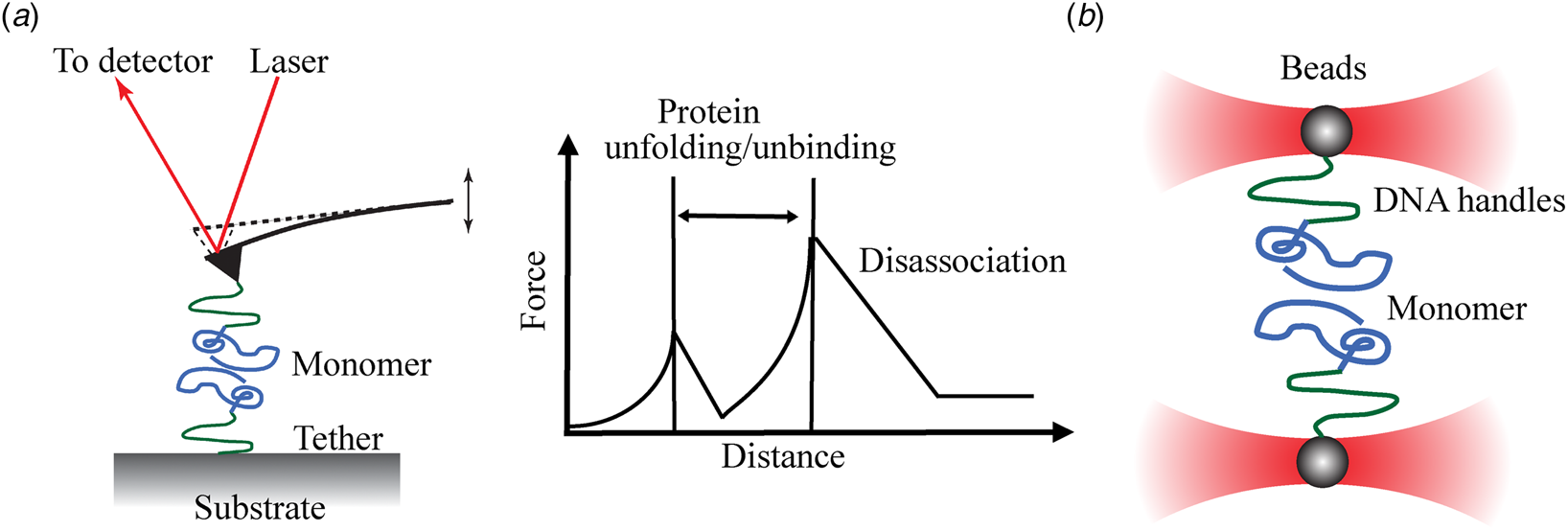
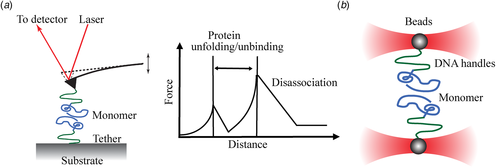
AFM is a type of scanning probe microscopy with a nanometer resolution, which mainly relies on Van der Waals interactions and Coulomb repulsions resulting in a weak deformation of the AFM micro-cantilever with respect to the sample surface (Binnig et al., Reference Binnig, Quate and Gerber1986). When the probe approaches and retracts from the substrate in the vertical direction, the changes of cantilever position will be systematically recorded and represented in a force–distance curve (Fig. 2a). These forces reflect the surface topography and structure of the biological sample. AFM enables the visualization of the microscopic surface of heterogeneous amyloid oligomers and fibrils under ambient atmospheric and liquid conditions, thereby provide information about parameters such as height, diameter and morphology (Adamcik et al., Reference Adamcik, Jung, Flakowski, De Los Rios, Dietler and Mezzenga2010; Adamcik and Mezzenga, Reference Adamcik and Mezzenga2012; Ruggeri et al., Reference Ruggeri, Benedetti, Knowles, Lashuel, Sekatskii and Dietler2018). For example, AFM imaging has been utilized to observe different shapes of amyloid aggregates under atmospheric conditions, including pore-like and spherical-shaped Aβ oligomers (Connelly et al., Reference Connelly, Jang, Arce, Capone, Kotler, Ramachandran, Kagan, Nussinov and Lal2012; Nirmalraj et al., Reference Nirmalraj, List, Battacharya, Howe, Xu, Thompson and Mayer2020). It was also found that the height of α-syn oligomers varies from 2 to 12 nm and is dependent on salt concentration (Hong et al., Reference Hong, Han, Fink and Uversky2011), whereas the height of primarily formed Aβ oligomers varies from 2 to 8 nm (Mastrangelo et al., Reference Mastrangelo, Ahmed, Sato, Liu, Wang, Hough and Smith2006). The height of α-syn fibrils in the air is 17% lower than those of the same fibrils imaged in liquids and the C-terminal tails of α-syn fibrils in liquids show different conformation in different ion concentrations (Sweers et al., Reference Sweers, Van Der Werf, Bennink and Subramaniam2012). Additionally, AFM allows to investigate the effect of lipid composition on amyloids binding to membranes. In the fluid phase, positively and neutrally charged lipid bilayers have stronger interference with Aβ42, compared to the negative and zwitterionic ones (Hane et al., Reference Hane, Drolle, Gaikwad, Faught and Leonenko2011). Lipid rafts cholesterol and ganglioside GM1 promote the harmful membrane interactions with Aβ (Azouz et al., Reference Azouz, Cullin, Lecomte and Lafleur2019). All these results demonstrate that conventional AFM has an irreplaceable advantage in providing morphological information of amyloids. However, traditional AFM imaging cannot show how two molecules interacting with each other affect the aggregation morphology. By tethering individual amyloid peptides/proteins to the substrate and probe, respectively, SM force AFM (SMF–AFM measures the internal mechanical features of small heterogeneous amyloid oligomers (dimers to tetramers). SMF–AFM mechanically unfolds the native molecule at a SM level with pN resolution (Rief et al., Reference Rief, Oesterhelt, Heymann and Gaub1997; Li et al., Reference Li, Zhang, Zhang, Shen, Liu, Gao and Zou1998; Hugel et al., Reference Hugel, Grosholz, Clausen-Schaumann, Pfau, Gaub and Seitz2001). The obtained force curves can be used to characterize the interaction forces between them or to reveal the unfolding pathway of oligomers (Yu et al., Reference Yu, Malkova and Lyubchenko2008, Reference Yu, Dee and Woodside2013; Trexler and Rhoades, Reference Trexler and Rhoades2010; Kim et al., Reference Kim, Palermo, Lovas, Zaikova, Keana and Lyubchenko2011). Lyubchenko's group has covalently immobilized the C-terminal of a series of cysteine-modified amyloid proteins on both probe and substrate surface (Krasnoslobodtsev et al., Reference Krasnoslobodtsev, Volkov, Asiago, Hindupur, Rochet and Lyubchenko2013; Maity and Lyubchenko, Reference Maity and Lyubchenko2020). The pulling strength/rupture forces of monomers between the probe and substrate can provide information about the spontaneous dissociation and the resulting off-rate constant, indicative of the degree of intermolecular interactions and the self-assembly of an amyloid protein dimer (Kransnoslobodtsev et al., Reference Kransnoslobodtsev, Shlyakhtenko, Ukraintsev, Zaikova, Keana and Lyubchenko2005). It has been reported that α-syn dimer has two different dissociation rates (0.25 and 13.2 s−1), implying there might be two distinct pathways of forming α-syn dimers (Yu and Lyubchenko, Reference Yu and Lyubchenko2009). For Aβ40 dimers, four close rupture forces (~120 pN) with different rupture positions from 25 to 45 nm were observed (Kim et al., Reference Kim, Palermo, Lovas, Zaikova, Keana and Lyubchenko2011). It suggested the formation of different transient misfolded states. Aβ42 and Aβ40 display similar rupture forces and rupture position when probed by SMF–AFM (Kim and Lyubchenko, Reference Kim and Lyubchenko2014). The approach further reveals a set of transients of Aβ dimers with different conformations, representing diverse misfolding pathways of Aβ dimers. In addition, the dimer lifetime was quantified by combining dynamics force spectroscopy. Compared to dynamic monomeric states, the dimer lifespan of α-syn and Aβ is unexpectedly high within the range of seconds (Yu et al., Reference Yu, Malkova and Lyubchenko2008; Kim et al., Reference Kim, Palermo, Lovas, Zaikova, Keana and Lyubchenko2011; Kim and Lyubchenko, Reference Kim and Lyubchenko2014), indicating that the stable dimers may act as nuclei in the aggregation process. Also, the amyloid dimer lifetime value is pH-dependent. α-syn has two different lifetimes at acid pH conditions (4 and 0.8 s at pH 2.7, 1.35 and 0.05 s at pH 3.7) (Yu et al., Reference Yu, Malkova and Lyubchenko2008), demonstrating two different pH-dependent α-syn aggregation processes. As for Aβ40 dimer, the lifetime is 0.46 s at pH 7.0 and 2.5 s at pH 2.7, respectively (Kim et al., Reference Kim, Palermo, Lovas, Zaikova, Keana and Lyubchenko2011). Aβ42 has slightly different lifetimes (0.1 s at pH 7.0 and 2.7 s at pH 2.7) (Kim and Lyubchenko, Reference Kim and Lyubchenko2014). The results suggest that experimental conditions play a vital role in the aggregation process. As protein interactions highly depend on their conformational changes, SMF–AFM is suitable for directly detecting the rupture forces to indicate conformational changes of amyloid proteins. It has been shown that the pulling strength of α-syn and Aβ is the weakest at neutral pH, indicating a random coil structure. At an acidic pH, a higher pulling strength revealed the rapid conformation conversion to a more ordered, predominantly β-sheet structure (McAllister et al., Reference Mcallister, Karymov, Kawano, Lushnikov, Mikheikin, Uversky and Lyubchenko2005). Furthermore, SMF–AFM was applied to study Aβ14–23 trimers, where a larger rupture force is required to unravel trimers, compared to that of dimers (Maity and Lyubchenko, Reference Maity and Lyubchenko2016). This explains the different folding arrangements within trimers and dimers. Yu et al. revealed a dramatic increase in rupture events of α-syn interactions in the presence of Al3+ and Zn2+, demonstrating the supportive effect of metal ions on α-syn misfolding. In addition, dopamine does not increase the rupture events and forces, which suggests dopamine may not involve in the α-syn dimerization (Yu et al., Reference Yu, Warnke and Lyubchenko2011). Based on these results, SMF–AFM can be used as an efficient tool to screen drugs or biomolecules that interfere with amyloid aggregation in the early stages of the aggregation. Another advantage is that SMF–AFM can be conducted in a broad pH distribution, which is important as it has been shown that acidic pH dramatically facilitates amyloid aggregation and amyloid monomers and oligomers in vivo normally occur in an acidic environment (Pasternak et al., Reference Pasternak, Callahan and Mahuran2004).
Schematic examples of the basic principles of SMF techniques. (a) One single amyloid protein is attached to the functionalized AFM tip and a mica surface separately. When the probe approaches and retracts from the substrate in the vertical direction, the force–distance curve that is systematically recorded reveals the protein unfolding pathways. (b) Two single amyloid proteins are tethered to two beads constricted in traps. SMF–OTs exert extremely small forces via a highly focused laser beam that generates a very strong electric field gradient.
As trimers, tetramers and larger oligomers also contribute to the heterogeneity of amyloid oligomers in the aggregation process, flexible nanoarrays (FNAs), an extension of SMF–AFM, were developed to investigate the intermolecular interactions of higher oligomeric structures (Tong et al., Reference Tong, Mikheikin, Krasnoslobodtsev, Lv and Lyubchenko2013; Krasnoslobodtsev et al., Reference Krasnoslobodtsev, Zhang, Viazovkina, Gall, Bertagni and Lyubchenko2015; Maity et al., Reference Maity, Viazovkina, Gall and Lyubchenko2017). In the FNA setup, amyloid proteins (dimer or monomer) are covalently immobilized within a highly flexible, linear polyethylene glycol-type polymer on which the tethered proteins can interact and form the higher complex (Maity et al., Reference Maity, Viazovkina, Gall and Lyubchenko2016). The two functionalized ends of the FNA polymer are linked to the AFM tip and substrate, respectively. As the synthesis process of the polymer can control the incorporation of the monomers, the attached amyloid molecules can be anchored to the desired positions within the polymer (Krasnoslobodtsev et al., Reference Krasnoslobodtsev, Zhang, Viazovkina, Gall, Bertagni and Lyubchenko2015). Then different scenarios can be studied. For example, a dimer construct can interact with another monomer, which is tethered to the flexible polymer, and this creates a trimer model to explore the dimer–monomer intermolecular interaction. Analysis of the dissociation force of Aβ14–23 trimers showed that the first rupture force of the monomer is ~48 ± 2.4 pN, which is slightly lower than the second step of rupturing the dimers (~53 ± 3.2 pN) (Maity et al., Reference Maity, Pramanik and Lyubchenko2018). This demonstrated that the dimers are at a more stable state than the dynamic monomers in the early stage of aggregation. Another example is where a dimer construct forms at the end of the flexible polymer, which allows dimer–dimer interactions to be studied (Maity et al., Reference Maity, Viazovkina, Gall and Lyubchenko2017). The results of the rupture force and lifetime of Aβ14–23 tetramers are close to those of trimers at the neutral pH. However, the acid condition affects oligomer behaviors with different rupture forces and lifetimes. At pH 3.7, the lifetime histogram of the tetramer and trimer has a broad distribution and each fits three Gaussian peaks. Different sets of lifetimes indicate different intermolecular interactions with distinct structures. A similar change due to an altered pH was found for the strengths of the bonds. These results are consistent with the known heterogeneity of amyloid oligomers and suggest they can follow different aggregation pathways that are pH-dependent. Similarly, the intermolecular interaction of higher-order oligomerization such as pentamers and hexamers might be studied. FNA allows dissociating the organized oligomers in a stepwise way and provides a good way to reveal the interaction of different states of oligomers, which can contribute greatly to the understanding of the mechanisms involved in amyloid aggregation.
SMF–OTs
Compared to SMF–AFM that probes biomolecules directly, SMF–OTs indirectly manipulate and trap sub-nanometer to micron-sized dielectric particles by generating a very strong electric field gradient via a highly focused laser beam that exerts extremely small forces. SMF–OTs use a high numerical aperture (>1) microscope objective to create a stable optical trap to stabilize the vicinal dielectric microparticles. The working principle is illustrated in Fig. 2b. A single molecule, such as a DNA strand, a peptide or protein, is attached to a microparticle (Jagannathan and Marqusee, Reference Jagannathan and Marqusee2013). SMF–OTs exert a force of up to 100 pN on particles with a 10−8 N force accuracy, a 10−4 s temporal resolution and 10−9 m spatial resolution. By adjusting the position of the traps, the force on the molecule can be indirectly obtained through the real-time measurement of three-dimensional displacement of the particles. In Woodside's group, the Syrian hamster PrP is linked to DNA handles and connected to the polystyrene beads (Yu et al., Reference Yu, Liu, Neupane, Gupta, Brigley, Solanki, Sosova and Woodside2012). A single PrP molecule is trapped and then the end-to-end extension is traced by exerting the denaturing tension on the PrP. The traps are taken apart at a constant rate to ramp up the force until the monomer PrP is totally stretched out. Under force-extension, PrP shows conformational fluctuations in the folding process with high precision. No partially folded intermediate is found to regulate the misfolding event during the native folding process. It suggests that the native state or the on-pathway intermediate is not an important factor for PrP misfolding. Instead, the unfolded states are found to involve three different misfolding off-pathways, which indicate these unfolded states serve as intermediates in the misfolding process. SMF–OTs allow the observation of the transient transitions from the unabiding structural fluctuations of the SM folding trajectory, which makes SMF–OTs a powerful tool to explore the different aspects of amyloid protein misfolding mechanisms.
Based on the above SMF–OTs setup, Neupane et al. (Reference Neupane, Solanki, Sosova, Belov and Woodside2014) found different intermediates with various sizes and stabilities during α-syn aggregation. They reported that many metastable α-syn structures are formed from small oligomers with a wide range of sizes. By monitoring the folding kinetics of a single α-syn monomer, small parts of discrete unfolding events were analyzed by fitting the force-extension curves. The change of contour length in the conformational transitions upon unfolding varies from 10 to 36 nm. These separate unfolding transitions are also observed in α-syn dimers and tetramers, indicating the formation of unfolding intermediates with several distinct structures. Furthermore, the unfolding forces which reflect the structural stability and the height of the energy barrier to unfolding were compared with the monomers, dimers and tetramers. It shows that the unfolding forces increase with the growing size of the oligomer, confirming that larger oligomers of α-syn have higher structural complexity.
Unlike a single amyloid monomer, it is difficult to manipulate one single fibril by an optical-trapping experiment as amyloid fibrils are highly flexible. Dong and coworkers introduced a new method to tether Sup35 PrP fibrils (Dong et al., Reference Dong, Castro, Boyce, Lang and Lindquist2010). They first attached the N-domain of prion monomer variant (NM), which only contains prion N and M regions, onto the glass. Then these tethered monomers exert the self-recognition feature to capture prion fibrils in which the attachment happens at the end of fibrils. The polystyrene bead was used to capture the other end of the fibrils. After applying normal pulling strength, prion fibrils showed resistance to disruption and maintained fibril integrity. When exerting much higher forces of up to 250 pN, some force-dropping events were monitored before the fibril ruptured and it suggests that dropping events result from the prion intrinsic fold properties rather than from the extrinsic surface attachments. SMF–OTs’ results support the β-helix model structure for prion fibrils rather than the β-pleated-sheet model. Based on this method, the mechanical properties of NM from prion fibrils were explored and showed that the persistence length of NM is much shorter than that of insulin and Aβ fibrils (Castro et al., Reference Castro, Dong, Boyce, Lindquist and Lang2011). This result means that NM fibrils are much less resistant to bending than the other amyloids. The formation of point defects (hinges) of the prion fibril structure greatly reduced the bending stiffness. Goldmann previously reported that actin becomes more susceptible to fragmentation when bending decreases (Goldmann, Reference Goldmann2000). Thus, SMF–OTs' results support the hypothesis that prion fibrils are not rigid enough to be fragmented, producing more seeds to function as protein-based elements of inheritance and leading to stronger prion phenotypes in vivo (Castro et al., Reference Castro, Dong, Boyce, Lindquist and Lang2011). Other mechanical parameters such as the axial and torsional stiffness are also deduced by the SMF–OTs method. These results provide meaningful guidance for designing novel amyloid-based nanomaterials including scaffolds and biosensors. Recently, Fränzl et al. (Reference Fränzl, Thalheim, Adler, Huster, Posseckardt, Mertig and Cichos2019) designed a thermophoretic trap to observe the single amyloid fibril growth over rather long periods of time. Analogous to the electric field gradient in the SMF–OTs, the temperature field gradient was created by heating an optically heated, 10-μm-sized chrome ring via a focused laser beam. The trapped single amyloid fibril showed the rare fibril fracture events, confirming the idea of existing secondary processes during the growth of amyloid fibrils. With a similar concept with SMF–OTs, MTs use the gradient of a magnetic field to manipulate paramagnetic beads (Strick et al., Reference Strick, Allemand, Bensimon, Bensimon and Croquette1996; Gosse and Croquette, Reference Gosse and Croquette2002). There are several reviews detailing the principles of MTs and their application at the SM level (Gosse and Croquette, Reference Gosse and Croquette2002; Neuman and Nagy, Reference Neuman and Nagy2008; Sarkar and Rybenkov, Reference Sarkar and Rybenkov2016). Though the technique of MTs is mature to handle, to the best of our knowledge, there is no report where MTs are applied to characterize amyloid proteins.
Single-nanopore electrical systems (SNS)
Single-nanopore electrical recording has been developing into a powerful SM approach to characterize the properties of amyloid proteins (such as structure, aggregation, abundance and interaction with lipid bilayer or other molecules) within an aqueous solution in real-time. There are three classes of nanopores: biological protein nanopores embedded in lipid layers (such as α-hemolysin (α-HL), aerolysin, mycobacterium smegmatis porin A, ClyA and DNA packaging motors), synthetic solid-state nanopores (usually in a SiNx, SiO2, Al2O3, TiO2 or graphene membrane) or a hybrid of these that consists of a protein channel set in the synthetic membrane. The nanopore system confines the disordered protein in a nanoscopic environment of the pores, providing a method to detect the dynamics of a single protein in nanoconfinement (Gurnev et al., Reference Gurnev, Yap, Pfefferkorn, Rostovtseva, Berezhkovskii, Lee, Parsegian and Bezrukov2014). Here, we mainly talk about the application of biological and solid nanopores for the detection of intrinsically disordered proteins.
Biological nanopores
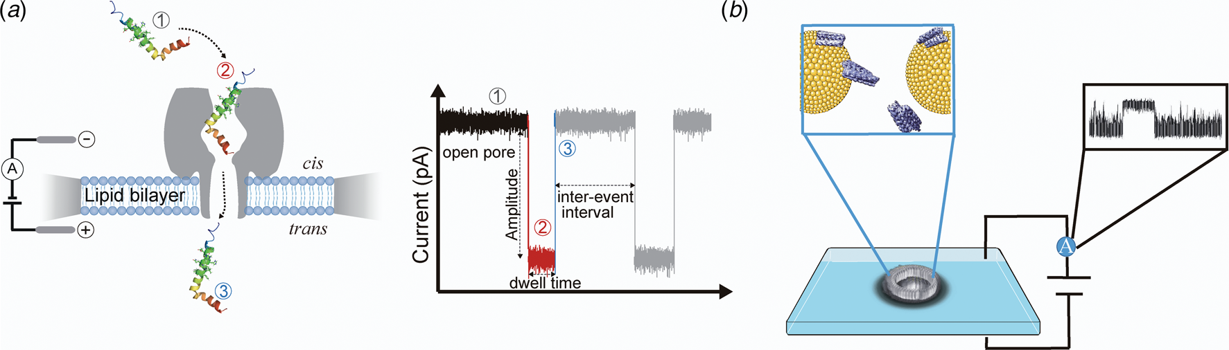
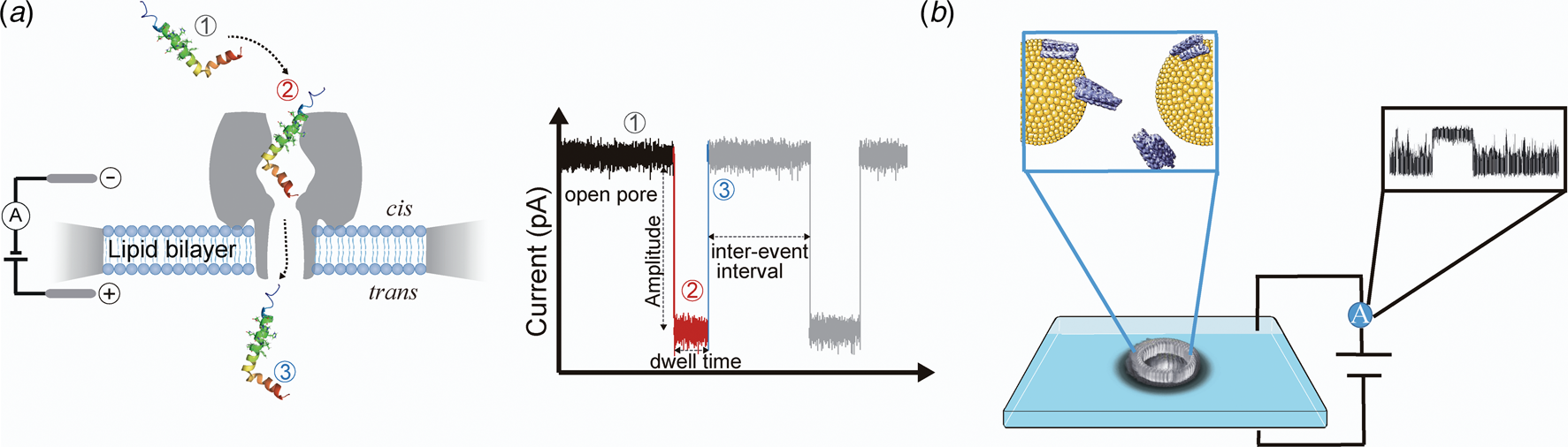
The transportation of ions or molecules into and out of cells depends on channels, which are protein pores in the cell membrane with a size range of 0.3–10 nm. The typical biological nanopore apparatus is composed of two chambers or reservoirs (cis and trans, as illustrated in Fig. 3a) immersed in an electrolyte solution separated by a lipid bilayer film that is formed across an orifice, and a nanopore set in the lipid bilayer. An electric potential applied between the two chambers generates an electric field across the nanopore, which drives charged ions or molecules to pass through the nanopore (Chen and Liu, Reference Chen and Liu2019). For small unfolded molecules, they are easy to translocate the pore, whereas molecules with large sizes, such as aggregates, fibrils or folded proteins, tend to simply clog the pore or bump into the pore and then diffuse away (Madampage et al., Reference Madampage, Tavassoly, Christensen, Kumari and Lee2012). By analyzing the amplitudes, durations and shapes of the events, the size, conformational changes and interactions with other molecules can be determined at a SM level. Recently, single-nanopore technologies have shown great potential for the characterization of individual amyloid proteins (Houghtaling et al., Reference Houghtaling, List and Mayer2018), which will be further discussed.
Illustration of single-nanopore measurements. (a) The process of single amyloid protein translocation the biological nanopore system (using α-HL as an example) (Left). (Right) Typical current trace of a single protein across a nanopore. After applying a constant voltage, an open pore current is recorded (①); once a single molecule has been captured and sliding inside the pore, the current value drops to a lower state (②); the current back to the open pore state when the molecule completely translocates the pore (③). (b) Solid-state nanopore setup with fluid access channels to a nanopore across a silicon nitride membrane. A typical current event is recorded. The components of (b) are from Servier Medical Art.
Zhao et al. (Reference Zhao, Jayawardhana, Wang and Guan2009) studied the translocation of Aβ10–20 and various short peptides through an engineered α-HL nanopore. Both the dwell time and the change in the amplitude of current blockades increase as the length of the sequence increases. A stronger binding affinity was observed between peptides and pores when more aromatic binding sites were engineered to be present inside the lumen of the pore. Based on the characteristic currents caused by different lengths and structures of the peptides, biological nanopores make it possible to study structural changes when Aβ peptides interact with small molecules. Wang et al. (Reference Wang, Ying, Li, Kraatz and Long2011) probed the different aggregation transitions of Aβ42 in the presence of an aggregation promoter (β-cyclodextrin (β-CD)) and an inhibitor (Congo red (CR)) using α-HL nanopores to differentiate between fibrils and oligomers. Two populations were observed of Aβ42-CR, namely, one current amplitude is the same as the events caused by Aβ42 alone, while the other current is slightly higher, indicating that after the incubation with the CR, a new structure emerged in the Aβ42 protein. For Aβ42-β-CD, barely any events were observed, and the current distribution is in a wide range, suggesting that the detected protein structure is bigger than the α-HL and thus cannot be captured effectively. Therefore, only bumping events were recorded as would be expected from large aggregates. Madampage et al. (Reference Madampage, Tavassoly, Christensen, Kumari and Lee2012) also obtained similar results when Aβ42 is aggregated and they investigated the effect of mutation on the conformational changes and aggregation propensities. Aβ40 translocation experiments showed recognizable events corresponding to α-helical structures whereas Aβ40 mutant protein, D23N, has typical folded β-sheet signals. Two mutants (A30P and A53T) but not the E46K mutant of α-syn caused more bumping events than wild-type α-syn, suggesting the two former mutations increase the propensity for misfolding and aggregation whereas the latter does not. Hu et al. (Reference Hu, Ying, Gu, Cao, Yan, Wang and Long2016) characterized the preliminary structural aspects during the aggregation process of Aβ25–35 and its reversed sequence Aβ35–25 in a time-dependent way. Their results showed that Aβ35–25 conforms to a random coil structure while Aβ25–35 comprises both random coil and β-sheet structures, which is consistent with the CD spectra measurements. As the nanopore-based detection is performed in aqueous solution, it is well-suited for studying the kinetic interactions between metals and Aβ proteins. Asandei et al. (Reference Asandei, Schiopu, Iftemi, Mereuta and Luchian2013) compared the Cu2+ binding affinity of Aβ16 derived from human and rat tissues. Compared to peptides from humans, Cu2+ binding of Aβ16 peptides from rats showed higher association and dissociation kinetics of the peptides reversible interaction with the pore (Asandei et al., Reference Asandei, Iftemi, Mereuta, Schiopu and Luchian2014). In addition, based on single-nanopore experiments the binding affinity of metals to Aβ16 obeyed the following order: Cu2+ > Zn2+ > Fe3+ > Al3+.
Gurnev et al. (Reference Gurnev, Yap, Pfefferkorn, Rostovtseva, Berezhkovskii, Lee, Parsegian and Bezrukov2014) utilized α-HL nanopore to explore how α-syn interacts with the lipid membranes at a SM level. It showed the N-terminus of α-syn first binds to the lipid membrane, then the C-terminus enters and finally passes through the nanopore. Additionally, their study of the interaction affinity between α-syn and voltage-dependent anion channels (VDACs) revealed a previously unknown mechanism of mitochondrial outer membrane permeability regulation induced by α-syn (Rostovtseva et al., Reference Rostovtseva, Gurnev, Protchenko, Hoogerheide, Yap, Philpott, Lee and Bezrukov2015). α-syn is able to block VDACs in a concentration and voltage-dependent manner and cross the mitochondrial outer membrane into the intermembrane space when the voltage is above 43 mV. Biological nanopore technologies have been demonstrated to hold the potential for applications in the design of drug screening assays and gaining a deeper understanding of the aggregation mechanisms of amyloid proteins when investigating their interactions with lipid membranes as well as biomolecules such as chaperones and metal ions at the early stage of aggregation.
Solid-state Nanopores
Although biological nanopores possess extremely high sensitivity and specificity to study structural variations of monomeric amyloid proteins, it remains a challenge to monitor the whole process of amyloid protein aggregation due to the small volume of a biological nanopore, their mechanically fragile nature and their poor chemical, thermal and mechanical stability under extreme solution conditions (Haque et al., Reference Haque, Li, Wu, Liang and Guo2013). To overcome these limitations, solid-state nanopores, fabricated from synthetic materials, are rapidly becoming an inexpensive and highly versatile alternative due to their tunable size and shape, ability to be fabricated as high-density arrays, and the possibility of integrating them with either an optical detection platform or nanofluidic or other nanodevices (Talaga and Li, Reference Talaga and Li2009). Different fabrication techniques have been reviewed by Chen and Liu (Reference Chen and Liu2019). Yusko and coworkers first reported the use of silicon nitride to detect amyloid proteins (Yusko et al., Reference Yusko, Johnson, Majd, Prangkio, Rollings, Li, Yang and Mayer2011). They coated a solid nanopore with a fluid lipid bilayer and controlled the translocation speed of proteins via increasing the viscosity of the coated lipid membrane. More importantly, this enables us to translocate Aβ oligomers and fibrils without clogging the nanopores. Four distinct Aβ clusters could be clearly distinguished via this solid nanopore, which represent spherical oligomers, protofibrils shorter than the effective pore length, protofibrils longer than effective pore length, and mature fibers (Yusko et al., Reference Yusko, Prangkio, Sept, Rollings, Li and Mayer2012). It proved the tremendous heterogeneity of the sizes of Aβ aggregates. Besides lipid-coated nanopores, Martyushenko et al. used quartz glass nanopores to observe the translocation events of lysozyme fibrils at a low pH and low-salt concentration. Due to the robustness of these solid-state nanopores over a period of 24 h, tracking the development of protein aggregation over a longer time period becomes possible (Martyushenko et al., Reference Martyushenko, Bell, Lamboll and Keyser2015). Yu et al. (Reference Yu, Lu, Xu, Li, Xu, Ying and Long2019) applied the glass nanopore to study the dynamic amyloidosis process of Aβ42 peptides. In addition to Aβ peptides, Zhao’s group identified four types of α-syn oligomers as intermediates during the aggregation process and found the aggregation rate of α-syn has been dramatically accelerated by the presence of negatively charged lipids (Hu et al., Reference Hu, Diao, Li, Tang, Li, Leitz, Long, Liu, Yu and Zhao2016). Combing MD simulation, Bu et al. clarified a molecular mechanism of N-terminal acetylation of α-syn in decelerating small oligomer formation (Bu et al., Reference Bu, Tong, Li, Hu, He, Zhao, Hu, Li, Shao, Liu, Zhao, Ji and Diao2017). However, there is a crucial question about the differences in protein adsorption at the solid–liquid interface without a lipid coating under such non-physiological conditions. To address this problem, Balme et al. (Reference Balme, Coulon, Lepoitevin, Charlot, Yandrapalli, Favard, Muriaux, Bechelany and Janot2016) characterized the adsorption influence of three proteins (avidin, lysozyme, and IgG) on silicon nitride nanopore surfaces. The data showed that only lysozyme has no specific adsorption and can be totally neglected. Five parameters of proteins could be analyzed simultaneously, namely the shape, volume, charge, rotational DC and dipole moment (Yusko et al., Reference Yusko, Bruhn, Eggenberger, Houghtaling, Rollings, Walsh, Nandivada, Pindrus, Hall, Sept, Li, Kalonia and Mayer2017). It demonstrated a solid-state nanopores system is a valuable approach for characterizing amyloid aggregates or fibrils.
SM dual measurements
Combining the benefits of two or more SMTs can provide additional information about the biological system under investigation. Multiparametric analysis can further deepen the collective understanding and possibly initiate different solutions to diagnose and treat amyloid-based diseases such as AD and PD.
The first example of a dual technique is the high-resolution ‘Fleezers’, which combines optical tweezers with fluorescence imaging to simultaneously obtain sub-nanometer displacements, sub-piconewton forces, and fluorescence signals. Marqusee's group introduced fluorescent imaging to visualize a single prion fibril, which was stretched by high force optical trapping (Jagannathan and Marqusee, Reference Jagannathan and Marqusee2013). After a single prion fibril was tethered to the glass surface via the self-templating feature, the imaging method allows us to observe the behaviors of the uniform and unbranched single fibrils under various optical forces. Simultaneous imaging contributes to confirming the force-dropping events originated from the breaking of the fibril itself. Moreover, the microstructures of the constructed single prion fibril generally show structural non-uniformity such as the inhomogeneous static curvature of local regions, despite the single fibril being obtained in a biologically homogeneous way (Castro et al., Reference Castro, Dong, Boyce, Lindquist and Lang2011). Together with TIRF imaging, SMF–AFM has been implemented to investigate intermolecular interactions of dimers or tetramers of the Aβ14–23 peptides (Maity et al., Reference Maity, Viazovkina, Gall and Lyubchenko2017). In an example of the trimer, the fluorescence burst occurs once the attached dimer binds to the monomer. The fluorescence duration that represents trimer stability can be monitored by using TIRF microscopy and the rupture force exerted by SMF–AFM would be subsequently recorded. Dimer–dimer interaction has been investigated in the same way as trimers to assemble tetramers. At neutral pH, the lifetime and rupture force of trimers are similar to these of tetramers, suggesting a similar structure and assembly process for trimers and tetramers (Maity et al., Reference Maity, Viazovkina, Gall and Lyubchenko2017).
Another dual-measurement experiment was carried out by combining the highly sensitive TCCD with TIRF microscopy to study chaperone interaction with amyloid proteins (Narayan et al., Reference Narayan, Orte, Clarke, Bolognesi, Hook, Ganzinger, Meehan, Wilson, Dobson and Klenerman2011). TCCD distinguishes the different species of oligomers by the coincident fluorescence bursts in solution, based on the principle that the numbers of oligomers contribute to sizes of oligomers with distinct fluorescence brightness signal. These isolated species are then deposited onto a surface for imaging by TIRF. For example, two chaperone proteins (clusterin and αB-crystallin) show strong interaction with small Aβ40 oligomers by forming a stable and long-life complex (Narayan et al., Reference Narayan, Orte, Clarke, Bolognesi, Hook, Ganzinger, Meehan, Wilson, Dobson and Klenerman2011, Reference Narayan, Meehan, Carver, Wilson, Dobson and Klenerman2012). By observing the persistent oligomer populations when the chaperone dissociates/degrades the fibrils, αB–crystallin–Aβ40 oligomer complex was found to have a 17 ± 2 h half time, which is much shorter than that of clusterin with 50 ± 10 h. It suggests that clusterin has a higher binding affinity to Aβ40 compared to αB–crystallin. Moreover, the complex of αB–crystallin formed in the disaggregation (degradation) is more stable than that formed in the early stage of the aggregation, suggesting a structural difference between the two oligomers. Not only to Aβ, clusterin and α2-macroglobulin also bind directly to α-syn oligomers. Each protein possesses different binding abilities, which was explained by the formation of two different types of α-syn oligomers in the process of aggregation (Whiten et al., Reference Whiten, Cox, Horrocks, Taylor, De, Flagmeier, Tosatto, Kumita, Ecroyd, Dobson, Klenerman and Wilson2018). Apart from chaperone proteins, the interaction between Aβ and the specific receptor-PrP on live hippocampal cells was also investigated using the TCCD–TIRF combination (Ganzinger et al., Reference Ganzinger, Narayan, Qamar, Weimann, Ranasinghe, Aguzzi, Dobson, Mccoll, St George-Hyslop and Klenerman2014). It indicated that mainly the small Aβ oligomers (dimers and trimers) bind to PrP. These examples prove that this combination of techniques provides a good way to visualize the amyloid protein interactions with other molecules at the SM level, in particular the inhibition process of chaperone proteins that appear to interact with the small oligomers. Furthermore, the protective role of the chaperone proteins reminds us that keeping a normal level of chaperone proteins in the brain is a potential therapeutic approach against the development of AD.
TIRF imaging has been integrated with single-channel electrical recording to detect SM diffusion on the lipid membrane. This combination was firstly developed by Toru and Toshio to detect the moving paths of single lipid molecules on an artificial lipid membrane (Ide and Yanagida, Reference Ide and Yanagida1999). To overcome the problem of unsupported bilayers which would result in high lateral mobility of lipids and short bilayer lifetime, glass, gels or polymers supporting substrates were designed (Wagner and Tamm, Reference Wagner and Tamm2001; Goennenwein et al., Reference Goennenwein, Tanaka, Hu, Moroder and Sackmann2003; Tanaka and Sackmann, Reference Tanaka and Sackmann2005). Currently, droplet/hydrogel interfaces have been utilized to study the membrane protein diffusion in combination with TIRF microscopy (Bayley et al., Reference Bayley, Cronin, Heron, Holden, Hwang, Syeda, Thompson and Wallace2008) whereas droplet interface bilayers are too far from the surface to allow visualization by TIRF microscopy. This combination provides a good example of how to possibly study protein diffusion in the lipid bilayers. Yet, the applications remain to be explored for amyloid proteins.
Comparisons of SM optical, force and electrical approaches
Besides the working principles and applications mentioned in the previous sections, we summarize and compare the benefits and limits of SM mechanical, electrical and optical approaches for amyloid protein studies in Table 1. Like other conventional biophysical methods, SM approaches possess inherent limits and only disclose a few aspects of the complexity of amyloid aggregation. Particularly, the fluorescent label attached to the amyloid peptide/protein may alter the traits of the peptide/protein and influence the detailed aggregation process. With real-time and label-free probing, single-nanopore electrical measurements are mainly limited by spatial resolution through the single nanopore and need sufficient data collection for statistically robust conclusions. In the SM mechanical measurement, high-resolution optical trapping is not strictly limited to any dielectric particle and therefore requests highly homogenous sample preparation, which remains challenging for amyloid peptides/proteins. The discrimination between interactions of the AFM cantilever with different domains of amyloid protein is still challenging. The stiffness of the AFM cantilever and optical trap may also limit SM studies of amyloid protein where particular care must be exercised under physiological conditions. Some of these limits shown in Table 1 may be overcome by SM dual measurements discussed in section ‘Comparisons of SM optical, force and electrical approaches’.
The benefits and limits of single-molecule methods and the main application on amyloid proteins

Contributions to the understanding of amyloid toxicity in lipid membranes
Using ensemble techniques, the toxicity on lipid membranes by amyloid proteins has been proposed to be due to the formation of a ring-like amyloid oligomer that forms a pore in lipid membranes (Quist et al., Reference Quist, Doudevski, Lin, Azimova, Ng, Frangione, Kagan, Ghiso and Lal2005). Various amyloid proteins, including Aβ40, α-syn, ABri, ADan, serum amyloid A and islet amyloid polypeptide (IAPP), show a pore-like morphology as revealed by AFM and electron microscopy (Lashuel et al., Reference Lashuel, Hartley, Petre, Walz and Lansbury2002). These results may explain the pore-like activity of amyloid proteins towards lipid vesicles and planar bilayers. In the early 1990s, Arispe and colleagues first incorporated freshly prepared Aβ into liposomes and observed voltage-dependent ion channel activity after the liposomes were fused with planar lipid 1-palmitoyl-2-oleoyl-sn-glycero-3-phosphoethanolamine bilayer (Arispe et al., Reference Arispe, Pollard and Rojas1993a, Reference Arispe, Rojas and Pollard1993b, Reference Arispe, Pollard and Rojas1996). The channels exhibited various fluctuations (Arispe et al., Reference Arispe, Pollard and Rojas1993a). Also, other freshly prepared amyloid proteins, including IAPP (Mirzabekov et al., Reference Mirzabekov, Lin and Kagan1996), β2-microglobulin (Hirakura and Kagan, Reference Hirakura and Kagan2001), and a PrP fragment (Lin et al., Reference Lin, Mirzabekov and Kagan1997), possess very similar characteristics in the measured currents as with the Aβ in the planar bilayers. These fluctuating and poor reproducible features of electrical signals cannot be attributed to the currents from individual events that may be explored by SM approaches.
The SM approaches mentioned in the previous sections, enable the time-resolved detection of individual amyloid protein interaction with lipid membranes, with a possibility to distinguish specific pore formation and step-wise membrane permeabilization. Both pore formation and step-wise membrane permeabilization, as schematically illustrated in Fig. 4 have been observed by different SM methods. For example, Hannestad et al. (Reference Hannestad, Rocha, Agnarsson, Zhdanov, Wittung-Stafshede and Höök2020) found that α-syn induces a step-wise disruption and asymmetric membrane deformation of DOPG (1,2-Dioleoyl-sn-glycero-3-phosphoglycerol). Aβ aggregates cause the leakage of the vesicle bilayer and rapidly cross the vesicle (Widenbrant et al., Reference Widenbrant, Rajadas, Sutardja and Fuller2006). As shown in steps 1 and 2 of Fig. 4, these illustrate the step-wise membrane permeabilization by local amyloid proteins rather than a specific pore structure. On the contrary, small amyloid oligomers may act as a preformed pore to insert into lipid membranes (as illustrated in step 3) (Ding et al., Reference Ding, Schauerte, Steel and Gafni2012). FRAP experiments suggested that A β monomers interact and diffuse within membrane surfaces to cause pore formation in lipid membranes (steps 1 and 4) (Schauerte et al., Reference Schauerte, Wong, Wisser, Ding, Steel and Gafni2010).
Illustrated mechanisms for amyloid toxicity in lipid membranes. Amyloid monomers or unstable oligomers interact with the polar head group of lipids and then thin the acyl chain layer in stepwise membrane permeabilization (steps 1–2). Alternatively,a specific pore forms in lipid membranes (steps 3–4).

To further understand the interaction of amyloid proteins with membranes, we may compare amyloid proteins with antimicrobial peptides (AMPs) (Last and Miranker, Reference Last and Miranker2013). Comparable to amphipathic AMPs, amyloid proteins show a strong binding preference to lipid membranes at a μM range (Matsuzaki, Reference Matsuzaki2007; Galvagnion et al., Reference Galvagnion, Buell, Meisl, Michaels, Vendruscolo, Knowles and Dobson2015). This binding results in packing defects and increases the surface pressure in lipid membranes (Li et al., Reference Li, Micic, Orbulescu, Whyte and Leblanc2012; Chaari et al., Reference Chaari, Horchani, Frikha, Verger, Gargouri and Ladjimi2013). For instance, the thinning of the outer acyl chain layer of lipid membranes has been observed in the presence of α-syn, IAPP and Aβ (Hebda and Miranker, Reference Hebda and Miranker2009; Hellstrand et al., Reference Hellstrand, Grey, Ainalem, Ankner, Forsyth, Fragneto, Haertlein, Dauvergne, Nilsson, Brundin, Linse, Nylander and Sparr2013; Li et al., Reference Li, Wan, Gao and Fang2016; Korshavn et al., Reference Korshavn, Satriano, Lin, Zhang, Dulchavsky, Bhunia, Ivanova, Lee, La Rosa, Lim and Ramamoorthy2017). Presumably, the thinning of lipid membranes decreases the entropy and tension of the head and acyl regions of lipid membranes. Therefore, amyloid proteins theoretically exert the thinning of the acyl region to reduce the tension of the bilayer. Upon individual amphipathic peptides/protein (such as amyloid protein or AMP) binding to the lipid membranes, its permeabilization occurs spontaneously, possibly, promoted by favorable but not necessarily conformational specific oligomeric pores. In summary, the hydrophilic part of the amyloid protein interacts with the head group of lipid membranes. Once the integrity of the lipid membrane is affected, specific oligomers form and interact with the lipid membrane followed by pore formation. However, if an asymmetric and unstable pore is formed, membrane permeabilization can be enlarged by extra amyloid proteins.
Application of SMTs for amyloidosis diagnosis
There is a need for the development of reliable biochemical markers that allow identifying the onset of amyloid diseases such as the progression from mild cognitive impairment (MCI) to AD dementia (Trojanowski et al., Reference Trojanowski, Vandeerstichele, Korecka, Clark, Aisen, Petersen, Blennow, Soares, Simon, Lewczuk, Dean, Siemers, Potter, Weiner, Jack, Jagust, Toga, Lee and & Shaw2010; Molinuevo et al., Reference Molinuevo, Blennow, Dubois, Engelborghs, Lewczuk, Perret-Liaudet, Teunissen and Parnetti2014). As the most reliable tool for the standard diagnostic procedures (Mattsson et al., Reference Mattsson, Andreasson, Persson, Carrillo, Collins, Chalbot, Cutler, Dufour-Rainfray, Fagan, Heegaard, Robin Hsiung, Hyman, Iqbal, Kaeser, Lachno, Lleó, Lewczuk, Molinuevo, Parchi, Regeniter, Rissman, Rosenmann, Sancesario, Schröder, Shaw, Teunissen, Trojanowski, Vanderstichele, Vandijck, Verbeek, Zetterberg and Blennow2013), CSF biomarkers (Aβ42, total tau (T-tau), phosphorylated tau (P-tau)) have tremendously contributed to the clinical diagnosis of MCI to AD due to the high sensitivity and specificity (Blennow and Hampel, Reference Blennow and Hampel2003; Olsson et al., Reference Olsson, Lautner, Andreasson, Öhrfelt, Portelius, Bjerke, Hölttä, Rosén, Olsson, Strobel, Wu, Dakin, Petzold, Blennow and Zetterberg2016). It is clear that in AD patients, the level of monomeric Aβ42 is normally reduced due to cortical deposits and the level of tau is increased due to cortical neuronal loss (T-tau) and tangle formation (P-tau) (Olsson et al., Reference Olsson, Lautner, Andreasson, Öhrfelt, Portelius, Bjerke, Hölttä, Rosén, Olsson, Strobel, Wu, Dakin, Petzold, Blennow and Zetterberg2016). Though the enzyme-linked immunosorbent assay (ELISA) has been widely used to determine CSF oligomer levels in AD (Savage et al., Reference Savage, Kalinina, Wolfe, Tugusheva, Korn, Cash-Mason, Maxwell, Hatcher, Haugabook, Wu, Howell, Renger, Shughrue and Mccampbell2014), the measured level still shows large variability between different studies and individual patients (Verwey et al., Reference Verwey, Van Der Flier, Blennow, Clark, Sokolow, De Deyn, Galasko, Hampel, Hartmann, Kapaki, Lannfelt, Mehta, Parnetti, Petzold, Pirttila, Saleh, Skinningsrud, Swieten, Verbeek, Wiltfang, Younkin, Scheltens and Blankenstein2009). This decreases the diagnostic accuracy of CSF biomarkers, thus hinders the use and integration of these biomarkers as the routine clinical diagnostics. To enhance the accuracy of CSF biomarkers, we propose to analyze the size distributions of these biomarkers and their response to toxicity(such as inflammation response, Ca2+ influx, neuronal death), in addition to the detection of the overall levels of CSF biomarker. Our suggestion is based on the increasing reports that show no significant difference in the total number of aggregated species among AD or PD and control CSF samples (Noguchi-Shinohara et al., Reference Noguchi-Shinohara, Tokuda, Yoshita, Kasai, Ono, Nakagawa, El-Agnaf and Yamada2009; Spies et al., Reference Spies, Melis, Sjögren, Rikkert and Verbeek2009; De et al., Reference De, Whiten, Ruggeri, Hughes, Rodrigues, Sideris, Taylor, Aprile, Muyldermans, Knowles, Vendruscolo, Bryant, Blennow, Skoog, Kern, Zetterberg and Klenerman2019). It indicates that the overall level of aggregates might remain constant over long periods of time. However, in different stages of neurodegenerative disease, aggregates do vary with structures or sizes, resulting in different toxicity. Detecting these structural specific and toxic various aggregates can improve the diagnostic accuracy as well as the utilization rate of biomarkers. It is laboratorial available since SMTs have ultra-sensitivity for identifying the heterogeneous oligomers and handling the small CSF volumes and low-working concentrations. Recently, with TIRF microscopy, De et al. (Reference De, Whiten, Ruggeri, Hughes, Rodrigues, Sideris, Taylor, Aprile, Muyldermans, Knowles, Vendruscolo, Bryant, Blennow, Skoog, Kern, Zetterberg and Klenerman2019) found that compared to the control CSF, CSF from patients with MCI have a higher amount of small Aβ42 aggregates (<50 nm) that could induce higher membrane permeabilization, while the CSF from AD patients show a larger number of longer and matured aggregates (size 40–200 nm) that could trigger higher inflammation response in microglial cells. Horrocks et al. (Reference Horrocks, Lee, Gandhi, Magdalinou, Chen, Devine, Tosatto, Kjaergaard, Beckwith, Zetterberg, Iljina, Cremades, Dobson, Wood and Klenerman2016) also showed no significant difference in total α-syn concentration in the CSF of PD patients and healthy controls via ELISA, nevertheless, with TIRF, they observed there is a different number distribution of aggregates with β-sheet structure. These results demonstrate that SMTs can assist in the feasibility studies to implement size distributions as complementary diagnostic criteria, besides the existing overall aggregate level in CSF biomarker.
Additionally, statistical significance is important for establishing any standard diagnostic criteria. High-throughput methods are required when handing multiple samples. Up to now, a high-throughput for determining oligomers via SMTs alone has not been reported, but combining FRET with high-throughput flow cytometry, Santos et al. (Reference Santos, Torkler, Nowak, Schlittig, Goerdes, Lauber, Trischmann, Schaupp, Penz, Tiller and Böhm2007) measured Aβ oligomers in 174 CSF samples of various neurological disorders but non-demented control patients. They confirmed the great variability of oligomer concentration. Another example to improve the time resolution of SMTs is combining FRET with fast-flow microfluidics (Horrocks et al., Reference Horrocks, Tosatto, Dear, Garcia, Iljina, Cremades, Dalla Serra, Knowles, Dobson and Klenerman2015), the measurement time of α-syn oligomers in vitro is tremendously decreased from 3 h to only 5 min. Although these methods have not yet been applied as clinical diagnostic tools, the enhanced efficiency could help further establish potential diagnostic criteria in a very efficient way.
It could be effective to implement CSF biomarkers as a strategy to perform the early clinical diagnosis. However, a lumbar puncture to collect CSF samples is an invasive procedure, which is the other main reason to prevent the broad dissemination of CSF biomarkers in the routine. A minimally invasive, cost-effective blood-based biomarker was suggested as an alternative to CSF analysis for prediction of the early stage of AD (Zetterberg and Burnham, Reference Zetterberg and Burnham2019). However, it has been reported that the plasma Aβ42 or Aβ40 level through ELISA between AD and controls has no significant difference (Fukumoto et al., Reference Fukumoto, Tennis, Locascio, Hyman, Growdon and Irizarry2003), while Janelidze et al. (Reference Janelidze, Stomrud, Palmqvist, Zetterberg, Van Westen, Jeromin, Song, Hanlon, Tan Hehir, Baker, Blennow and Hansson2016) found the reduced plasma Aβ42 and Aβ40 in AD dementia stages, compared with cognitively healthy control individuals. The inconsistent data could attribute to, at least in part, the limits of sensitivity of analytical methods. Additionally, the blood–brain barrier also leads to very low Aβ concentration in blood (35 pg ml−1), compared to that in CSF (350 pg ml−1) (Hampel et al., Reference Hampel, O'bryant, Molinuevo, Zetterberg, Masters, Lista, Kiddle, Batrla and Blennow2018). Thus, ultrasensitive analytical methods are needed to improve the accuracy of blood-based biomarkers for early-stage AD prediction. Recently, Tiiman et al. (Reference Tiiman, Jelić, Jarvet, Järemo, Bogdanović, Rigler, Terenius, Gräslund and Vukojević2019) identified the Aβ aggregates in blood serum from AD patients and found a higher concentration and larger size of ThT-reactive amyloidogenic nanoplaque compared to healthy controls through the SM FCS-ThT method. This result demonstrated the feasibility of detecting the actual amyloid states in the blood that corresponds to the fibrils in the brain. Though only big aggregates (40-mer) were detected in this method, SMTs could be further applied to determine the size of smaller oligomers in the blood. Plasma Aβ40/Aβ42 ratio was also reported as a possible surrogate biomarker of cortical Aβ deposition (Fandos et al., Reference Fandos, Pérez-Grijalba, Pesini, Olmos, Bossa, Villemagne, Doecke, Fowler, Masters, Sarasa and Group2017), yet no SMTs have been applied to characterize the levels. For mild PD clinical prediction, assessing plasma/serum α-syn monomer concentration shows differences between PD patients and controls (Shi et al., Reference Shi, Zabetian, Hancock, Ginghina, Hong, Yearout, Chung, Quinn, Peskind, Galasko, Jankovic, Leverenz and Zhang2010). Thus, the accurate determination of oligomers of plasma α-syn seems more promising. As the ultrasensitive methods, SMTs can contribute to driving this process. Recently, the presence of α-syn in saliva and tears has been proposed as the new potential biofluid biomarkers for PD diagnosis (Devic et al., Reference Devic, Hwang, Edgar, Izutsu, Presland, Pan, Goodlett, Wang, Armaly, Tumas, Zabetian, Leverenz, Shi and Zhang2011; Maass et al., Reference Maass, Rikker, Dambeck, Warth, Tatenhorst, Csoti, Schmitz, Zerr, Leha, Bähr and Lingor2020). Although their clinical feasibilities need more verification, SMTs provide the possibility to detect the low concentrations of heterogeneous amyloid aggregates.
Summary and future perspectives
SMTs have been increasingly employed to characterize individual heterogeneous amyloid aggregates. In the context of neurodegenerative diseases, we have discussed how the different optical, electrical and mechanical SM methods provide great insights into the physical–chemical behaviors of amyloid proteins in heterogenic samples. The working principles, benefits and limitations of the different available techniques were explained and the outcomes of experiments performed by others were summarized. As ideal complementary techniques to the regularly used bulk techniques and overcome some of their limitations, SMTs determine the size distribution and transient aggregates to correspond to the toxicity in different stages of neurodegenerative diseases. In CSF samples from MCI patients, the toxicity is mainly due to aggregates with a size <50 nm, which disturbs the Ca2+ homeostasis. On the contrary, in AD samples, mainly aggregates between 40 and 200 nm are responsible for the increased inflammatory response (De et al., Reference De, Whiten, Ruggeri, Hughes, Rodrigues, Sideris, Taylor, Aprile, Muyldermans, Knowles, Vendruscolo, Bryant, Blennow, Skoog, Kern, Zetterberg and Klenerman2019). As for CSF sampled from PD patients, it was shown that oligomers and short protofibrils with the size <250 nm increase reactive oxygen species and finally induce highly cytotoxicity to neuronal cells (Cremades et al., Reference Cremades, Cohen, Deas, Abramov, Chen, Orte, Sandal, Clarke, Dunne, Aprile, Bertoncini, Wood, Knowles, Dobson and Klenerman2012; Horrocks et al., Reference Horrocks, Lee, Gandhi, Magdalinou, Chen, Devine, Tosatto, Kjaergaard, Beckwith, Zetterberg, Iljina, Cremades, Dobson, Wood and Klenerman2016). Conventional gel chromatography method can also be used to measure the oligomer size distribution, but with larger ambiguity, as elution fractions do not match the individual oligomers precisely and it cannot determine larger oligomers (>10-mers) (Ding et al., Reference Ding, Wong, Lee, Gafni and Steel2009). Additionally, the SM fluorescence and nano-pore systems monitor the structural conversion of oligomers in a more dynamic process that barely be observed by the bulk method, like CD spectroscopy. A very typical example is the finding of the conformational convention of initially formed α-syn oligomer type A to stable and more compact proteinase-K-resistant oligomer type B through FRET (Cremades et al., Reference Cremades, Cohen, Deas, Abramov, Chen, Orte, Sandal, Clarke, Dunne, Aprile, Bertoncini, Wood, Knowles, Dobson and Klenerman2012). This result demonstrates the important role of structural conversion in α-syn aggregation. SMTs can also identify dynamic small fibril formation including α-syn, tau and Aβ (<250 nm) (Horrocks et al., Reference Horrocks, Lee, Gandhi, Magdalinou, Chen, Devine, Tosatto, Kjaergaard, Beckwith, Zetterberg, Iljina, Cremades, Dobson, Wood and Klenerman2016), however, for the larger fibrils and long-time observation, the traditional methods for kinetics such as the fluorescence assay (ThT assay) might be more suitable. For observing the shape (height or diameter) of the fibrils or big aggregates, conventional AFM exerts more advantages, as the structures of samples are stable and large enough to monitor by the bulk method. Nevertheless, SMF–AFM focuses on the intermolecular interaction between two monomers or dimers, contributing to explaining the initial folding and unfolding process at the SM level. Additionally, by characterizing the oligomers binding to membranes, SMTs provide a series of evidence to manifest that the permeabilization of cellular membrane induced by amyloid oligomers might be the main reason for disrupting cellular homeostasis. For example, by measuring the Ca2+ influx, smaller amyloid oligomers are demonstrated to cross the neuronal and CSF membrane (Drews et al., Reference Drews, Flint, Shivji, Jönsson, Wirthensohn, De Genst, Vincke, Muyldermans, Dobson and Klenerman2016, Reference Drews, De, Flagmeier, Wirthensohn, Chen, Whiten, Rodrigues, Vincke, Muyldermans, Paterson, Slattery, Fox, Schott, Zetterberg, Dobson, Gandhi and Klenerman2017). This supports the hypothesis that the damaged membrane facilitates the transport of the small molecules across the membrane. However, it remains unclear whether these species assemble onto the surface of membranes to make the tightly packed lipids thinner or they incorporate the membrane by forming a transmembrane pore. By combining FRAP with the conductivity experiment, these toxic oligomers are shown to have detectable conductance with the value proportional to the size of oligomers (Schauerte et al., Reference Schauerte, Wong, Wisser, Ding, Steel and Gafni2010). The small oligomers from tetramers to octamer might be the origin of forming the pores. Nevertheless, the reported step-wise structural changes and asymmetric membrane leakage induced by α-syn on vesicles do not support the pore model (Hannestad et al., Reference Hannestad, Rocha, Agnarsson, Zhdanov, Wittung-Stafshede and Höök2020) and the measurement of lower membrane fluidity induced by hIAPP oligomers is evidence of membrane thinning (Knight et al., Reference Knight, Hebda and Miranker2006). All these results demonstrate SMTs are very helpful tools to gain a deeper understanding of the membrane-involved toxicity induced by amyloid proteins at the SM level.
In summary, to provide more institutive application of SMTs on different amyloid aggregation processes, we illustrated Fig. 5 to show where certain SMTs have the most beneficial use. Monomeric amyloid proteins diffuse, unfold and assemble into oligomers over the lag phase as characterized by TIRF, FRAP, SMF–OTs and other SMTs. Cellular membranes contribute to the complex behaviors of amyloid proteins, which can be revealed by FRAP and nanopores. In the presence of lipid membranes, cellular toxic oligomers subsequently aggregate into amyloid protofibrils and fibrils that kinetically and mechanically can be investigated by SMF–OTs, TIRF and many other SMTs (see Fig. 5). Small molecules as well as lipids that may react with amyloid proteins and affect binding kinetics and interaction complex stoichiometry and other physical–chemical factors have been discussed in the application part. All of these demonstrate that SMTs enhance our understanding of the complicated interaction mechanism of transient, heterogeneous amyloid aggregates with lipids at the SM level, enabling us to put forward the diagnosis for AD and PD. Despite distinct clinical symptoms behind each disease, amyloid proteins share many above-mentioned structural and dynamic features, possibly damage cells and cause inflammation or other immune responses in a similar manner. We anticipate that these amyloid protein-related responses will be further explored by SMTs in the future. Overall, electrical, optical and mechanical SM methods have been indispensable in characterizing the physical–chemical complexity of amyloid proteins and contributed tremendously to the knowledge and detailed understanding of amyloid assembly and toxicity.
Illustrated amyloid fibrillation pathway and SM studies of amyloidogenesis. Oligomers are formed from disordered amyloid monomers in the lag phase, subsequently assembling into protofibrils and growing into mature fibrils. This process and relevant aggregates can be determined by several physical–chemical parameters, such as diffusion rate, folding kinetics, fibrillation kinetics and binding kinetics, which are characterized by SM electrical, optical and mechanical approaches.
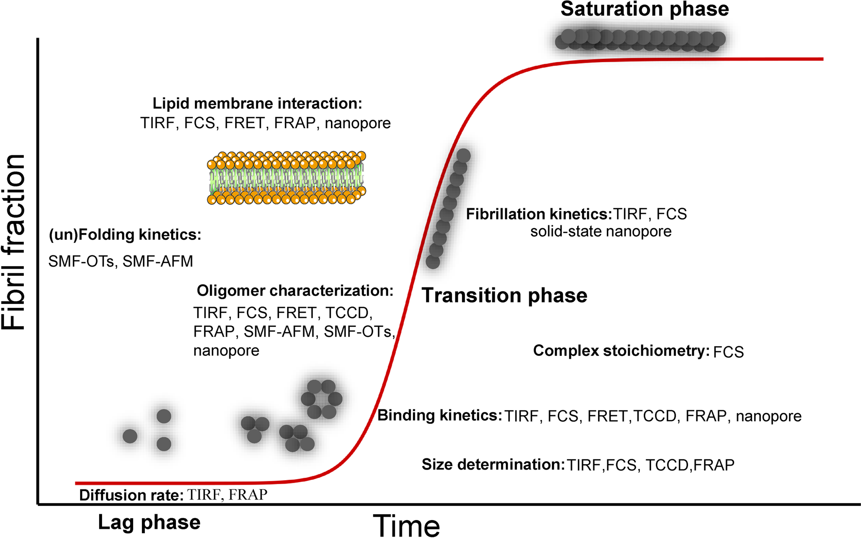
In the near future, technological and analytical developments in SM methods as well as combined computational modeling may permit a complete understanding of the complexity of amyloid protein assembly and further shed light on other applications such as the structure determination. One important future application of SM approaches will be to characterize amyloid aggregates of ex vivo samples providing an important determinant for the toxicity of biological samples and the diagnosis and subsequent therapy of neurodegenerative diseases.
Acknowledgements
We thank Prof. Guy Lippens (Université de Toulouse, France) for invaluable insights as well as critical advice on the manuscript. We acknowledge funding from the Swiss National Science Foundation project 310030_197626 (J.L), and the Brightfocus foundation A20201759S (J.L.).