Introduction
For the last 2.5 billion years, the Earth has been protected from harmful ultraviolet (UV) radiation by the ozone shield (Kastings & Siefert Reference Kasting and Siefert2002). However, the first 1 billion years of life's evolution occurred under a nitrogen and carbon dioxide atmosphere which does not have the same ability to absorb UV radiation (Cockell & Horneck Reference Cockell and Horneck2001). Organisms attempting to survive on the Earth's surface prior to the rise in oxygen would receive a radiation dose up to 1000 times more damaging to DNA than today (Cockell & Horneck Reference Cockell and Horneck2001). Indeed, it was originally proposed that the intense UV radiation flux experienced on the early Earth might have prevented the colonization of the land masses (Berkner & Marshall Reference Berkner and Marshall1965).
In high-UV environments on Earth today a number of survival strategies are observed. One such strategy is the matting habitat, whereby organisms achieve protection within thick laminated structures such as stromatolites. This has long been recognized as a potential means for early surface-dwelling organisms to have been protected from early intense UV radiation (Margulis et al. Reference Margulis, Walker and Rambler1976; Westall et al. Reference Westall, de Ronde, Southam, Grassineau, Colas, Cockell and Lammer2006). Alternatively, organisms commonly grow in the interior of rocky substrates or under them (Friedmann Reference Friedmann1980). These organisms are known as endoliths and hypoliths, respectively.
The depth of penetration of UV radiation into a rock will depend on the substrate. One rock substrate suitable for colonization is crystalline rocks shocked by asteroid and comet impacts, including gneisses, which become more porous as a result of the intense pressures and temperatures of impact shock (Cockell et al. Reference Cockell, Lee, Osinski, Horneck and Broady2002). Cockell et al. (Reference Cockell, Lee, Osinski, Horneck and Broady2002) calculated that organisms at 2 mm depth in porous impact-shocked gneiss under these early Earth radiation conditions would only encounter a maximum of one tenth of the DNA damage encountered on the surface of the Earth today, whilst still receiving sufficient photosynthetically active radiation for (anoxygenic or oxygenic) photosynthesis.
Previous work has only tested the protection afforded by rocks to UV radiation for a short period (Cockell et al. Reference Cockell, Rettberg, Horneck, Scherer and Stokes2003) and only using the present-day terrestrial flux. One location in which the putative early earth UV radiation flux can be simulated is low Earth orbit (LEO) (Rettberg et al. Reference Rettberg, Horneck, Strauch, Facius and Seckmeyer1998), where cut-off filters can be used to attenuate the extraterrestrial UV flux to simulate early Earth UV fluxes.
In the study presented here, we used the long-term exposure facility, EXPOSE-R, aboard the International Space Station to test the ability of an impact shocked endolithic substrate to provide adequate protection for phototrophs under a simulated worst-case prediction for early Earth's UV radiation regime. Here oxygenic phototrophs are studied. There are numerous lines of evidence which suggests oxygenic photosynthesis had evolved well before the atmosphere became oxygenated on a large scale (Buick Reference Buick2008). Therefore oxygenic photosynthesis would almost certainly have existed before UV-protection from the ozone shield was achieved. However, the principle we demonstrate is equally applicable to anoxygenic photosynthesizers.
Materials and methods
Organism and substrate selection
In this exposure experiment impact-shocked gneiss from the Haughton Impact Crater in the Canadian High Arctic was selected as a test substrate. A detailed outline of the geology of the Haughton Impact Structure from which the rocks were obtained can be found in Osinski et al. (Reference Osinski, Lee, Spray, Parnell, Lim, Bunch, Cockell and Glass2005). For a rock to be colonized it has to be suitably porous. Sandstones or vesicular volcanic rocks are porous under normal geological conditions making them good candidates for colonization (Cockell & Osinski Reference Cockell and Osinski2007). Crystalline rocks are generally low porosity and a poor prospect for colonization. However, asteroid and comet impacts, widely considered to be a purely destructive force, have the ability to alter crystalline rocks in a way which increases the porosity, and therefore the availability of microhabitats within the rock structure (Cockell et al. Reference Cockell, Lee, Osinski, Horneck and Broady2002, Reference Cockell, Rettberg, Horneck, Scherer and Stokes2003; Fike et al. Reference Fike, Cockell, Pearce and Lee2002; Pontefract et al. Reference Pontefract, Osinski, Lindgren, Parnell, Cockell and Southam2012). Cockell et al. (Reference Cockell, Lee, Osinski, Horneck and Broady2002) describe a 25 times increase in the porosity of gneiss (a crystalline, low-porosity metamorphic rock) which had been highly shocked by impact compared to nearby lightly shocked or non-shocked gneiss of the same parent material. They observe that this increased porosity allows bands of phototrophic cyanobacteria to colonize below the surface of the rock in a high-radiation natural environment.
The exposed land on the early Earth, whilst containing some sedimentary lithologies, would most likely have been predominantly comprised of crystalline rocks (Moorbath Reference Moorbath2005). In the period between 4.1 and 3.8 billion years ago, asteroid impacts were many orders of magnitude more common than today (Chyba & Owen Reference Chyba, Owen and Gehrels1994). Thus, when life appeared, impacts would have been widespread and could have altered the crystalline surface to provide suitable habitats in which the first microbes could survive under the harsh UV conditions encountered on the Earth's land masses.
Since asteroid and comet impacts are ubiquitous throughout the universe, we can extend our hypothesis that impact-shocked rocks provide important habitats to any rocky planet which lacks atmospheric oxygen and is therefore subject to intense UV radiation exposure. This could be particularly important on planets where the geology is dominated by low-porosity crystalline rocks rather than sedimentary lithologies and thus where potential endolithic habitats would be in short supply.
Further details on the shocked gneiss can be found in Fike et al. (Reference Fike, Cockell, Pearce and Lee2002) and Cockell et al. (Reference Cockell, Lee, Osinski, Horneck and Broady2002). Cockell et al. (Reference Cockell, Lee, Osinski, Horneck and Broady2002) discuss the improvements for life which resulted from the impact which altered the gneiss used here to its current state.
The polyextremotolerant cyanobacterium Chroococcidiopsis sp.029 was selected as a model organism. Chroococcidiopsis is one of the most tolerant to extremes of all of the known cyanobacteria. It is remarkably versatile with strains having been described from a wide range of extreme habitats such as hot springs (Geitler Reference Geitler1933), hypersaline (Dor et al. Reference Dor, Carl and Baldinger1991) and freshwater (Komarek & Hindak Reference Komarek and Hindak1975) environments, hot and cold deserts (Friedmann Reference Friedmann1980; Friedmann & Ocampo-Friedmann 1985; Budel & Wessels Reference Budel and Wessels1991) and within lichens as cyanobionts (Budel & Henssen Reference Budel and Henssen1983). In the most extreme hot, cold, arid and saline habitats on Earth, it is generally found to be the dominant cyanobacterium (Friedmann & Ocampo-Friedman Reference Friedmann and Ocampo-Friedmann1995). Chroococcidiopsis commonly adopts an endolithic lifestyle. The rocks it inhabits act as a shield from harmful environmental conditions (Friedmann Reference Friedmann1980).
The long-term survival of Chroococcidiopsis aboard the ISS was demonstrated as part of the ESA EXPOSE-E mission (Cockell et al. Reference Cockell, Rettberg, Rabbow and Olsson-Francis2011). Cells of Chroococcidiopsis sp. 029 were used to artificially augment a natural phototroph biofilm which was exposed to space conditions. These cells were shown to be viable after 534 days in LEO exposed to the full extraterrestrial UV radiation spectrum. This survival was attributed to the high numbers of Chroococcidiopsis cells relative to the abundance of other species in the natural phototroph community and protection of live cells by dead cells under a biofilm of cells, which was not a monolayer. In the experiment reported here, the exposure time was extended (22 months compared to 18 for EXPOSE-E) and pure cultures of Chroococcidiopsis used in sample preparation. The samples on glass discs have both high numbers of Chroococcidiopsis cells and a thin layer of cells so we will be able to test if these are the only attributes contributing to survival in the previous EXPOSE-E experiment.
Sample preparation
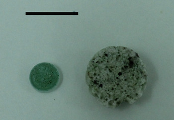
Chroococcidiopsis sp. CCMEE 029 was obtained from the Culture Collection of Microorganisms from Extreme Environments (CCMEE) established by E. Imre Friedmann and now maintained at the University of Rome ‘Tor Vergata’. Cells were cultured in BG-11 media as described previously (Cockell et al. Reference Cockell, Schuerger, Billi, Friedmann and Panitz2005). An aliquot of cells (~1.5×10⁶ cells) were transferred evenly onto the surface of 0.5 cm-diameter sterile glass discs or 1 cm-diameter discs of impact-shocked gneiss (Fig. 1). The impact-shocked gneiss was 5 mm thick, a thickness within which visible light transmission in the majority of the substrate is sufficient to support photosynthetic growth in natural communities that inhabit these rocks (Cockell et al. Reference Cockell, Lee, Osinski, Horneck and Broady2002).
Image displaying the impact-shocked gneiss (right) and borosilicate glass (left) used as substrates for the experiment.

Exposure conditions
The International Space Station (ISS) orbits the Earth at an altitude of around 450 km, in a region termed ‘low Earth orbit’ (LEO). In this region, our organisms are exposed to a variety of extremes: space vacuum, intense radiation bombardment from both solar and galactic sources as well as extreme and variable temperatures (Horneck et al. Reference Horneck, Klaus and Mancinelli2010).
The samples were fixed into the European Space Agency Expose-R facility in March 2009. The technical specifications of this facility have been described previously by Rabbow et al. (Reference Rabbow2009). The EXPOSE facility, run by the European Space Agency (ESA), is a multi-user facility which was designed to host medium- to long-term experiments (around 1.5 years) aboard the ISS. It is comprised of three trays containing hundreds of individual compartments in which samples are housed. The trays can be vented to the space environment or sealed and pressurized with defined gases to simulate an atmosphere. A variety of filters are used to control the wavelength and intensity of radiation the samples are exposed to. A range of bacteria, cyanobacteria, fungi, plants and some invertebrates have previously been shown to survive exposure to the full range of space conditions or selected parameters of it (summarized in Horneck et al. Reference Horneck, Klaus and Mancinelli2010).
A total of 36 glass discs and 12 rock discs were tested. Half of the glass discs were housed in containers which were vented to allow exposure to the vacuum of space whilst half were in sealed containers filled with argon gas. These experiments therefore investigate the effects of UV radiation alone, but do not take into account any potential confounding effects of interactions between UV radiation and atmospheric components that might have been present in the early Earth atmosphere such as carbon dioxide. Twelve glass discs in each condition were exposed to UV radiation whilst the rest were kept dark. MgF2 or Suprasil windows were used which let through UV>110 nm or >170 nm, respectively. In the Suprasil windows further cut-off filters were used to reduce the final UV cut-off to >200 nm. Neutral density filters were used to control radiation dose to 0.01, 1 or 100% of the total radiation (no neutral density filter). Three glass discs in each container (sealed or vented) were exposed to 100% UV>200 nm, two exposed to 1% UV>200 nm and three exposed to 0.01% UV>200 nm. These conditions were repeated for UV>110 nm to investigate the effects of very short wavelengths of UV radiation.
The UV conditions encountered by each sample type are summarized in Table 1. For the glass discs we report the averages across the two compartments (vented or sealed) as this should not influence the UV flux. The method of calculating UV fluences throughout the experiment are discussed in detail in Rabbow et al. (Reference Rabbow, Rettberg, Barczyk, Bohmeier, Parpart, Panitz, Horneck, Burfeindt, Molter, Jaramillo, Pereira, Weiß, Willnecker, Demets, Dettmann and Reitz2014, this issue).
Average radiation doses experienced by each sample type during the experiment

All of the samples on rock discs were housed in vented containers. Six rock discs were exposed to 100% of the UV radiation >110 nm and the remaining six kept dark. These rocks were exposed to 80 nm (110–190 nm) of UVC radiation not expected to have been encountered on the early Earth and therefore experienced a UV spectrum on their surface more severe than the worst-case early Earth spectrum. Identically prepared control samples were kept dark in the laboratory for the duration of the experiment. The experiment was conducted for 22 months (10 March 2009–21 February 2011).
Raman spectroscopy
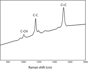
Raman spectroscopy was used to investigate the destruction of biomolecules on exposure to the extreme conditions in LEO. We used the presence or absence of carotenoids as a proxy for biomolecule destruction in our experiments. Carotenoids are a group of coloured pigments which are ubiquitous in nature, particularly in photosynthetic organisms like cyanobacteria and plants. Excitation of a carotenoid containing sample at 514 nm will reveal a characteristic spectrum (Fig. 2), where the Raman shifts relate to the stretching of the C=C and C–C bonds and to the bending of the C–CH groups within the conjugated molecule (Jorge Villar & Edwards Reference Jorge-Villar and Edwards2006).
Raman spectra taken from Chroococcidiopsis cells displaying the typical carotenoid signature where peaks correspond to the stretching of the C=C and C–C bonds and the bending of the C–CH group.

The observed Raman peaks arise from a resonance effect which causes an amplification of the band intensities above the background. This has proved to be extremely effective in the analysis of biological samples which are complex, of low concentration or which, as is common in cyanobacteria, are prone to being obscured by the significantly stronger fluorescence emission excited by visible laser wavelengths. The technique is particularly suited to the analysis of organic materials in mineral matrices, is also non-destructive and is therefore excellent for studies such as these where samples are extremely precious. It has been used on numerous occasions to detect cyanobacterial biomarkers from extreme environments (e.g. Jorge Villar & Edwards Reference Jorge-Villar and Edwards2006; Wang et al. Reference Wang, Hao, Huang, Chen, Li, Hu and Liu2010; Vitek et al. Reference Vítek, Edwards, Jehlicka, Ascaso, De los Ríos, Valea, Jorge-Villar, Davila and Wierzchos2010; Cockell et al. Reference Cockell, Rettberg, Rabbow and Olsson-Francis2011).
A Renishaw inVia laser Raman microscope (Renishaw, UK) was used and samples were excited at a wavelength of 514 nm. The laser was typically operated at 5% power with each spectrum being an average of ten acquisitions. Data were analysed using the commercial WiRE 3.2 software package (Renishaw, UK)
Before analysis of the rock discs, a cross-section was obtained with a sterile chisel. A positive or negative result for the carotenoid spectra both on the UV-exposed surface of the disc and the interior was recorded. Selection of the spot on which to sample within the rock was guided by the location of patches of cells as, owing to irregularities in pore spaces, they are not homogeneously distributed throughout the rock. For samples which were highly fluorescent at this level, the laser power was reduced to 1% to ensure that the signal was not hidden by the attendant fluorescence generated. Control spectra were also obtained from a segment of rock on which there were no cells and from dried cells of Chroococcidiopsis sp.029 on BG-11 agar.
One glass disc from each condition was probed in triplicate with a positive or negative result for the typical carotenoid spectra recorded.
Both the glass and the rock used for this experiment were investigated in an identical manner to the experimental samples to ensure that the substrate did not have an independent signal which could confuse the results.
Amplification of 16S rRNA genes from Chroococcidopsis on glass discs
To assess the effect of direct UV radiation exposure on DNA preservation, 16S rRNA was amplified from the glass discs in UV radiation exposed and dark conditions. Cells were recovered from glass discs by vortexing the discs in 200 μl of sterile MilliQ water. Cells were lysed by cryogenic grinding to a fine powder following sequential freeze–thawing cycles in liquid N2.
Polymerase chain reaction (PCR) was performed using cyanobacteria specific primers, forward primer CYA106F and reverse primer CYC781R (Nubel et al. Reference Nubel, Garcia-Pichel and Muyzer1997). CYA718 was an equimolar mixture of CYA781R(a) and CYA781R(b). The primers were synthesized commercially (Sigma-Aldrich, UK).
PCR amplifications were performed with an Eppendorf epgradient S mastercycler. Touchdown PCR was performed (Korbie & Mattick Reference Korbie and Mattick2008), to maximize sensitivity and specificity. 100 μl PCR reactions contained 50 pmoles of each primer, 25 nmol each of dNTP (Roche, Penzberg, Germany), 200 μg of BSA (New England Biolabs, Herts, UK), 10 μl of 10×PCR buffer and 20 μl of DNA sample. The MgCl2 concentration present in the reaction mixture was supplemented to give a final concentration of 2.5 mM. Reactions were started by the addition of 4 U of SuperTaq DNA polymerase (Cambio, Ltd, Cambridge, UK) after an initial denaturation step (5 min at 94 °C), at 80 °C. The first incubation cycle was 1 min at 94 °C, 1 min at 65 °C and 1 min at 72 °C. In the second cycle, the annealing temperature was decreased by 1–64 °C, in the third cycle by 1–63 °C, and so on in the same stepwise manner until the annealing temperature was 54 °C (after 12 cycles). Twenty-five further cycles were performed at this annealing temperature followed by a final elongation step of 10 min at 72 °C. The results shown are of a minimum of two independent amplifications. The amplification products were resolved on a 1% TAE agarose gel (40 mM Tris-acetate, 1 mM EDTA, pH 8.0) stained with SYBR Safe DNA gel stain (Invitrogen).
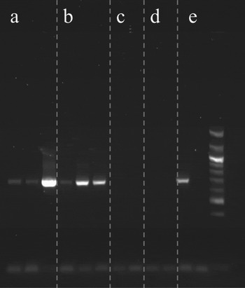
The PCR aims only to provide a qualitative assessment of DNA destruction. Since each sample was prepared identically, a negative result indicates DNA destruction compared to the control samples which consistently display a band.
Post-flight culturing
On return, one-third of each rock disc was placed in 100 ml of BG-11 media with triplicate cultures for each exposure condition. Subsequent growth was identified using bright field microscopy. Since the rock could not be broken into segments with adequate accuracy and cells were unevenly distributed throughout the pore space, calculation of the exact number of cells on each fragment of rock used for the inoculation was not possible. Therefore, only a positive or negative result for growth could be obtained. For the glass discs, one disc for each exposure condition was added to fresh BG-11 media.
Scanning electron microscopy
Both rock discs and glass discs were imaged using scanning electron microscopy. The rock discs were coated in gold before imaging using a Philips XL30CP scanning electron microscope (SEM) (Philips, UK) operated at 1 mbar pressure. Images were obtained using the absorbed current detector (AEI) at a voltage of 20 kV.
Observations on the glass discs were carried out using a CamScan MX2500 SEM (CamScan, UK) operated in controlled pressure mode (Envac, 30 Pa) and coupled to energy dispersive X-ray analysis (EDX) with Noran Vantage system and Vista software. Images were recorded at a working distance of 20 mm using the AEI at a voltage of 20 kV.
Results
Post-flight culturing
Within 4 weeks of inoculation of the fragments of rock discs, numerous 0.5–1 mm green specks were observed on the rock fragments. After 2 months, growth was clearly observed in the media of all experimental samples whether stored in the laboratory, kept dark in LEO or UV exposed in LEO. Chroococcidiopsis cells were confirmed under bright field microscopy. Some variability in the growth rates and concentrations of cells between conditions was observed but this was not quantifiable due to the necessary inaccuracies in the number of cells used for inoculation.
It was found that no samples on glass discs, whether in LEO or stored in the lab, had remained viable for the duration of the experiment.
Raman spectroscopy
In our samples, we found that glass discs which had been inoculated with cells of Chroococcidiopsis and either stored under laboratory conditions or kept dark whilst in the EXPOSE-R facility clearly exhibited the characteristic carotenoid bands described above. This indicates that despite the death of the cells during the period of desiccation the biomolecular carotenoids had not undergone degradation.
In the UV-exposed samples on glass discs the carotenoid peaks were only detectable at a very low level in one sample which had been exposed to 0.01% of the incoming radiation >110 nm in a vented container. All other UV-exposed samples tested exhibited no spectral peaks. Variations in the background fluorescence emission intensity were observed in several of these UV-exposed samples.
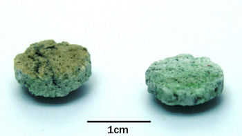
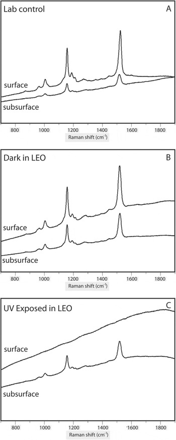
Rock discs which had been exposed to UV in LEO exhibited a browning of the surface which was not observed in rock discs which had been kept dark (Fig. 3). The spectra obtained from cells in the rock discs are displayed in Fig. 4. For cells on rock discs stored in the lab or kept dark in LEO the carotenoid signal was detected both on the surface and in the subsurface (Fig. 4(a) and (b)). In the rock discs it was found that cells on the UV-exposed surfaces of the rocks had experienced similar destruction to carotenoids as that exhibited by cells on glass UV-exposed discs (Fig. 4(c)). Cells on the surface of the rocks imaged through the Raman microscope had turned brown during exposure and they did not exhibit any characteristic Raman peaks when probed (Fig. 4(c)). However, below the surface in the cleaved samples, green flecks were observed which when probed at 514 nm exhibited the typical carotenoid Raman spectral signals (Fig. 4(c)). This demonstrates that the rocks were effective in shielding the cells housed internally from 100% exposure to the full extraterrestrial radiation dose >110 nm. We have determined that whilst some fluorescence contribution from glassy minerals in the rocks is present there is no interference that would confuse the interpretation of these characteristic carotenoid spectral signals.
Image showing colour change in UV-exposed rocks (left) compared to rock kept dark in LEO (right).

Raman shift at 514 nm of the patches of cells in the surface and subsurface of rock discs from each condition. The spectral bands indicative of carotenoids is present in the surface and subsurface of the laboratory controls and the dark samples from low Earth orbit as well as the subsurface of the UV-exposed rocks. The brown cells on the surface of the UV-exposed rocks do not show any distinct peaks but exhibit an increased fluorescence.

Polymerase chain reaction
The results of the PCR reactions are displayed in Fig. 5. 16S rRNA was successfully amplified from all of the cells on glass discs which had been kept in dark conditions, but not on any exposed to UV radiation. There was some variation between the intensity of the bands that were detected. The use of the PCR aimed to provide a proxy for DNA damage to the samples. The successful imaging of bands gives a qualitative assessment of the destruction of DNA since all discs were originally inoculated with the same numbers of cells. For this reason normalization of the DNA was not required.
Amplification of 16S rRNA genes from Chroococcidiopsis on glass discs exposed to various space conditions. (a) Amplification of genes from cells on three glass discs kept dark in sealed containers, (b) amplification of genes from cells on three glass discs kept dark in vented containers, (c, d) no gene bands observed from cells on UV-exposed glass discs in sealed (c) or vented, (d) containers, (e) positive control (left), negative control (middle) and marker lane (right).

Scanning electron microscopy
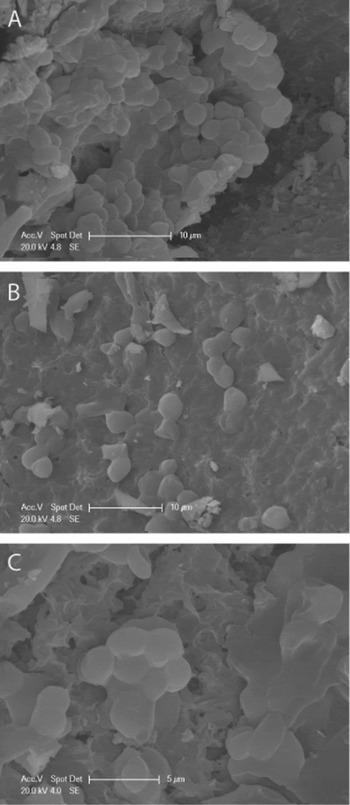
Figure 6 displays SEM images of cells in the pore space of the rocks in the control (a), dark (b) and UV exposed (c) samples. It was observed in the UV-exposed rocks that morphologically intact cells were present even in pores directly exposed to the surface. This could suggest that UV bleaching of the cells had occurred and the biomolecules destroyed whilst the cells still maintained their shape.
SEM images of the rock discs show that cell morphology observed in the control was preserved during exposure to space conditions, both dark and UV radiation exposed (a) rock control disc kept dark in the lab for the duration of the experiment, (b) rock disc kept dark in LEO and(c) rock disc exposed to 100% UV>110 nm in LEO.

SEM images of the glass discs reveal that samples which had been kept dark, despite being dead on their return to Earth, had also retained their morphology (not pictured). This was also the case for the glass discs exposed to UV radiation within vented containers but intact cells were not observed on glass discs which had been kept sealed.
Discussion
In this study, we used the UV radiation conditions found in low Earth orbit (LEO) to determine if an endolithic lifestyle could provide suitable shelter for phototrophs on the anoxic early Earth, or on other anoxic planets, which receive a higher UV radiation dose than the Earth's surface today. Impact shocked gneiss was chosen as the rock substrate to determine whether new habitats created by impact events could provide adequate protection from the higher UV radiation.
Phototrophs have the requirement for photosynthetically active radiation (PAR) for growth, meaning that they must be exposed to sunlight with the concomitant exposure to UV radiation. Therefore, simply growing at a depth where UV radiation is completely extinguished is not an option.
We report that organisms within impact-shocked gneiss exposed to the intense UV radiation environment in LEO for 22 months were viable on their return to Earth. This reveals that it would be possible for cyanobacteria to persist in a desiccated state for almost 2 years in these rocks under the worst-case UV radiation conditions.
The complete loss of viability in cells on all of the glass discs regardless of exposure conditions (and in controls) could be a result of extreme desiccation of the thin layer of cells on the glass disc. Cells within clumps inside the rocks might have been protected by the extracellular polysaccharide of other cells as observed with other cyanobacteria (Tamaru et al. Reference Tamaru, Takani, Yoshida and Sakomoto2005) or have had reduced rates of desiccation when they were prepared. The thin layer of cells desiccated onto the flat glass surface will have dried out quickly. Inside the rocks, cells contained within the liquid would have pooled in pore spaces allowing cells to desiccate more slowly, where it would form thick clumps as seen in Fig. 6(a).
Our results on the UV-exposed glass discs show that direct exposure to the worst-case early Earth UV radiation conditions over a long period will have a destructive effect on biomolecules, destroying both essential pigments and breaking up DNA even when this was attenuated to only 0.01% of the incoming UV. Our PCR and Raman spectroscopy results demonstrate that this damage was not as extensive in discs which had been kept dark compared to those exposed to UV radiation. This supports the general consensus that UV radiation exposure is the most destructive aspect of exposure to space conditions (Horneck et al. Reference Horneck, Klaus and Mancinelli2010). This also emphasizes the low survivability of photosynthetic life on the surface of the early Earth in the absence of active repair.
The positive detection of carotenoid signatures observed by Raman spectroscopy in cells exposed to 0.01% UV radiation of >110 nm in vented containers suggests that the radiation dose received at this level may have been close to a threshold level at which biological molecules can survive.
We also observed that UV radiation exposure, death and biomolecule destruction does not necessarily destroy the morphology of cells. SEM images of the rock discs showed that the morphology of some cells within the rocks remained intact regardless of exposure condition or proximity to the surface. The morphology could also be discerned from cells on glass discs from all conditions in vacuum but not on discs housed in argon gas. This could suggest that space vacuum might be advantageous to the preservation of morphology under extreme UV radiation stress. With only one sample available for this imaging analysis this result would require further confirmation; however, this could suggest that the presence of a thick atmosphere may be detrimental to biomarker preservation and detection on a planetary surface with a high-UV flux.
The extent of carotenoid destruction on directly exposed cells (either on the glass discs or the surface of the rocks) compared to those within the impact-shocked rocks highlights the importance of a shielding mechanism in high-UV radiation conditions whether on the early Earth or other rocky planetary surfaces. These data show that impact-shocked rocks provide protection against biomolecule destruction and ultimately loss of cell viability.
We conclude that the protection afforded to organisms within impact-shocked rocks is adequate to preserve viable cyanobacterial cells in a desiccated state for at least 22 months under a UV flux at least equal to the worst-case scenario on the early Earth. This result could extend to other terrestrial-type rocky planets lacking a sufficient atmospheric UV radiation shield. Cells actively growing, unlike the desiccated cells we studied here, would have the potential to actively repair UV radiation-induced damage (assuming the dose is sublethal), suggesting that our results are conservative. This work highlights the potential of impact craters and endolithic habitats as protective habitats on rocky planets with a high-UV radiation flux and it empirically demonstrates that phototrophic microorganisms could have colonized early land masses under a worst-case UV radiation flux even without a matting ability.
Acknowledgements
The authors thank Angela Dawson for her contribution to the PCR, Peter Chung for assistance with the Raman spectroscopy and Lore Troalen for assistance with the SEM on the glass discs. The PhD studentship for C. Bryce is funded by EPSRC.