Significance outcomes
∙ Immune system dysregulation and inflammation have a role in bipolar disorder and carcinoembryonic antigen stimulates monocytes and macrophages and cause the production of endothelial adhesion molecules and proinflammatory cytokines.
∙ Our study suggests that increased CEA is observed in individuals with bipolar disorder.
Limitations
In addition to measurement of CEA, not measurement of proinflammatory and anti-inflammatory marker is one of the limitation of our study.
Introduction
Carcinoembryonic antigen (CEA) is an oncofetal glycoprotein, molecular weight 180 kDa, that is widely used as a tumour marker in adenocarcinomas, especially in colorectal cancer (Reference Hammarstrom1). However, it is also expressed in normal mucosal cells (Reference Yalta, Yilmaz and Turgut2). CEA also increases at some non-neoplastic conditions, such as chronic inflammatory disease, hepatic and renal failure, aging and smoking (Reference Ruibal Morell3–Reference Witherspoon, Shuler, Alyea and Husserl5). In addition, increased CEA levels have been shown in hypothyroidism, chronic obstructive pulmonary disease and obesity (Reference Amino, Kuro and Yabu6–Reference Park, Choi, Jang, Jun and Kang8).
The function of CEA has not been fully illuminated, but previous studies have reported that monocytes and macrophages are stimulated by CEA to trigger the production of endothelial adhesion molecules and proinflammatory cytokines (Reference Ganguly, Yeltsin and Robbins9,Reference Aarons, Bajenova and Andrews10).
Bipolar disorder (BD), previously known as manic-depressive illness, has been ranked seventh among the worldwide. BP has a chronic and remitting nature characterised by periods of depression and periods of hypomania or mania in many patients (Reference Steinan, Krane-Gartiser, Langsrud, Sand, Kallestad and Morken11).
There are two types of BD. BD type I disorder has been defined as the presence of at least one manic episode; BD type II disorder has been defined as presence of at least one hypomanic episode along with the presence of one or more major depressive episodes (12).
The accepted prevalence of BD ranges from 0.5% to 1.5%, but these rates may be as high as 10%, depending on which diagnostic criteria are adopted in recent epidemiological studies (Reference Ghouse, Sanches, Zunta-Soares, Swann and Soares13).
Evidence from clinical studies showed that immune system dysregulation and inflammation have a role in BP (Reference Goldstein, Kemp, Soczynska and McIntyre14). It is reasonable to suspect that elevated inflammatory cytokines partially contribute to the cognitive impairments in BD (Reference Lotrich, Butters, Aizenstein, Marron, Reynolds and Gildengers15).
Progressive structural, functional and biochemical changes in BD might be related to neurotransmitter changes, neurotrophic alterations, oxidative stress and inflammation. Mitochondrial dysfunction, free radical production, neurodegeneration and alterations of several neurotransmitters can occur due to the effects of inflammatory changes in BD (Reference Berk, Kapczinski and Andreazza16,Reference Maes, Lin and Verkerk17). Inflammatory biomarkers have been considered as one of the main pillars of a multifactorial approach for estimation of BD in an at-risk population (Reference Brietzke, Mansur, Soczynska, Kapczinski, Bressan and McIntyre18).
Preclinical studies suggest that mood stabilisers such as lithium and valproic acid play a role in the modulation of neuroinflammatory processes, and this indicates a role for neuroinflammation in BD (Reference Kang, Kim and Kim19–Reference Xuan, Long and Li21).
It is shown that inflammatory processes played a role in the aetiology of BD. In addition, it is considered that CEA also increases the proinflammatory cytokines. Thus, we thought that CEA levels could be changed as an inflammatory marker in BD.
The association between elevated CEA and BP has not previously been examined. Our aim in this study was to evaluate CEA levels as a marker of inflammation in BD.
Methods
This study was approved by the Dicle University Ethical Committee. A total of 44 out-patients who agreed to participate in the study with type I BD, diagnosed according to the DSM-IV criteria, having been in a euthymic phase for at least 3 months, and followed by the Dicle University, Department of Psychiatry Mood Disorder Unit were enrolled. Patients were between 18–65 years of age.
Patients admitted in the Unit with a history of drug and alcohol abuse, chronic systemic diseases such as diabetes mellitus, morbid obesity, cancer, hypertension, etc., severe head injury or seizure disorders, or pregnant were excluded from the study. Patients who have comorbid psychiatric disorders and mental retardation were excluded from the study.
The control group consisted of 45 healthy subjects. The control group was selected from healthy individuals who donate blood to the blood bank. The case and control groups had similar distributions in age, sex, body mass index and smoking status.
Hamilton Depression Scale, the Young Mania Rating Scale and Clinical Global Impression Scale (CGI) were used in the patient group (Reference Akdemir, Turkcapar, Orsel, Demirergi, Dag and Ozbay22,Reference Karadag, Oral and Yalçın23).
Blood samples were obtained from the subjects in the morning after an overnight fast, centrifuged within 30 min at 2500×g at 4°C for 10 min. Serum aliquots were prepared and stored at −80°C until analysis and the assays for variables analyzed in the current study were performed on the same day
CEA values were measured in Dicle University, Medical Faculty Central Laboratory using electrochemiluminescence immunoassay (Roche Cobas e601). Assay precision was evaluated by testing two control concentrations of commercially available quality control materials.
Statistical analysis
SPSS® for Windows 15.0 statistical program was used for statistical analysis. The Kolmogorov–Smirnov test was used to confirm that data within the ranges of normal distribution in both groups. A parametric test was employed for the variables in the normal distribution. The significance of differences between groups was estimated by Student’s t-test. χ2 test was used when comparing proportions. Differences were accepted as significant when p<0.05. Bivariate comparisons were examined via Spearman correlation coefficients and values were corrected for ties.
Results
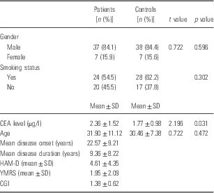
The mean age was 31.90±11.12 and 30.46±7.38 years for patients and controls, respectively. The mean serum CEA level in BD was 2.36±1.52 µg/l and in control was 1.77±0.98 µg/l. CEA levels were significantly increased in euthymic BD patients when compared with controls (p=0.031). Furthermore, while 49.4% of patients and 50.6% of control group were smokers, there was no significant difference between the smoking status of patient and control groups (p=0.302) (Table 1). There were no correlations between mean CEA levels and age, onset of disease and disease duration (p>0.05). The sociodemographic variables are shown in Table 1.
Sociodemographic variables and biochemical measurements of patients and controls

CEA, carcinoembryonic antigen; CGI, Clinical Global Impression Scale; HAM-D, Hamilton Depression Scale; YMRS, Young Mania Rating Scale.
In all, 88.6% (n=39) of patients were using antipsychotics, 93% (n=41) were using mood stabilisers, and 20.5% (n=9) were using the anti-depressant; 4.5% (n=2) of our patients were using first-generation antipsychotics, 75% (n=33) second-generation antipsychotics, and 9% (n=4) first- and second-generation combined antipsychotics drugs. Only six patients (13.6%) were using one psychiatric drug and 38 patients (86.4%) were using combined drugs. Control group was not using any drug. There was no statistically significant relationship between CEA levels and using these drugs (p>0.05).
Discussion
In this study we investigated the CEA levels in euthymic bipolar patients and this is the first study to examine CEA levels at BD.
An oncofetal glycoprotein CEA, which is a member of the immunoglobulin superfamily, is a cell surface adhesion molecule. CEA is overexpressed in adenocarcinoma, especially in colorectal cancer. It is also expressed in normal mucosal cells. Elevated CEA levels can be seen in pancreatitis, inflammatory bowel disease, cirrhosis, biliary obstruction, peptic ulcer disease, hypothyroidism and cigarette smoking. The serum concentration is <2.5 and <5 µg/l in non-smokers and smokers, respectively (Reference Fletcher24,Reference Bast, Bates and Bredt25).
Previous studies have suggested that macrophages or monocytes may be stimulated by CEA via binding to the CEA receptor. It results in the up-regulation of endothelial adhesion molecules and the production of proinflammatory cytokines such as reactive oxygen species, interleukin-1b (IL-1b), tumour necrosis factor-α (TNF-α), IL-6, nitric oxide (NO) and eicosanoids (Reference Ganguly, Yeltsin and Robbins9,Reference Aarons, Bajenova and Andrews10).
CEA may increase the release of inflammatory cytokines and activate endothelium, supporting the expression of adhesion molecules and inflammatory events. The relationship between CEA and high-sensitivity C-reactive protein (hs-CRP) have been found in patients with colorectal cancers (Reference Aarons, Bajenova and Andrews26,Reference Minami, Furui and Kanematsu27).
Inflammation plays a role in the pathogenesis of atherosclerosis, insulin resistance and glucose intolerance (Reference No, Yang, Hyun, Yeon and Choi28). Bae et al. (Reference Bae, Shim, Lee and Shin29) reported that CEA and hs-CRP level was related with arterial stiffness. In addition, in another study serum levels of CEA, could be influenced by high glucose levels and HbA1C in diabetic patients (Reference Kim, Kim, Suh, Lee, Lew and Lee30). These data indicate serum CEA may be associated with inflammation.
Studies suggest that inflammation may be involved in the pathophysiology of BD. In the periphery and in the brain, chronic, mild inflammation occurs during BD (Reference Goldstein, Kemp, Soczynska and McIntyre14,Reference Hamdani, Tamouza and Leboyer31). Kunz et al. showed that involvement of inflammatory mechanisms is a component of the pathophysiology of BD. They observed that the anti-inflammatory IL-10 have increased in euthymic patients in their study (Reference Kunz, Cereser and Goi32).
Previous studies reported that proinflammatory cytokines are increased in BD (Reference Brietzke and Kapczinski33,Reference O’Brien, Scully, Scott and Dinan34). Some recent studies have reported that levels of the proinflammatory cytokine TNF-α and IL-6 are increased in acutely manic or depressed patients with BD. TNF-α levels were increased in both manic and depressed BD patients but while IL-6 levels were higher in depressive BD patients in a study, in another study it was higher in manic BD patients. There were no euthymic patients in their study(Reference O’Brien, Scully, Scott and Dinan34,Reference Ortiz, Goin, Gomez de Alzaga, Hammarstrom and Jimenez de Asua35). Kim et al. (Reference Kim, Jung, Myint, Kim and Park36) studied only in manic BD patients and they found that TNF-α and IL-6 levels were increased.
In our study, we investigated CEA levels in patients with BD and found them to be higher than in controls (p=0.031). This supports other studies showing that BD is associated with inflammation. Our study’s effect size was 70.1%.
Andreazza et al. (Reference Andreazza, Kauer-Sant’anna and Frey37) in their meta-analysis study reported that oxidative stress markers and NO activity are increased in individuals with BD. CEA may cause production of oxidative stress markers and NO. Dickerson et al. (Reference Dickerson, Stallings, Origoni, Vaughan, Khushalani and Yolken38) showed that elevated levels of C-reactive protein, which plays a central role in the inflammatory process, are associated with lower cognitive functioning in individuals with BD.
Kauer-Sant’Anna et al. found increased TNF-α and IL-6 levels in those with early-stage BD and an even greater increase in those with late-stage BD; they reported that levels of TNF-α and IL-6 were compared between patients at early and late stages of illness, TNF-a showed a significant increase at the later stage but there was a significant decrease in IL-6 in the later stage. These changes in cytokine levels may play a role in the pathophysiology of BD (Reference Kauer-Sant’Anna, Kapczinski and Andreazza39).
In our study, all patients were euthymic. In euthymic phase of BD neuropsychological disturbances, such as loss of functionality and impairment of cognitive functions continue (Reference Martínez-Arán, Vieta and Reinares40). This indicates that the disease is associated with chronic immune activation and inflammatory syndrome. CEA may be the cause of the increased production of proinflammatory cytokines.
In our study patients were using antipsychotics, mood stabilisers and anti-depressant drugs. To the best of our knowledge, there was not any study in literature about the effect of these drugs on CEA levels.
Cigarette smoking increases serum CEA levels without evidence of malignant diseases (Reference Stevens and Mackay41). Fukuda et al. (Reference Fukuda, Yamakado and Kiyose4) concluded that the serum CEA level was influenced by smoking, especially in males.
In our study, although the number of patients who smoke was high, there was no significant difference at smoking status when compared with the control group (p=0.302).
In addition to measurement of CEA, not measurement of proinflammatory and anti-inflammatory marker is one of the limitation of our study. It will be considered in further research.
In conclusion, increased CEA is observed in individuals with BD. CEA levels may be a secondary marker for diagnosing BD. Further studies with larger samples are needed to delineate this relationship.
Acknowledgements
The authors are grateful to Dicle University DUBAP for their assistance in the English editing of this manuscript. Author contributions: I.K. wrote the whole version of the manuscript and made the labaratory assesments, M.B. and A.A. performed clinical assessment of the patients, M.C.K and M.G made statistical analysis and interpretation of data, l.C designed the study methods and helped to write the final version of the manuscript to be published.
Financial Support
None.
Conflicts of Interest
None.
Ethical Standards
The authors assert that all procedures contributing to this work comply with the ethical standards of the relevant national and institutional committees on human experimentation and with the Helsinki Declaration of 1975, as revised in 2013 and approved by the local ethics committee.