Introduction
Giant cell reparative granuloma is an uncommon, non-neoplastic tumour typically occurring in the mandible or maxilla.Reference Dimitrakopoulos, Lazaridis, Sakellariou and Asimaki1 Giant cell reparative granuloma involving the temporal bone is extremely rare, and there have been no previous reports of hearing preservation following resection of this tumour in the temporal bone.Reference Menge, Maier, Feuerhake, Kaminsky and Pfeiffer2
We report a case of extensive giant cell reparative granuloma of the temporal bone which was successfully resected, with hearing preservation, using a transmastoid approach. Follow-up magnetic resonance imaging (MRI) performed one year after surgery showed no evidence of tumour recurrence.
Case report
A 37-year-old man presented to our department with a one-year history of right-sided hearing loss, complicated by a three-month history of aural fullness and otalgia. He had no history of previous trauma.
Clinical examination on presentation showed that the anterior wall of the right external auditory canal was swollen, blocking the canal and preventing visualisation of the tympanic membrane (Figure 1a).
(a) Otoscopic view of the right ear. The anterior wall of the external auditory canal is smoothly swollen, preventing examination of the tympanic membrane. (b) Pre-operative pure-tone audiogram showing conductive hearing loss with an air–bone gap of more than 30 dB.

Pure-tone audiometry revealed a conductive hearing loss with an air–bone gap of more than 30 dB (Figure 1b).
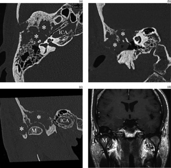
Computed tomography (CT) and MRI demonstrated an osteolytic tumour lesion in the right temporal bone, which surrounded the mandibular condyle and infiltrated the middle fossa plate (Figure 2).
(a) Axial and (b) coronal computed tomography (CT) scans showing tumour in the right temporal bone and extending into the middle-ear cavity. The external auditory canal is completely blocked by the tumour. The ossicular chain is surrounded by the tumour but does not seem to have been eroded by it. (c) Coronal CT scan of the right mandibular condyle, showing no invasion by the tumour. However, the glenoid fossa and zygomatic arch do show erosion, extending medial to the condyle but not reaching the horizontal segment of the internal carotid artery. (d) Coronal magnetic resonance imaging scan showing a ‘pepper’ shadow in the right temporal bone, although the tumour region is not clear (certainly compared with the CT appearance). Both the dura mater and the mandibular condyle appear intact. * = tumour; ICA = internal carotid artery; M = mandible

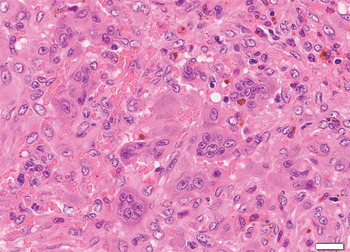
Open biopsy was performed via simple mastoidectomy. Histopathological examination of the surgical specimen revealed massive fibroblastic proliferation with numerous heavily haemosiderin-laden macrophages and several multinucleated giant cells (Figure 3). Mitotic figures and necrosis were absent. Histopathological appearance was consistent with a diagnosis of giant cell reparative granuloma.
Photomicrograph of the biopsy specimen, showing multinucleated giant cells irregularly distributed in a fibroblast-rich stroma with haemorrhage and haemosiderin deposits. Mitotic figures are absent. Bar = 20 µm. (H&E; original magnification ×400)

Tumour resection was performed via a transmastoid approach with combined pre- and post-auricular skin incisions (Figure 4), followed by meatoplasty. The glenoid fossa was completely drilled and the zygomatic arch was partially drilled under retraction of the mandibular condyle, because of suspected bony invasion. The mandibular condyle was preserved. Mastoidectomy was then performed with removal of the bony external auditory canal, accessing the middle-ear cavity. The attic and part of the mesotympanum also showed continuous involvement with tumour tissue, but these areas were only attached to the ossicles and showed no erosion. The lower side of the tumour protruding to the tympanum was thus dissected carefully, with preservation of the ossicular chain. Tumour invasion was suspected in the antero-superior part of the external meatal skin, which was removed, necessitating meatoplasty. The middle fossa plate showed partial infiltration by tumour but the dura of the middle fossa remained intact, without leakage of cerebrospinal fluid; therefore, duraplasty was not needed. Following tumour removal, dead space in the temporal bone was filled using a free muscular flap.
Diagram showing the combined pre- and post-auricular skin incisions, performed to promote hearing preservation.

The post-operative course was uneventful. Facial nerve function was intact. The tympanic membrane could be seen through the external auditory canal, and post-operative pure-tone audiometry revealed preserved hearing (Figure 5a and 5b).
(a) Otoscopic view of the right external auditory canal taken one year after surgery. The external auditory canal is wider and the tympanic membrane is visible, although middle-ear effusion is apparent through the tympanic membrane. (b) Post-operative pure-tone audiogram showing persistent conductive hearing loss, although the air–bone gap has decreased compared with the pre-operative audiogram.

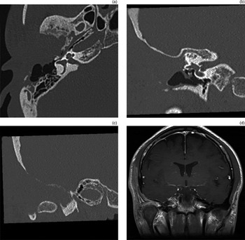
One year after surgery, follow-up CT and MRI showed no evidence of tumour recurrence (Figure 6). At the time of writing, the intention was to monitor the patient with clinical and radiological investigations at regular, six-month intervals.
Radiological images taken one year after surgery. (a) Axial and (b) coronal computed tomography (CT) scans showing that the right temporal bone tumour has been removed; the resultant dead space has been filled with a free muscle flap and shows no bony structures, and the middle-ear cavity and eustachian tube are aerated. (c) Coronal CT scan showing the right mandibular condyle preserved in situ. (d) Coronal magnetic resonance imaging scan showing the muscle flap obliterating the dead space cavity superior and medial to the right mandibular condyle.

Discussion
Giant cell reparative granuloma is an uncommon, benign, osteolytic lesion typically occurring in the mandible or maxilla. Numerous cases have been reported since Jaffe first described giant cell reparative granuloma of the jaw in 1953, but giant cell reparative granuloma of the temporal bone remains quite rare.Reference Dimitrakopoulos, Lazaridis, Sakellariou and Asimaki1, Reference Jaffe3 In 1974, Hirschl and Katz described the first case of giant cell reparative granuloma originating in the temporal bone.Reference Hirschl and Katz4 This tumour can arise at any age, and does not appear to have any clear gender predilection.Reference Liu, Zhong, Liu, Han, Yang and Jiang5 Symptoms include hearing loss, local swelling, tinnitus, a palpable mass, pain, vertigo and facial weakness. Conductive hearing loss is the most common symptom. Although giant cell reparative granuloma is regarded as a benign entity, the tumour can be locally aggressive, and surgical excision is recommended whenever possible. In cases where complete resection is not possible, post-operative radiotherapy has been advocated.Reference Ung, Li, Keith and McKenna6–Reference Alappat, Pillai, Prasanna and Sambasivan8 In their study of patients with giant cell reparative granuloma of the temporal bone who were followed up after treatment, Boedeker et al. reported an estimated recurrence rate of 12.5 per cent.Reference Boedeker, Kayser, Ridder, Maier and Schipper9
We report herein a case of giant cell reparative granuloma of the temporal bone which was treated surgically via a transmastoid approach, with hearing preservation. The main method used to access lesions around the glenoid fossa is the infrafossa type B approach developed by Fisch and Mattox.Reference Fisch, Mattox, Fisch and Mattox10 However, this procedure includes transection of the external auditory canal, followed by its suturing. Since pre-operative hearing loss in our patient was mild, we selected a transmastoid approach with combined pre- and post-auricular skin incision, in order to maximise hearing preservation. The bulk of the tumour surrounded the mandibular condyle, with extension to the tympanum, but had not eroded the ossicular chain. Total resection was achieved with preservation of the ossicular chain. If the tumour had extended more aggressively and eroded the ossicular chain and/or the inner ear, hearing preservation after total resection would have been much more difficult to achieve, if not impossible. In order to optimise hearing preservation, surgery should proceed as soon as possible after diagnosis of giant cell reparative granuloma of the temporal bone. Although complete resection is often a surgical challenge given the complexity and number of vital structures around the skull base, it is associated with a recurrence rate of only 10 per cent, compared with recurrence of up to 50 per cent following simple curettage.Reference Ung, Li, Keith and McKenna6, Reference Ciappetta, Salvati, Bernardi, Raco and Di Lorenzo7, Reference Auclair, Cuenin, Kratochvil, Slater and Ellis11
Histologically, giant cell reparative granuloma of the temporal bone should be differentiated from a number of other lesions containing giant cells, such as fibrous dysplasia, chondroblastoma, osteosarcoma and giant cell tumour.Reference Sakamoto, Hayashida and Sugasawa12–Reference Bertoni, Unni, Beabout and Ebersold14 The primary difference between giant cell tumour and giant cell reparative granuloma is the clinical prognosis. Giant cell tumour is a true neoplasm with a higher incidence of recurrence and potential for malignant transformation and metastatic spread.Reference Ung, Li, Keith and McKenna6 In contrast, malignancy and metastasis have never been reported in cases of giant cell reparative granuloma.Reference Ung, Li, Keith and McKenna6, Reference Boedeker, Kayser, Ridder, Maier and Schipper9, Reference Sharma, Verma, Pawar, Dev, Devadas and Shiv15 Differentiation of giant cell reparative granuloma from giant cell tumour is important, given the latter's far worse prognosis.Reference Hirschl and Katz4, Reference Boedeker, Kayser, Ridder, Maier and Schipper9 On histological examination, the typical appearance of giant cell tumour is of larger multinucleated giant cells within a background of mononuclear giant cells, with the latter commonly containing mitotic figures. The present case was histopathologically distinguishable from giant cell tumour on the basis of fibroblastic proliferation, fewer giant cells and absence of mitotic activity.
• Giant cell reparative granuloma typically occurs in the mandible and maxilla
• It is extremely rare in the temporal bone
• A case of the latter is presented, with successful total resection via a transmastoid approach
• This is the first report of such treatment with hearing preservation
• Combined pre- and post-auricular skin incisions were used
Conclusion
Giant cell reparative granuloma is an uncommon, non-neoplastic tumour typically occurring in the mandible and maxilla. Appearance in the temporal bone is extremely rare, and there have been no previous reports of surgical resection of these latter tumours with hearing preservation. We report a case of extensive giant cell reparative granuloma of the temporal bone, successfully resected via a transmastoid approach, with hearing preservation. A follow-up MRI scan one year after surgery showed no evidence of tumour recurrence.