Introduction
The development of new regenerative therapies adapted to the reproductive needs of women has been challenging in the last years (Schenke-Layland & Brucker, Reference Schenke-Layland and Brucker2015). Stem cell-based strategies for ovarian regeneration and oocyte production have been proposed as future clinical therapies for treating infertility in women (Volarevic et al., Reference Volarevic, Bojic, Nurkovic, Volarevic, Ljujic and Arsenijevic2014), as well as to increase multiplication of genetically superior animals or endangered species (Singhal et al., Reference Singhal, Singhal, Malik, Singh, Kumar and Kaushik2015). Some studies have been conducted to identify, characterize, and differentiate cells from different sources (Zomer et al., Reference Zomer, Vidane, Gonçalves and Ambrósio2015). The cells that can potentially be used for clinical studies include embryonic stem cells (ESC) (Keefer et al., Reference Keefer, Panta, Blomberg and Talbot2007), stem cells isolated from adult tissues, such as mesenchymal stem cells (MSC) (Ratajczak et al., Reference Ratajczak, Zuba-Surma, Kucia, Poniewierska, Suszynska and Ratajczak2012), and induced pluripotent stem cells (iPS), which are adult somatic cells reprogrammed to pluripotency (Angelos & Kaufman, Reference Angelos and Kaufman2015).
The generation of pluripotent stem cells (iPS) by reprogramming somatic cells was reported by Takahashi & Yamanaka (Reference Takahashi and Yamanaka2006) and these cells have the capacity to generate all cell types of adult tissues (Hirschi et al., Reference Hirschi, Li and Roy2014). Pluripotency induction was performed by using a combination of four reprogramming factors, including OCT4 (Octamer binding transcription factor-4), SOX2 (Sex determining region Y)-box 2, KLF4 (Kruppel Like Factor-4) and c-MYC (Takahashi & Yamanaka, Reference Takahashi and Yamanaka2006; Singh et al., Reference Singh, Kalsan, Kumar, Saini and Chandra2015). REX-1 is another pluripotent cell marker that is directly regulated by NANOG and SOX-2 (Shi et al., Reference Shi, Wang, Pan, Geng, Guo and Pei2006). Conventional reprogramming techniques depend on the stable integration of these genes, by using viral vectors, such as retroviruses (Takahashi & Yamanaka, Reference Takahashi and Yamanaka2006) and lentiviruses (Picanço-Castro et al., Reference Picanço-Castro, Russo-Carbolante, Reis, Fraga, de Magalhães and Orellana2011), but can introduce the current risk of insertional mutagenesis (Diecke et al., Reference Diecke, Jung, Lee and Ju2014). A chemical reprogramming process is a promising strategy for iPS generation and many small molecules, like 5-Aza, have been identified and used to replace exogenous transcription factors (Lin & Wu, Reference Lin and Wu2015). These small molecules promote chemical reprogramming by remodeling the epigenome and facilitating the activation of pluripotency-related genes (Zhang et al., Reference Zhang, Li, Laurent and Ding2012). 5-Azacytidine, a DNA methyltransferase inhibitor, can improve reprogramming efficiency by activating the expression of silent genes and altering the differentiation state of cells (Constantinides et al., Reference Constantinides, Jones and Gevers1977; Jones, Reference Jones1985). Taylor & Jones (Reference Taylor and Jones1979) used 5-Aza to convert a mesenchymal cell line into muscle cells, adipocytes and chondroblasts. More recently, Pennarossa et al. (Reference Pennarossa, Maffei, Campagnol, Tarantini, Gandolfi and Brevini2013, Reference Pennarossa, Maffei, Campagnol, Rahman, Brevini and Gandolfi2014) reprogrammed human and pig dermal fibroblast into insulin-secreting cells by a brief exposure to 5-Aza. However, the use of 5-Aza to reprogram bovine skin fibroblast and to differentiate these cells into bovine germ cells has not yet been reported.
Studies in human species have shown that the efficiency of differentiating iPS cells into female germ cells is between a low 1.0 and 2.0% (Ishii, Reference Ishii2014; Eguizabal et al., Reference Eguizabal, Montserrat, Vassena, Barragan, Garreta and Garcia-Quevedo2011). The formation of iPS-derived germ cells requires a strategy that involves differentiating iPS cells into primordial germ cells (PGCs), and subsequently directing the PGCs to undergo meiosis to form functional gametes (Park et al., Reference Park, Galic, Conway, Lindgren, Van Handel and Magnusson2009; Ishii, Reference Ishii2014). Several studies have indicated that the efficiency of PGC differentiation can be increased by the addition of bone morphogenetic proteins (BMP4, -7 and -8b) (human: Kee et al., Reference Kee, Gonsalves, Clark and Reijo Pera2006, and murine: Panula et al., Reference Panula, Medrano, Kee, Bergström, Nguyen, Byers, Wilson, Wu, Simon, Hovatta and Reijo Pera2011). Günesdogan et al. (Reference Günesdogan, Magnúsdóttir and Surani2014) reported that BMPs signals through a receptor complex including BMP receptor type II and ALK3/6, which results in SMAD1/5 phosphorylation, to form a complex with SMAD4 and translocate to the nucleus to control target gene expression. Mice carrying null mutations for BMP4, BMP8B, SMAD1 and SMAD5 genes had impaired PGC development (Hayashi et al., Reference Hayashi, Kobayashi, Umino, Goitsuka, Matsui and Kitamura2002). Ying & Zhao (Reference Ying and Zhao2001) demonstrated that mutation in BMP2 gene affects the size of the PGC-founding population rather than PGC proliferation and/or survival. In humans, Kee et al. (Reference Kee, Gonsalves, Clark and Reijo Pera2006) showed that BMP4 increased the expression of the germ cell-specific markers VASA and SCP3 during differentiation of hES cells. These authors also described that BMP7 and BMP8b have additive effects on germ cell induction when added together with BMP4. West et al. (Reference West, Roche-Rios, Abraham, Rao, Natrajan and Bacanamwo2010) reported that BMP4 increased the expression of pre-migratory (OCT4, NANOG, cKIT) and post-migration (DAZL, VASA) markers in PGC differentiated from ESC. Studies have also shown that follicular fluid contains many bioactives factors (Bertoldo et al., Reference Bertoldo, Nadal-Desbarats, Gérard, Dubois, Holyoake and Grupen2013) that induce PGC differentiation in vitro. Dyce et al. (Reference Dyce, Wen and Li2006) showed that porcine follicular fluid induced of germ cell formation and supported the expression of PGC markers, like OCT4, GDF9B, VASA and DAZL. In human species, follicular fluid triggered the development of putative stem cells from the ovarian surface epithelium and induced the expression of several genes related to oocytes (Virant-Klun et al., Reference Virant-Klun, Skutella, Kubista, Vogler, Sinkovec and Meden-Vrtovec2013).
The aims of this study were to investigate the expression of pluripotency markers (OCT4, NANOG, REX, SOX2) in bovine fibroblast treated with different concentration of 5-Aza. It also evaluates the effects of BMP2, BMP4 or follicular fluid on expression of mRNA of markers for germ cells (VASA, DAZL, cKIT), meiosis (SCP3) and oocytes (ZPA and GDF9) during culture of 5-Aza treated fibroblasts in vitro.
Materials and Methods
Skin fibroblasts
All biological material was collected from animals at the local abattoir. Primary bovine skin fibroblast cultures were established from fresh biopsies from fetal ear skin. Fragments of tissues were washed twice in saline solution (0.9% NaCl) that contained antibiotics (100 IU/ml penicillin and 100 mg/ml streptomycin) (Sigma, St. Louis, MO, USA) and then, transported within 1 h to the laboratory in this same solution.
In the laboratory, fragments of tissues of approximately 2 mm3 were transferred into 24-well culture dishes (Corning, Lowell, MA, USA) that contained 1 ml of culture medium. The basic culture medium consisted of DMEM (pH 7.2–7.4) supplemented with 20% FBS (Sigma), 2 mM glutamine (Sigma) and antibiotics (100 IU/ml penicillin and 100 mg/ml streptomycin) (Sigma). Cells were cultured at 38.5°C in 5% CO2 in air in a humidified incubator. After 7 days, fibroblasts started to grow out of the tissue fragments and, after removal of the tissues, the cells were passaged twice a week in a 1:3 ratio. The experiments were performed with cells from at least three different fetuses.
Treatment of skin fibroblasts with 5-Aza and cell viability
Dermal fibroblasts (1.5 × 10⁵ cells/well) were cultured in 0.1% gelatin (Sigma) precoated 4-well multidish (Nunc) and exposed to 0.5, 1.0 or 2.0 μM of 5-Aza (Sigma) for 18 h, 36 h or 72 h (Pennarossa et al., Reference Pennarossa, Maffei, Campagnol, Tarantini, Gandolfi and Brevini2013). The basic culture medium consisted of DMEM (pH 7.2–7.4) supplemented with 20% FBS (Sigma), 2 mM glutamine (Sigma) and antibiotics (100 IU/ml penicillin and 100 mg/ml streptomycin) (Sigma). Cells were cultured at 38.5°C in 5% CO2 in air in a humidified incubator. At the end of the culture period, the proportion of living and dead cells was assessed with calcein AM (Molecular Probes) and ethidium homodimer-1 (Molecular Probes). Calcein AM (4 µM) and ethidium homodimer-1 (2 µM) were added to wells and incubated protected from light for a period of 15 min at room temperature. Determination of calcein and ethidium fluorescence at excitation/emission wave lengths of 488/568 nm was performed using an inverted microscopy (NIKON, Eclipse, TS100). Besides morphological evaluation, from each treatment, samples of cells were collected and stored at –80°C until RNA extraction to analyze the expression of markers for pluripotency (OCT4, NANOG, SOX2 and REX).
Influence of follicular fluid and BMPs on the differentiation of germ cells
After treatment with 5-Aza (concentration and time of incubation determined previously), the bovine fibroblast cells were cultured in DMEM/F12 medium supplemented with 0.1 mM β-mercaptoethanol (Sigma), 2 mM glutamine (Sigma), 1 mM sodium pyruvate (Sigma), 1 mM Non-Essential Amino Acids (Sigma), antibiotics (100 IU/ml penicillin and 100 mg/ml streptomycin) (Sigma), 20% Knockout Serum Replacement (KSR) (Life Technologies, Grand Island, NY, USA). For the treatments, this medium was supplemented with 10 ng/ml BMP2 (R&D Systems, Minneapolis, MN, USA), or 10 ng/ml BMP4 (R&D Systems) or 5% follicular fluid (Dyce et al., Reference Dyce, Liu, Tayade, Kidder, Betts and Li2011). Concentrations of BMP2 and BMP4 were chosen according to results of Rossi et al. (Reference Rossi, Cunha, Portela, Passos, Costa, Silva, Saraiva, Peixoto, Donato, van den Hurk and Silva2016) and Park et al. (Reference Park, Woods and Tilly2013), respectively. Cells were cultured at 38.5°C in 5% CO2 in a humidified incubator. Every 2 days, the culture medium was replaced with fresh medium. After 7 and 14 days of culture, morphological analysis was performed and cellular viability was determined by immunofluorescence analysis (calcein AM and ethidium homodimer-1) as described previously. Culture time was chosen according to Panula et al. (Reference Panula, Medrano, Kee, Bergström, Nguyen, Byers, Wilson, Wu, Simon, Hovatta and Reijo Pera2011). These authors cultured induced pluripotent stem cells in presence of BMPs for 14 days and differentiated them in germ cells. Besides morphological evaluation, from each treatment, samples of cells were collected and stored at –80°C until RNA extraction to analyze the expression of markers for germ cells (VASA, DAZL and cKIT), and oocytes (GDF9, SCP3 and ZPA).
RNA extraction and cDNA synthesis
Isolation of total RNA was performed using the Trizol® Plus purification kit (Invitrogen, São Paulo, Brazil). According to the manufacturer's instructions, 800 µl of Trizol solution was added to each frozen samples and the lysate was aspirated through a 20-gauge needle before centrifugation at 10,000 g for 3 min at room temperature. Thereafter, all lysates were diluted 1:1 with 70% ethanol and subjected to a mini-column. After binding of the RNA to the column, DNA digestion was performed using RNase-free DNase (340 Kunitz units/ml) for 15 min at room temperature. After washing the column three times, the RNA was eluted with 30 µl RNase-free water. The RNA concentration was estimated by reading the absorbance at 260 nm and was checked for purity at 280 nm in a spectrophotometer (Amersham Biosciences, Cambridge, UK). For each sample, RNA concentrations were adjusted and used to synthesize cDNA. Before the reverse transcription reaction, samples of RNA were incubated for 5 min at 70°C and then cooled in ice. The reverse transcription was performed in a total volume of 20 µl composed of 10 µl of sample RNA, 4 µl reverse transcriptase buffer (Invitrogen, São Paulo, Brazil), 8 units RNase Out, 150 units of reverse transcriptase Superscript III, 0036 U random primers, 10 mM DTT and 0.5 mM of each dNTP (Invitrogen, São Paulo, Brazil). The mixture was incubated at 42°C for 1 h, subsequently at 80°C for 5 min, and finally stored at –20°C. The negative control was prepared under the same conditions, but without the addition of reverse transcriptase.
Real-time polymerase chain reaction (PCR)
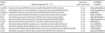
Quantification of mRNA was performed using GoTaq® qPCR Master Mix. PCR reactions were composed of 1 μl cDNA as a template in 7.5 μl of GoTaq® qPCR Master Mix (Promega Corporation, Madison, WI, USA), 5.5 µl of ultra-pure water, and 0.5 μM of each primer. The primers were designed by using the PrimerQuestSM program (http://www.idtdna.com). Primers used in this study are shown in Table 1. The specificity of each primer pair was confirmed by melting curve analysis of PCR products. The thermal cycling profile for the first round of PCR was: initial denaturation and activation of the polymerase for 10 min at 95°C, followed by 40 cycles of 15 s at 95°C, 30 s at 58°C, and 30 s at 72°C. The final extension was for 10 min at 72°C. All reactions were performed in StepOne Real-Time PCR (Applied Biosystems, Foster, CA, USA). Relative quantifications of mRNA were carried out using the comparative threshold (Ct) cycle method. The delta-delta-Ct method was used to transform the Ct values into normalized relative expression levels (Livak & Schmittgen, Reference Livak and Schmittgen2001).
Primer pairs used in real-time PCR for quantification of markers of pluripotency, germ cells and oocytes genes expressed in cells cultured

Statistical analysis
Levels of mRNA for pluripotency (SOX2, NANOG, OCT4, REX), germ cells (VASA, DAZL, cKIT) and oocytes (ZPA, GDF9, SCP3) genes were analyzed by using the non-parametric Kruskal–Wallis test and Dunn's test for post hoc pair-wise comparisons (P < 0.05). Data were expressed as mean ± s.e.m.
Results
Cellular morphology and viability after treating fibroblasts with 5-Aza
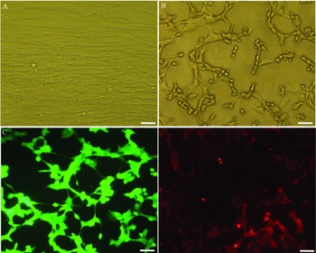
After the exposure to 2.0 µM 5-Aza for 72 h, cell phenotype changed and fibroblast elongated morphology (Fig. 1 A) was replaced by an oval or round shape (Fig. 1 B). Figure 1(C) show that the great majority of the cells remained viable (calcein AM) after 5-Aza treatment, and low number of cells positive for ethidium homodimer-1 was observed (Fig. 1 D).
Morphology of bovine fibroblasts before (A) and after exposure to 5-Aza (B). Fluorescence staining shows viable cells positive for calcein AM (C) and non-viable cells positive for ethidium homodimer-1 (D). Scale bar = 50 µm.

Expression of mRNA for markers of pluripotency after treating fibroblasts with 5-Aza
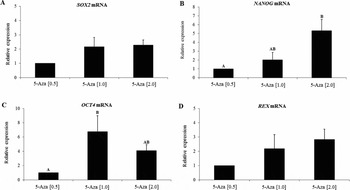
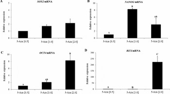
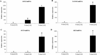
After treatment cells with 5-Aza for 18 h, the levels of mRNA for SOX2 (Fig. 2 A) and REX (Fig. 2 D) was not influenced by any of the concentrations tested. However, NANOG expression was higher (P < 0.05) after treatment with 2.0 μM 5-Aza, when compared with 0.5 μM (Fig. 2 B). Cells treated with 1.0 μM 5-Aza increased the levels of mRNA for OCT4, when compared with those treated with 0.5 μM 5-Aza (Fig. 2 C). After 36 h of 5-Aza treatment, the expression of mRNA for SOX2 (Fig. 3 A) did not change (P > 0.05) in any of the concentrations tested. The levels of mRNA for NANOG (Fig. 3 B) and OCT4 (Fig. 3 C) was increased (P < 0.05) in cells cultured with 1.0 and 2.0 μM 5-Aza, respectively, when compared with those treated with 0.5 μM 5-Aza. In addition, expression of mRNA for REX (Fig. 3 D) was increased (P < 0.05) in cells cultured 1.0 or 2.0 μM of 5-Aza when compared with those cultured with 0.5 μM 5-Aza. After a culture period of 72 h, the levels of mRNA for SOX2 (Fig. 4 A), NANOG (Fig. 4 B), OCT4 (Fig. 4 C), and REX (Fig. 4 D) was significantly higher (P < 0.05) in cells cultured in the presence of 2.0 μM 5-Aza, when compared with 0.5 and 1.0 μM 5-Aza.
Levels of mRNA for SOX2 (A), NANOG (B), OCT4 (C) and REX (D) in fibroblasts cultured for 18 h in different concentrations of 5-Aza (0.5, 1.0 or 2.0 µM). (A,B)Different letters denote significant differences among treatments (P < 0.05).

Levels of mRNA for SOX2 (A), NANOG (B), OCT4 (C) and REX (D) in fibroblasts cultured for 36 h in different concentrations of 5-Aza (0.5, 1.0 or 2.0 µM). (A,B,C)Different letters denote significant differences among treatments (P < 0.05).

Levels of mRNA for SOX2 (A), NANOG (B), OCT4 (C) and REX (D) in fibroblasts cultured for 72 h in different concentrations of 5-Aza (0.5, 1.0 or 2.0 µM). (A,B,C)Different letters denote significant differences among treatments (P < 0.05).

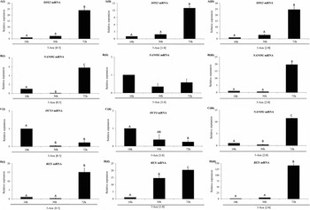
Regarding the effects of culture period, the presence of 0.5 (Fig. 5 Ai), 1.0 (Fig. 5 Aii) or 2.0 µM (Fig. 5 Aiii) 5-Aza in culture medium for 72 h increased (P < 0.05) the expression of SOX2 mRNA, when compared with those seen after 18 h or 36 h of culture. Fibroblasts cultured with 5-Aza (0.5 μM or 2.0 μM) for 72 h had higher levels (P < 0.05) of mRNA for NANOG than those cultured for 18 and 36 h (Fig. 5Bi and 5Bii). However, no effect of culture period on expression of NANOG was seen in cells cultured in presence of 1.0 μM 5-Aza (Fig. 5 Bii).
Levels of mRNA for of pluripotency genes, (A) SOX2, (B) NANOG, (C) OCT4 and (D) REX, in fibroblasts cultured for 18 h, 36 h or 72 h with different concentrations of 5-Aza (0.5, 1.0 or 2.0 µM). (A,B,C)Different letters denote significant differences among treatments (P < 0.05).

A reduction in OCT4 expression was seen when increasing the incubation time from 18 to 72 h (Fig. 5 Ci and 5Cii) in cells cultured with 0.5 and 1.0 μM 5-Aza. In contrast, fibroblasts cultured with 2.0 μM 5-Aza for 72 h had higher levels of OCT4 mRNA than those cultured for 18 and 36 h (Fig. 5 Ciii). The mRNA levels for REX in fibroblasts cultured either with 0.5 μM or 2.0 μM 5-Aza for 72 h were higher 9 (P < 0.05) than those observed after 18 and 36 h (Fig. 5 Di and 5Diii). Cells cultured with 1.0 μM 5-Aza had a progressive and significant increase in REX expression after increasing culture period from 18 to 36 and 72 h (Fig. 5 Dii).
Cellular morphology and viability after culturing in differentiation medium
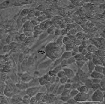
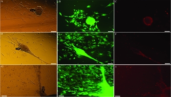
The above cited results showed that cells cultured for 72 h in medium supplemented with 2.0 μM 5-Aza had higher levels of mRNA for pluripotency markers. Thus, cells submitted to this treatment were cultured in differentiation medium supplemented with BMP2, BMP4 or follicular fluid for 7 or 14 days. After 7 days of culture in differentiation medium, some subpopulations of the cells became morphologically different from the starting cultures, increased their volume, assumed a round shape and formed aggregates (Fig. 6). These cells increased they diameters, and gradually became structures similar to oocytes after 14 days of culture in medium supplemented with BMP2 (Fig. 7 A–C), BMP4 (Fig. 7 D–F) or follicular fluid (Fig. 7 G–I).
Cells morphologically different from the starting cultures, with increased volume and round shape (arrow) after 7 days of culture. Scale bar = 50 µm.

Morphology of bovine fibroblasts exposed to 5-Aza and cultured in differentiation medium for 14 days. Fibroblast cultured for 14 days in medium supplemented with BMP2 (line 1), BMP4 (line 2) and follicular fluid (line 3), (A, D, G). Fluorescence staining shows viable cells positive for calcein AM (B, E, H) and non-viable cells positive for ethidium homodimer-1 (C, F, I). Scale bar = 100 µm. Arrows show oocyte-like structures formed after 14 days of culture.

Expression of mRNA for markers of germ cells and oocytes after culturing cells in differentiation medium
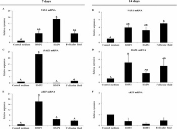
After 7 days of culture, BMP4 stimulated an increase (P < 0.05) in the expression of VASA when compared with control medium, but BMP2 or follicular fluid had no effect (Fig. 8 A). After 14 days of culture, follicular fluid increased (P < 0.05) the expression of VASA, compared with culture medium (Fig. 8 B). DAZL expression was increased (P < 0.05) after culture in medium containing BMP2, when compared with other treatments (day 7, Fig. 8 C) and to control group (day 14, Fig. 8 D). BMP2 increased (P < 0.05) the levels of mRNA of cKIT after 7 days of culture (Fig. 8 E) when compared with other treatments. Expression of cKIT was not influenced by treatments after culture for 14 days (Fig. 8 F).
Levels of mRNA for markers of germ cells: VASA (A, B), DAZL (C, D), and cKIT (E, F) in cells cultured for 7 days (A, C, E) or 14 days (B, D, F) in control medium alone or supplemented with BMP2, BMP4 and follicular fluid. (A,B)Different letters denote significant differences among treatments (P < 0.05).

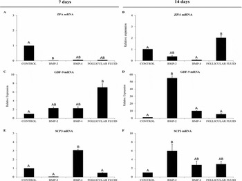
Regarding the expression of oocyte markers, BMP2 reduced (P < 0.05) ZPA expression after 7 days of the culture, when compared with the control group (Fig. 9 A), but follicular fluid increased (P < 0.05) its expression after 14 days, when compared with cells cultured in control medium alone or with BMP4 (Fig. 9 B). Follicular fluid increased (P < 0.05) mRNA levels for GDF9 (Fig. 9 C) after 7 days in relation to control group, while BMP2 increased (P < 0.05) GDF9 expression after 14 days (Fig. 9 D). BMP4 increased (P < 0.05) SCP3 mRNA expression after 7 days (Fig. 9 E), while BMP2 increased (P < 0.05) SCP3 expression after 14 days, when compared with control medium (Fig. 9 F).
Levels of mRNA for markers of oocytes [ZPA (A, B), GDF9 (C, D), SCP3 (E, F)] in cells cultured for 7 days (A, C, E) or 14 days (B, D, F) in control medium alone or supplemented with BMP2, BMP4 and follicular fluid. (A,B)Different letters denote significant differences among treatments (P < 0.05).

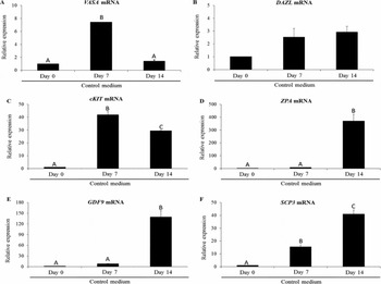
Figures 10, 11, 12 and 13 shows the effects of culture period (0, 7 and 14 days) on expression of makers for germ cells and oocytes. For cells cultured in control medium, VASA expression was increased (P < 0.05) after 7 days, but not after 14 days (Fig. 10 A). DAZL expression was not changed after 7 or 14 days (Fig. 10 B). An increase in mRNA levels for cKIT was observed after 7 or 14 days of culture (Fig. 10 C). Expression of ZPA (Fig. 10 D) and GDF9 (Fig. 10 E) were increased (P < 0.05) after 14 days culture when compared with 0 and 7 days. A progressive and significant increased in SCP3 expression was observed when increasing culture time from 0 to 7 and 14 days (Fig. 10 F).
Levels of mRNA for markers of germ cells [VASA (A), DAZL (B) and cKIT (C)] and oocytes [ZPA (D), GDF9 (E) and SCP3 (F)] after culture cells for 0 (5-Aza), 7 or 14 days in control medium. (A,B,C)Different letters denote significant differences among treatments (P < 0.05).

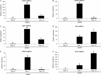
Levels of mRNA for markers of germ cells [VASA (A), DAZL (B) and cKIT (C)] and oocytes [ZPA (D), GDF9 (E) and SCP3 (F)] after culture cells for 0 (5-Aza), 7 or 14 days in medium supplemented with 10 ng/ml of BMP2.

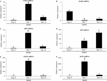
Levels of mRNA for markers of germ cells [VASA (A), DAZL (B) and cKIT (C)] and oocytes [ZPA (D), GDF9 (E) and SCP3 (F)] after culture cells for 0 (5-Aza), 7 or 14 days in medium supplemented with 10 ng/ml of BMP4. (A,B,C)Different letters denote significant differences among treatments (P < 0.05).

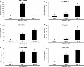
Levels of mRNA for markers of germ cells [VASA (A), DAZL (B) and ckit (C)] and oocytes [ZPA (D), GDF9 (E) and SCP3(F)] after culture cells for 0 (5-Aza), 7 or 14 days in medium supplemented with 5% follicular fluid. (A,B,C)Different letters denote significant differences among treatments (P < 0.05).

Regarding the cells cultured in medium supplemented with BMP2, an increase (P < 0.05) in mRNA levels for VASA (Fig. 11 A), DAZL (Fig. 11 B), cKIT (Fig. 11 C) and GDF9 (Fig. 11 E) was observed after 7 and 14 days, but the levels of these mRNAs after 14 days were lower than those seen after 7 days. After 14 days, the levels of mRNA for ZPA (Fig. 11 D) and SCP3 (Fig. 11 F) were higher (P < 0.05) than those observed after 0 and 7 days.
For cells cultured in presence of BMP4, an increase in mRNA levels for VASA (Fig. 12 A), cKIT (Fig. 12 C), GDF9 (Fig. 12 E) and SCP3 (Fig. 12 F) was observed after 7 and 14 days of culture, but a reduction in expression of these mRNAs was seen when increasing culture time from 7 to 14 days. A reduction in DAZL expression (Fig. 12 B) was also observed after 7 and 14 days culture. In contrast, a progressive and significant increase in mRNA levels for ZPA (Fig. 12 D) was observed when increasing culture time from 0 to 7 and 14 days.
Cells cultured in medium containing follicular fluid had increased (P < 0.05) levels of mRNA for VASA after 7 and 14 days, but a reduction in its expression was seen when increasing culture time from 7 to 14 days (Fig. 13 A). The levels of mRNA for DAZL were increased (P < 0.05) after 14 days of culture (Fig. 13 B). In addition, a progressive and significant increase in mRNA levels for cKIT (Fig. 13 C), ZPA (Fig. 13 D) and SCP3 (Fig. 13 F) was observed when increasing culture time from 0 h to 7 or 14 days. The levels of mRNA for GDF9 were increased after 7 or 14 days (Fig. 13 E).
Discussion
This study shows for the first time that 5-Aza induces expression of genes related with pluripotency in cultured bovine fibroblasts. The 5-Aza is able to modify cell morphology (in tumour-derived cell lines; Taylor & Jones, Reference Taylor and Jones1979; Enjoji et al., Reference Enjoji, Nakashima, Honda, Sakai and Nawata1997) and embryonic and adult cells (Taylor & Jones, Reference Taylor and Jones1982). Pennarossa et al. (Reference Pennarossa, Maffei, Campagnol, Tarantini, Gandolfi and Brevini2013) reported that 5-Aza induced conversion of human skin fibroblasts into insulin-secreting cells. 5-Aza is a chemical derivative of the DNA nucleoside cytidine that causes DNA demethylation or hemi-demethylation, binds covalently and irreversibly to DNA methyltransferase 1 (DNMT1), and regulate gene expression by relaxing chromatin structure. Consequently, 5-Aza allows transcription factors to bind to the promoter regions of genes associated with cell pluripotency (Federation et al., Reference Federation, Bradner and Meissner2014). Previous studies have reported that 5-Aza alter cellular phenotype, gene expression and promotes the generation of iPS in mouse (Hou et al., Reference Hou, Li, Zhang, Liu, Guan and Li2013). Efficient reprogramming methods have been explored as the first report of the generation of human induced pluripotent stem cells (Takahashi & Yamanaka, Reference Takahashi and Yamanaka2006). Besides 5-Aza, a growing number of compounds have been identified that can functionally replace reprogramming transcription factors, enhancing efficiency of iPS generation and accelerating the reprogramming process (Zhang et al., Reference Zhang, Li, Laurent and Ding2012). Among these compounds, it can be highlighted N-phthalyl-l-tryptophan (RG108), valproic acid (VPA), trichostatin A (TSA), sodium butyrate (NaB) and other molecules (Federation et al., Reference Federation, Bradner and Meissner2014).
In this study we demonstrate that exposure of bovine skin fibroblasts to 2.0 μM of 5-Aza for 72 h lead to highest expression of mRNA for SOX2, NANOG, OCT4 and REX. These genes are considered the main pluripotency markers and their expression supported the pluripotency of iPS in goat species (Singhal et al., Reference Singhal, Singhal, Malik, Singh, Kumar and Kaushik2015). Exposure of porcine fetal fibroblasts to 5-Aza reduced the expression of DNA methyltransferase 1 (DNMT1) and altered the expression of genes involved in imprinting (IGF2) and apoptosis (BAX, BCL2L1) (Mohana Kumar et al., Reference Mohana Kumar, Jin, Kim, Song, Hong and Balasubramanian2006). In addition, Pennarossa et al. (Reference Pennarossa, Maffei, Campagnol, Tarantini, Gandolfi and Brevini2013, Reference Pennarossa, Maffei, Campagnol, Rahman, Brevini and Gandolfi2014) exposed human and pig dermal fibroblasts to 5-Aza for 18 h and differentiated them into endocrine pancreatic cells. Oct4, a regulator of pluripotency, is expressed in pluripotent stem cells, in iPS and in germ cells during embryogenesis (Takahashi & Yamanaka, Reference Takahashi and Yamanaka2006; Sterneckert et al., Reference Sterneckert, Hoing and Schöler2012). It has been demonstrated that germline stem cells derived from ovaries also expressed OCT4 (Pacchiarotti et al., Reference Pacchiarotti, Maki, Ramos, Marh, Howerton and Wong2010). The transcription factor NANOG is essential for the establishment of pluripotency during the derivation of ESC and iPS. However, NANOG is not essential to maintain pluripotency (Marthaler et al., Reference Marthaler, Tubsuwan, Schmid, Poulsen, Hyttel and Nielsen2016). The repression of OCT4 or SOX2 in ESCs promotes cellular differentiation (Masui et al., Reference Masui, Nakatake, Toyooka, Shimosato, Yagi and Takahashi2007), besides that, OCT4 and SOX2 proteins dimerize on DNA to activate their target genes in order to specify pluripotency (Remenyi et al., Reference Remenyi, Lins, Nissen, Reinbold, Schöler and Wilmanns2003). NANOG directly transactivates REX promoter and positively regulates REX expression. OCT4 and SOX2 can either activate or repress the REX promoter, depending on the cellular environment (Son et al., Reference Son, Choi, Han and Cho2013). REX is a stringent pluripotency marker that specifies a subpopulation of undifferentiated ESC (Kopper et al., Reference Kopper, Giladi, Golan-Lev and Benvenisty2010).
Culture of 5-Aza treated fibroblasts in differentiation medium supplemented with BMP2 and BMP4 induced the expression of mRNA for germ cell and oocyte markers. In vivo and in vitro evidences demonstrate that BMP4 induce the differentiation of PGC in mouse developing embryos (Ohinata et al., Reference Ohinata, Ohta, Shigeta, Yamanaka and Wakayama2009). In vitro differentiation of human PGCs was increased by addition of BMP4, BMP7 and BMP8b in co-culture with human or mouse fetal gonad stromal cells (Bucay et al., Reference Bucay, Yebra, Cirulli, Afrikanova, Kaido and Hayek2009; Park et al., Reference Park, Galic, Conway, Lindgren, Van Handel and Magnusson2009). Several studies reported PGC absence in BMP4 mutant mouse embryos and a reduction of these cells in BMP2 and BMP8 mutant embryos (Ying & Zhao, Reference Ying and Zhao2001; Kishigami & Mishina, Reference Kishigami and Mishina2005). Additionally, BMP4 promotes mammalian oogonial stem cell differentiation in humans (Yu et al., Reference Yu, Vu, Cho, Guo and Chen2014), buffaloes (Shah et al., Reference Shah, Saini, Ashraf, Singh, Manik and Singla2015), and goats (Singhal et al., Reference Singhal, Singhal, Malik, Singh, Kumar and Kaushik2015). BMP2 also plays a crucial role in the differentiation of PGCs (Pera et al., Reference Pera, Andrade, Houssami, Reubinoff, Trounson and Stanley2004).
The presence of follicular fluid in differentiation medium influences the expression of mRNA for germ cell and oocyte markers in cultured 5-Aza-treated fibroblasts. The follicular fluid is an ultrafiltrate of plasma that has been modified by secretion and uptake of specific components by the cells within the follicle itself (Rodgers & Irving-Rodgers, Reference Rodgers and Irving-Rodgers2010). The follicular fluid may contain several proteins, such as BMP that are involved in the formation of germ cells (Dyce et al., Reference Dyce, Zhu, Craig and Li2004; Reference Dyce, Wen and Li2006; Linher et al., Reference Linher, Dyce and Li2009). In porcine species, follicular fluid successfully simulate differentiation of oocyte during culture of stem cells obtained from skin, adipose and ovarian tissues (Dyce et al., Reference Dyce, Wen and Li2006; Reference Dyce, Liu, Tayade, Kidder, Betts and Li2011; Song et al., Reference Song, Kumar, Kang, Lee, Kim and Ock2011), and this cells expressed DAZL, VASA and SCP3 (Dyce et al., Reference Dyce, Liu, Tayade, Kidder, Betts and Li2011). Follicle-like structures were also successfully differentiated in vitro from hESCs in the presence of follicular fluid (Aflatoonian et al., Reference Aflatoonian, Ruban, Jones, Aflatoonian, Fazeli and Moore2009). Cheng et al. (Reference Cheng, Chen, Yu, Zheng and Wang2012) promoted differentiation of human amniotic fluid stem cells into oocyte-like cells after culturing in presence of porcine follicular fluid.
Our results indicate that BMP2, BMP4 or bovine follicular fluid influences the expression of markers for germ cells (VASA, DAZL and cKIT), meiosis (SCP3) and oocyte (GDF9 and ZPA). Studies indicated that VASA is expressed in post-migratory PGCs until the post-meiotic stage of oocytes (Castrillon et al., Reference Castrillon, Miao, Kollipara, Horner and DePinho2003). DAZL is also considered to be essential for PGC development, as DAZL knockout mice lack a germ cell population (Kee et al., Reference Kee, Angeles, Flores and Nguyen2009). DAZL and VASA have been used as markers for germ cell differentiation in various species: human: Chen et al., Reference Chen, Kuo, Chien, Shun, Yao and Ip2007), murine: (Niikura et al., Reference Niikura, Niikura and Tilly2009), and porcine (Dyce et al., Reference Dyce, Wen and Li2006). Clark et al. (Reference Clark, Bodnar, Fox, Rodriquez, Abeyta and Firpo2004) indicated that ckit is one of the most used markers for PGCs, and this receptor is important for migration and survival of PGC (Cai et al., Reference Cai, Rothbart, Lu, Xu, Chen and Tripathy2013). GDF9 is expressed specifically in oocytes and have been used as oocyte marker after its differentiation from stem cells (Dyce et al., Reference Dyce, Wen and Li2006; Linher et al., Reference Linher, Dyce and Li2009). GDF9 was expressed in populations of cultured mouse ESC with an oocyte-like phenotype (Salvador et al., Reference Salvador, Silva, Kostetskii, Radice and Strauss2008). SCP3 expression is frequently used as a meiotic marker (Lin et al., Reference Lin, Chen, Yu, Yu, Lin and Wu2016). The zona pellucida glycoprotein is only expressed in oocytes (Lefièvre et al., Reference Lefièvre, Conner, Salpekar, Olufowobi, Ashton and Pavlovic2004) and earlier studies reported ZP-like structures surrounding OLCs differentiated in vitro (Hu et al., Reference Hu, Lu, Cao, Deng, Li and Wan2015). Singhal et al. (Reference Singhal, Singhal, Malik, Singh, Kumar and Kaushik2015) recently showed that expression of germ cell markers, like zona pellucida and VASA, was stimulated by BMP4.
Regarding the effect of culture time on cellular differentiation, cells cultured for 7 days had an increase in the expression of germ cell markers VASA and cKit, emphasizing the formation of these cells after 7 days of culture. Interestingly, depending on the culture medium, after 14 days of culture, the expression of oocytes markers (GDF9, ZPA and SCP3) was increased, which shows that this culture time was able to induce the formation of oocyte-like cells in vitro. Previously, Panula et al. (Reference Panula, Medrano, Kee, Bergström, Nguyen, Byers, Wilson, Wu, Simon, Hovatta and Reijo Pera2011) reported the iPSs culture for 14 days in differentiation medium formed meiotic cells, with extensive synaptonemal complexes, and post-meiotic haploid cells.
In conclusion, 2.0 μM 5-Aza induces the expression of mRNAs for SOX2, OCT4, NANOG and REX in bovine skin-derived fibroblasts cultured for 72 h. The presence of BMP2, BMP4 or follicular fluid in differentiation medium induces cellular morphological changes and promotes expression of markers for germ cells (VASA, DAZL and cKIT), meiosis (SCP3) and oocyte (GDF9 and ZPA). This method can be effective to produce oocytes from somatic cells and can thus contribute, in the future, for the development of new biotechnologies to solve fertility problems in human species, and to increase the reproductive potential of genetically superior animals or endangered species.
Acknowledgements
This work was financially supported by CNPq (Grant No. 478198/2013-2), and the authors thank the members of the Laboratory of Animal Reproduction of the Biotechnology Nucleus of Sobral.
Conflict of interest
The authors declare that there is no conflict of interest that would prejudice the impartiality of this scientific work.