INTRODUCTION
Cryptosporidium parvum is an intracellular apicomplexa protozoan parasite, which primarily infects gastrointestinal epithelial cells of many vertebrates, including humans, and causes a diarrhoeal illness, cryptosporidiosis (Tzipori and Ward, Reference Tzipori and Ward2002). Infections in immunocompetent individuals are normally asymptomatic or characterized by self-limiting diarrhoea, but it can develop into severe chronic, prolonged watery diarrhoea which is frequently fatal among immunocompromised patients, such as people with acquired immunodeficiency syndrome (Tzipori and Ward, Reference Tzipori and Ward2002). As effective therapeutic or preventive interventions for cryptosporidiosis are not yet available, the development of effective anticryptosporidial drugs is urgently required. However, the poor experimental tractability of the protozoan parasite is one of the main obstacles to the development of effective vaccines or therapeutics.
Many protozoan parasites have been regarded as anaerobes, but these parasites have been recognized to be aerotolerant or microaerophilic, consuming oxygen to a certain extent (Mehlotra, Reference Mehlotra1996). Therefore, parasitic protozoa develop antioxidant systems to protect themselves from endogenous oxidative stress, as well as oxidative killing by host immune effector cells (Mehlotra, Reference Mehlotra1996; Nickel et al. Reference Nickel, Rahlfs, Deponte, Koncarevic and Becker2006). Due to their essential roles in parasite physiology, antioxidant enzymes have been regarded as attractive drug targets for parasitic protozoa (Flohé et al. Reference Flohé, Hecht and Steinert1999; Andricopulo et al. Reference Andricopulo, Akoachere, Krogh, Nickel, McLeish, Kenyon, Arscott, Williams, Davioud-Charvet and Becker2006; Boucher et al. Reference Boucher, Brzozowski, Brannigan, Schnick, Smith, Kyes and Wilkinson2006; Nickel et al. Reference Nickel, Rahlfs, Deponte, Koncarevic and Becker2006; Wilkinson et al. Reference Wilkinson, Prathalingam, Taylor, Ahmed, Horn and Kelly2006; Na et al. Reference Na, Kang, Kim and Sohn2007). The genome of Cryptosporidium also suggests that the parasite is capable of both aerobic and anaerobic metabolisms (Xu et al. Reference Xu, Widmer, Wang, Ozaki, Alves, Serrano, Puiu, Manque, Akiyoshi, Mackey, Pearson, Dear, Bankier, Peterson, Abrahamsen, Kapur, Tzipori and Buck2004; Heiges et al. Reference Heiges, Wang, Robinson, Aurrecoechea, Gao, Kaluskar, Rhodes, Wang, He, Su, Miller, Kraemer and Kissinger2006), but it has been proposed that Cryptosporidium has a poor capacity to scavenge reactive oxygen species (ROS) compared with other protozoan parasites, which makes this parasite potentially more susceptible to ROS (Entrala et al. Reference Entrala, Mascaro and Barrett1997). Indeed, ROS was observed to have protective roles in experimental C. parvum infection in the neonatal mouse (Leitch and He, Reference Leitch and He1999), which inversely suggests the essential role of an antioxidant system for the survival and pathology of the parasite. However, the antioxidant system of C. parvum is poorly understood and only a few enzymes, which are involved in the antioxidant defence mechanism, have been characterized in the parasite (Kang et al. Reference Kang, Cheun, Kim, Moon, Park, Kim, Sohn and Na2008; Joung et al. Reference Joung, Yoon, Choi, Kim, Park and Yu2011). Therefore, understanding the nature and mechanisms of the antioxidant system of Cryptosporidium would be important to gain an insight into the biology of the parasite, as well as being important for the development of therapeutic drugs.
Glutathione peroxidase (GPx; EC 1.11.1.9) is one of the most important endogenous antioxidant enzymes and mediates antioxidant defence reactions by catalysing the reduction of organic hydroperoxides and hydrogen peroxide to water and oxygen using glutathione (GSH) as an electron donor (Herbette et al. Reference Herbette, Roeckel-Drevet and Drevet2007; Toppo et al. Reference Toppo, Vanin, Bosello and Tosatto2008). Global distribution of the enzyme in all oxygen-consuming organisms suggests a key protective role of the enzyme in cells/organisms from oxidative damage. The enzymes belonging to the GPx family share similar structural and enzymatic features with one another, and are classified into two large subfamilies of selenium-dependent GPx (Se-GPx) and selenium-independent GPx (NS-GPx). A cysteine residue encoded by UGU, which is located in the active site, is a specific feature of NS-GPxs, whereas Se-GPxs possess a seleno-cysteine (SeCys) residue encoded by UGA. The presence of a SeCys insertion sequence (SECIS) motif, a characteristic stem loop structure located downstream from the UGA codon which recognizes the UGA codon as a signal to insert a SeCys residue, in Se-GPxs is another important structural feature discriminating Se-GPxs from NS-GPxs (Herbette et al. Reference Herbette, Roeckel-Drevet and Drevet2007; Toppo et al. Reference Toppo, Vanin, Bosello and Tosatto2008).
The pivotal physiological roles of GPxs in parasitic protozoa have been acknowledged, and the enzymes have been postulated as potential chemotherapeutic drug targets (Wilkinson et al. Reference Wilkinson, Meyer and Kelly2000; Sztajer et al. Reference Sztajer, Gamain, Aumann, Slomianny, Becker, Brigelius-Flohé and Flohé2001; Wilkinson and Kelly, Reference Wilkinson and Kelly2003). In this study, we have identified a gene encoding GPx of C. parvum (CpGPx) and characterized biochemical properties of the recombinant enzyme.
MATERIALS AND METHODS
Identification and cloning of the gene encoding CpGPx
The gene encoding a CpGPx (Gene ID: cgd3_460) was identified by data-mining the Cryptosporidium Genomics Resources (CryptoDB, http://cryptodb.org). The open reading frame of CpGPx was amplified with forward (5′-ATGGGGAATTTTTTAGCATCCACAAAGATA-3′) and reverse (5′-TTAAGAACCTTGTTCACTTTGTTTTTGCTT-3′) primers with Ex Taq DNA polymerase (Takara, Otsu, Japan) and C. parvum Iowa II genomic DNA. The amplified PCR product was gel-purified, ligated into the T&A cloning vector (Real Biotech Corporation, Banqiao City, Taiwan), and transformed into Escherichia coli DH5α. The nucleotide sequence of cloned gene was confirmed by automated DNA sequencing. Analysis of the primary structure of the deduced amino acid sequences was conducted with DNASTAR (DNASTAR, Madison, WI, USA), TMHMM (http://www.cbs.dtu.dk/services/TMHMM-2.0), and Signal P (http://www.cbs.dtu.dk/services/SignalP). TargetP1.1 (http://www.cbs.dtu.dk/services/TargetP) and PSORT (http://www.psort.org) were used to predict protein localization. SECISearch 2.19 (http://genome.unl.edu/SECISearch) was used for in silico detection of the SECIS motif within the 3′-untranslated region (UTR) of CpGPx.
Expression and purification of recombinant CpGPx
To express the recombinant CpGPx, the full-length gene of CpGPx was amplified using the primers 5′-GGATCCATGGGGAATTTTTTAGCATCCACA-3′, which contained a 5′ Bam HI site, and 5′-CTGCAGTTAAGAACCTTGTTCACTTTGTTT-3′, which harboured a 5′ Pst I site. The PCR product was purified, ligated into the T&A cloning vector (Real Biotech Corporation), and transformed into E. coli DH5α. The resulting plasmid DNA was digested with the appropriate restriction enzymes and ligated into the pQE-30 expression vector (Qiagen, Valencia, CA, USA), which was then transformed into E. coli M15 [pREP4] cells (Qiagen). Selected clones were grown and induced with 1 mm isopropyl-1-thio-β-D-galactopyranoside (IPTG) at 37 °C for 3 h. The cells were harvested by centrifugation at 12 000 g for 15 min at 4 °C, and suspended in native lysis buffer (50 mm NaH2PO4, 300 mm NaCl, 10 mm imidazole, pH 8·0), sonicated on ice, and then centrifuged at 4 °C for 20 min at 12 000 g . The recombinant CpGPx was purified by nickel-nitrilotriacetic acid (Ni-NTA) affinity chromatography (Qiagen), and the purification and purity of the recombinant protein was determined by sodium dodecyl sulphate-polyacrylamide gel electrophoresis (SDS-PAGE).
Enzyme assay
GPx activity was assayed by monitoring the glutathione reductase-catalysed nicotinamide adeninedinucleotide phosphate (NADPH; Sigma, St. Louis, MO, USA) oxidation at 37 °C in 200 μL of assay buffer, which contained 0·3 mm NADPH, 0·1% Triton, 5 mm EDTA, 3 mm glutathione (GSH; Sigma), glutathione reductase (1·2 units mL‒1, Sigma), 73 mm cumene hydroperoxide (Cumene-OOH; Sigma) and 0·1 m Tris-HCl (pH 7·6). The level of NADPH oxidation was measured at 340 nm for 5 min by using Multiscan FC spectrophotometer (Thermo, Vantaa, Finland). All the reagents were prewarmed to room temperature before performance of the assay. As a negative control, bovine serum albumin (BSA, Sigma) was used in the reaction mixture. One unit of GPx activity was defined as the amount of enzyme required to cause the oxidation of 1 nm of NADPH per min under the above assay condition.
Production of antibody for CpGPx (anti-CpGPx) and Western blot analysis
Specific antibody against recombinant CpGPx was produced by immunizing BALB/c mice with the purified recombinant protein (50 μg) three times at 2-week intervals. Two weeks after the final inoculation, the mice were euthanased and the sera were collected. The immunoglobulin G (IgG) fraction was isolated from the sera with a Protein G-Sepharose column (Amersham Biosciences, Piscataway, NJ, USA). The specificity of anti-CpGPx was determined by Western blot analysis. The oocysts of C. parvum (Iowa strain) passaged in calves were purchased from Waterborne Inc. (P102C; New Orleans, LA, USA). The oocysts (107) were excysted by incubation in excystation medium (0·5% trypsin, pH 2·5) for 30 min at 37 °C. The excysted sporozoites were immediately collected, rinsed five times with sterile phosphate buffered saline (PBS; pH 7·4), and suspended in sterile PBS which contained complete protease inhibitor cocktail (Roche, Mannheim, Germany). The excysted sporozoites were confirmed by examination under microscopy. The sporozoites were sonicated on ice using 5-s pulses at 50% output power for up to 3 min in a W-380/385-series SONICATOR Ultrasonic Liquid Processor (Misonix Inc., Farmingdale, NY, USA). The soluble extract of C. parvum sporozoites (20 μg) was collected by centrifugation and subjected to SDS–PAGE, and the proteins were transferred to a nitrocellulose membrane (Bio-Rad, Hercules, CA, USA). The membrane was blocked with PBS supplemented with 0·05% Tween 20 (PBST, pH 7·4) and 3% skim milk for 1 h. The membrane was then incubated with anti-CpGPx which was diluted 1:1000 in PBST at room temperature for 2 h. After several washes with PBST, the membrane was incubated with 1:1000 diluted horseradish peroxidase-conjugated goat anti-mouse IgG (Sigma). The blot was visualized with 4-chloro-1-naphthol (Sigma) and the reaction was stopped by washing the membrane with distilled water.
Biochemical properties of recombinant CpGPx
The optimal pH for the maximum activity of CpGPx was assessed in sodium acetate (pH 5·0–5·5), sodium phosphate (pH 6·0–6·5), Tris-HCl (pH 7·0–8·5) and glycine–NaOH (pH 9·0). Recombinant CpGPx was added to each pH buffer supplemented with all chemicals and enzymes and the enzyme activity was measured as described above. For each pH, the appropriate blank was separately measured as a control. The pH stability of CpGPx was also examined at pH 5·0, 6·0, 7·0, 8·0 or 9·0 by incubating the enzyme at 37 °C in the appropriate buffers. Substrate specificity of CpGPx was determined its relative activity against different hydroperoxide substrates, cumene-OOH, t-butyl hydroperoxide (t-butyl-OOH) or hydrogen peroxide (H2O2). For inhibitor test, CpGPx was preincubated with different concentrations (0·01 to 10 mm) of potassium cyanide (KCN) or sodium azide (NaN3) in 50 mm Tris–HCl (pH 7·5) at room temperature for 1 h. The residual enzyme activity of each sample was measured using cumene-OOH as a substrate. All assays were carried out triplicate and the mean and standard deviation (s.d.) were calculated.
Fast protein liquid chromatography (FPLC)
To determine the molecular structure of CpGPx, gel filtration chromatography was performed with a Superdex 200 HR 10/30 column using an Äcta FPLC system (GE Biosciences, Pittsburgh, PA, USA). The column was equilibrated and eluted with 20 mm Tris–HCl (pH 8·0) containing 0·15 m NaCl at a flow rate of 0·5 mL min−1. Recombinant CpGPx was loaded onto the column, and fractions (each 0·5 mL) were collected. The collected fractions were analysed by both enzyme assay and SDS-PAGE analysis. The column was calibrated with the following molecular weight markers (Sigma): blue dextran (Mr 2000 kDa), β-amylase (Mr 200 kDa), alcohol dehydrogenase (Mr 150 kDa), BSA (Mr 66 kDa), carbonic anhydrase (Mr 29 kDa) and cytochrome c (Mr 12·4 kDa). The K av value for each protein was calculated according to the equation: K av = (V e−V 0)/(V t−V 0), where V e is the elution volume of the protein, V 0 is the elution volume of blue dextran and V t is the total bed volume.
Protective role of CpGPx from oxidative damage
The protective role of CpGPx on DNA and protein from oxidative damage was analysed. pET-45b plasmid (Novagen; La Jolla, CA, USA) was incubated with different concentrations (0, 0·1 and 0·2%) of H2O2 in the presence or absence of different amounts of recombinant CpGPx at 37 °C for 2 h and was analysed by agarose gel electrophoresis. For the protein assay, BSA (10 μg) was incubated with different concentrations (0, 0·1 and 0·2%) of H2O2 in the presence or absence of different amounts of recombinant CpGPx at 37 °C for 2 h, and was then analysed by SDS-PAGE.
Expression profile of CpGPx
The expression pattern of CpGPx in different developmental stages of C. parvum was analysed by in vitro cultivation followed by semi-quantitative reverse transcription PCR (RT–PCR). For in vitro cultivation assay, the oocysts of C. parvum (107) were bleached with 1:20 diluted household bleach for 30 min at room temperature to sterilize the parasites, and were then washed three times with sterile phosphate buffered saline (PBS, pH 7·4) as described previously (Borowski et al. Reference Borowski, Thompson, Armstrong and Clode2010). Sterilized oocysts were incubated in 0·2 μm membrane-filtered excystation medium (0·5% trypsin, pH 2·5) for 30 min at 37 °C. The excysted sporozoites were collected, rinsed five times with sterile PBS, and then were suspended in serum-free RPMI medium. The excysted sporozoites were confirmed by microscopic examination. The sporozoites of C. parvum were experimentally infected to human ileocaecal epithelial cells (HCT-8, ATCC CCL-244; American Type Culture Collection, Rockville, MD, USA) by adding 105 sporozoites to HCT-8 cells which were grown to a 70% confluent monolayer in 6-well plates, and the cells were then incubated at 37 °C with 5% CO2 for 1 h. The supernatant was discarded and the cells were washed with PBS several times to remove any uninfected C. parvum sporozoites. After adding fresh RPMI medium with 10% fetal bovine serum to each well, the cells were incubated at 37 °C in 5% CO2 for 6, 12, 24, 36, 48 and 60 h, respectively. The cells were collected at each indicated time point and infection was quantified by an enzyme-linked immunosorbent assay, as described previously (Wanyiri et al. Reference Wanyiri, O'Connor, Allison, Kim, Kane, Qiu, Plaut and Ward2007). Total RNAs were isolated from each sample using Trizol reagent (Invitrogen, Carlsbad, CA, USA) and semi-quantitative RT–PCR was performed with equal amounts of total RNA (1 μg each) and CpGPx specific primers. The RNAs purified from excysted sporozoites were also included. PCR products were analysed on 1·5% agarose gel with ethidium bromide staining. Cryptosporidium parvum heat shock protein 70 (CpHSP70; cdg2_20), which is reported to be constitutively expressed in different developmental stages of C. parvum (Shahiduzzaman et al. Reference Shahiduzzaman, Dyachenko, Obwaller, Unglaube and Daugschies2009), was included as a standard control. The COWP6 (cdg4_3090), a major oocyst wall protein expressed in oocyst of C. parvum (Chatterjee et al. Reference Chatterjee, Banerjee, Steffen, O'Connor, Ward, Robbins and Samuelson2010), and enolase (cdg5_1960), the expression of which is spiked in immature meronts (Mauzy et al. Reference Mauzy, Enomoto, Lancto, Abrahamsen and Rutherford2012), were also analysed to verify the development of the parasite.
RESULTS
Molecular characterization of CpGPx
The gene encoding CpGPx consisted of 657 bp that encoded 218 amino acid residues with a predicted molecular mass of 24·8 kDa. The deduced amino acid sequence alignment of CpGPx and several GPxs from other organisms is shown in Fig. 1. CpGPx showed 11·5–26·3% sequence identity to those from the GPx superfamily of human, and was more related to GPx4 (phospholipid hydroperoxide GPx; PHGPx) than to any of the other GPx types. CpGPx showed higher sequence identity to GPxs from plants and yeast than those of mammals. Neither potential signal peptide sequence nor putative glycosylation site was found in the sequence of CpGPx. The three amino acid residues, Cys51, Gln86 and Trp145, which form the catalytic triad of GPxs (Toppo et al. Reference Toppo, Vanin, Bosello and Tosatto2008), were well conserved in CpGPx. As plant GPx-like proteins, CpGPx contained the standard codon for cysteine (UGU), rather than the opal UGA codon for SeCys at the catalytic site (Fig. 1). In silico prediction analysis of the SECIS motif within the 3′-UTR of CpGPx revealed that no SECIS motif was found in the sequence (data not shown), which suggested its selenium-independent nature. Prediction analysis for cellular localization of CpGPx implied that this enzyme is most likely to be localized in cytosol (data not shown). The PGGG motif and oligomerization interface, which are associated with a tetrameric structure formation and are well-conserved in tetrameric GPxs (Toppo et al. Reference Toppo, Vanin, Bosello and Tosatto2008), were not identified in CpGPx. However, GPxs of Cryptosoporidium and Plasmodium species had a short insert, consisting of 14–15 amino acids, in the corresponding region of the oligomerization interface in mammalian tetrameric GPxs. Phylogenetic analysis revealed that CpGPx was closely related to GPxs of other sporozoan parasites, including Cryptosporidium spp., Plasmodium spp., Toxoplasma gondii and Perkinsus marinus (Fig. 2).
Multiple sequence alignment analysis. The deduced amino acid sequence of CpGPx was aligned with those of GPxs from Cryptosporidium hominis (XP_668297), Cryptosporidium muris (XP_002139111), Plasmodium falciparum (XP_001350528), Plasmodium knowlesi (XP_002260396), Human GPx1 (NP_000572), Human GPx2 (NP_002074), Human GPx3 (NP_002075), Human GPx4 (NM_002085), Human GPx5 (NP_001500), Human GPx6 (NP_874360), Human GPx7 (NP_056511), Human GPx8 (NP_001008398), Arabidopsis thaliana (AJ000470), Zea mays (AA582602), and Saccharomyces cerevisiae (P38143). Gaps are introduced into the sequences to maximize alignment. The asterisks indicate the catalytic triad (Cys, Gln and Trp). The amino acid residues involved in binding of the GSH molecule are marked with closed circles. The PGGG motif and oligomerization interface are indicated by bold line and double line on the sequence, respectively. The shading represents the degree of identity among the sequences: black (100%), dark grey (50–92%) and light grey (15–49%). Sequence identity of CpGPx to other GPxs is represented on the right. The abbreviation ‘U’ represents seleno-cysteine (SeCys).

Phylogenetic analysis. The phylogenetic tree was constructed with the neighbour-joining method using the MEGA4 program. Numbers on the branches indicate bootstrap proportions (1000 replicates).

Expression and biochemical properties of CpGPx
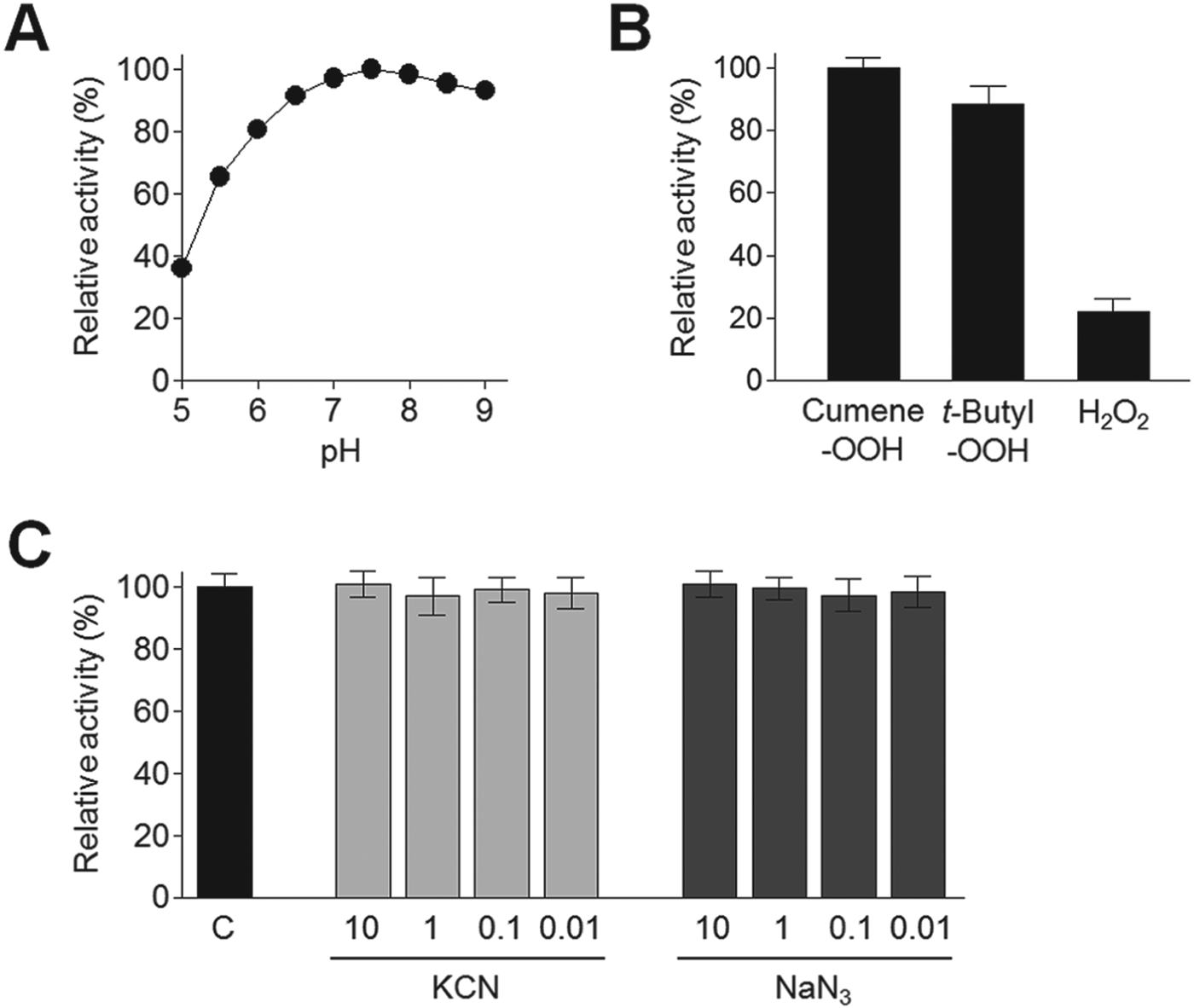
Recombinant CpGPx was successfully expressed in E. coli as a soluble protein of about 25 kDa, which coincided with an estimated molecular mass (Fig. 3A). To confirm the expression of CpGPx in C. parvum, we performed Western blot analysis using anti-CpGPx and a soluble extract of C. parvum sporozoites. Anti-CpGPx was specifically reacted with a protein band having an approximate molecular mass of 25 kDa, which coincided well with the size of recombinant CpGPx, suggesting that CpGPx is functionally expressed in C. parvum sporozoites (Fig. 3B). CpGPx was proven to be monomeric when subjected to Superdex 200 HR 10/30 gel filtration under non-reducing condition. It eluted in the fractions corresponding to the approximate molecular mass of 25 kDa, whereas no activity could be detected in the fractions corresponding to the molecular mass of dimeric or tetrameric forms (data not shown). Recombinant CpGPx was active over a wide pH range with a maximum activity at pH 7·5 (Fig. 4A) and was stable under physiological conditions. The recombinant enzyme showed the activity of 84·3±5·8 units mg‒1 protein for cumene-OOH. It showed substrate preference against organic hydroperoxides, such as cumene-OOH and t-butyl-OOH, but it showed low activity against inorganic hydroperoxide, H2O2 (Fig. 4B). The activity of CpGPx was not inhibited by either KCN or NaN3 (Fig. 4C). CpGPx was not inhibited by KCN, even though the enzyme was preincubated with high concentration of KCN (20 mm) for up to 3 h (data not shown).
Expression and purification of recombinant CpGPx. (A) Expression and purification. A full-length gene of CpGPx was ligated into pQE-30 expression vector and transformed into E. coli M15 [pREP4] cells. Expression of recombinant protein was induced with 1 mm IPTG. Proteins were analysed by SDS–PAGE and stained with Coomassie blue. Lane 1, E. coli lysate control; lane 2, IPTG-induced E. coli lysate; lane 3, recombinant CpGPx purified with Ni-NTA affinity chromatography; (B) Western blot analysis. Recombinant CpGPx and the soluble extract of C. parvum sporozoites were separated on SDS-PAGE and the proteins were transferred to nitrocellulose membrane. The blot was probed with anti-CpGPx. Lane 1, recombinant CpGPx (5 μg); lane 2, soluble extract of C. parvum sporozoites (20 μg).

Biochemical properties of CpGPx. (A) Optimal pH. Enzyme activity was assayed in different pH buffers ranging from pH 5·0–9·0 by using cumene-OOH as a substrate. For each pH, the appropriate blank was separately measured as the control group. Maximal activity was represented as 100%. Three independent assays for each pH were carried out and the mean value was calculated. (B) Substrate specificity. Enzyme activity of CpGPx for different hydroperoxide substrates, cumene-OOH, t-butyl-OOH or H2O2, was assayed. Maximal activity observed for cumene-OOH was represented as 100%. All the assays were carried out triplicate and the mean and standard deviation (s.d.) were calculated. (C) Inhibition assay. CpGPx was preincubated with different concentrations of KCN or NaN3 in 50 mm Tris-HCl (pH 7·5) at room temperature for 20 min. The residual enzyme activity of each sample was measured using cumene-OOH as a substrate. C, control CpGPx without pretreatment of any chemical. All the assays were carried out triplicate and the mean and standard deviation (s.d.) were calculated.

Protective role of CpGPx from oxidative damage
To analyse the protective role of CpGPx towards oxidative damage of biomolecules, we assayed the protection activity of CpGPx on DNA and protein from oxidative damage induced by H2O2. Recombinant CpGPx effectively protected plasmid DNA from H2O2-induced oxidative damage in a dose-dependent manner (Fig. 5A). The H2O2-untreated plasmid DNA showed a major single band corresponding to the supercoiled form (S form). Treatment with H2O2 resulted in oxidative damage of plasmid DNA, which was evaluated by assessing the shift in the mobility of plasmid DNA, which resulted from cleavage into a nicked circular form (N form), in proportion to H2O2 concentration. Treatment of CpGPx inhibited the conversion of the S form into the N form of plasmid DNA in the presence of H2O2 in a dose-dependent pattern, which indicated that CpGPx protected plasmid DNA against oxidative damage. Incubation of BSA with H2O2 also induced oxidative damage of the protein, which resulted in degradation of BSA into various sizes of smaller fragments (Fig. 5B). Treatment of CpGPx also effectively inhibited H2O2-induced damage of BSA in a dose-dependent manner.
Protection of DNA and protein from oxidative damage by CpGPx. (A) DNA cleavage assay. pET-45b plasmid was incubated with different concentrations of H2O2 in the presence or absence of different concentrations of recombinant CpGPx at 37 °C for 2 h and was analysed by agarose gel electrophoresis. N, nicked plasmid; S, supercoiled plasmid; (B) Protein damage assay. BSA (10 μg) was incubated with different concentrations of H2O2 in the presence or absence of different concentrations of recombinant CpGPx at 37 °C for 2 h and analysed by SDS–PAGE.

Expression profile of CpGPx in different developmental stages of C. parvum
To analyse the expression pattern of CpGPx in different developmental stages of C. parvum, we performed in vitro cultivation of the parasite using HCT-8 cells as host cells followed by semi-quantitative RT-PCR analysis using total RNA collected at different time points after infection. The amplification of CpGPx transcripts demonstrated that the gene was transcribed throughout all of the developmental stages of the parasite examined in this study (Fig. 6). To validate the development of the parasite in the cultivation system, we also performed semi-quantitative RT-PCR analysis for three C. parvum genes, including HSP70, enolase and COWP6, the expression patterns of which have been previously demonstrated (Shahiduzzaman et al. Reference Shahiduzzaman, Dyachenko, Obwaller, Unglaube and Daugschies2009; Chatterjee et al. Reference Chatterjee, Banerjee, Steffen, O'Connor, Ward, Robbins and Samuelson2010; Mauzy et al. Reference Mauzy, Enomoto, Lancto, Abrahamsen and Rutherford2012). All tested control genes were confirmed to be expressed at corresponding developmental stages.
Expression profile of CpGPx in the developmental stages of C. parvum. The sporozoites of C. parvum were experimentally infected to HCT-8 cells and the cells were collected at each indicated time point after infection. Total RNAs were isolated from each sample and semi-quantitative RT–PCR was performed with equal amounts of total RNA (1 μg each) and specific primers. PCR products were analysed on a 1·5% agarose gel with ethidium bromide staining. CpHSP70, C. parvum heat shock protein 70 (cdg2_20); enolase, C. parvum enolase (cdg5_1960); COWP6, C. parvum oocyst wall protein 6 (cdg4_3090); PC, positive control (cloned gene); NC, control HCT-8 cells without infection with C. parvum sporozoites; Sp, sporozoites.

DISCUSSION
Sequence analysis of CpGPx suggested that the enzyme is a member of cytosolic NS-GPx family. Immunoblot analysis using soluble extract of C. parvum sporozoites and anti-CpGPx antibody also suggests that the enzyme is a cytosolic protein which is functionally expressed in sporozoites of the parasite. Protozoan GPxs have been acknowledged to be classified into two subclades which cluster in the same branches of the monomeric forms of the human GPx family, including GPx-4, GPx-7 and GPx-8 (Toppo et al. Reference Toppo, Vanin, Bosello and Tosatto2008). CpGPx also showed higher sequence identity with monomeric GPxs than with tetrameric GPxs of mammals. Absences of the PGGG motif and oligomerization interface, which are involved in the formation of tetrameric structures (Toppo et al. Reference Toppo, Vanin, Bosello and Tosatto2008), in CpGPx also imply that the enzyme has a monomeric structure. Both PAGE analysis under non-reducing conditions and gel filtration analysis of recombinant CpGPx also support the notion that CpGPx is a monomeric protein. Sequence and phylogenetic analyses of CpGPx also revealed that CpGPx showed higher sequence identity with plant and yeast GPxs than with animal GPxs. However, this is not surprising given that close association and common ancestry of apicomplexa protozoa parasites with plants, cyanobacteria, and algae have been previously suggested (Wilson et al. Reference Wilson, Williamson and Preiser1994; Stack et al. Reference Stack, Lowther, Cunningham, Donnelly, Gardiner, Trenholme, Skinner-Adams, Teuscher, Grembecka, Mucha, Kafarski, Lua, Bell and Dalton2007; Yu et al. Reference Yu, Zhang and Zhu2010; Kang et al. Reference Kang, Ju, Sohn and Na2011).
Biochemical properties of CpGPx also propose that this enzyme is a member of the NS-GPx family of enzymes. One of the major functional differences between Se-GPxs and NS-GPxs is that Se-GPxs catalyse the reduction of both organic and inorganic peroxides like H2O2, whereas NS-GPxs react preferentially with organic peroxides (Tang et al. Reference Tang, Gounaris, Griffiths and Selkirk1995; Sun et al. Reference Sun, Song, Yan, Xu and Li2012). CpGPx showed a substrate preference against organic hydroperoxide, such as cumene-OOH and t-butyl-OOH, rather than inorganic hydroperoxide, H2O2, which is coincided with previous results from NS-GPx family enzymes of other organisms (Tang et al. Reference Tang, Gounaris, Griffiths and Selkirk1995; Wilkinson et al. Reference Wilkinson, Meyer and Kelly2000; Jones et al. Reference Jones, Reavy, Smant and Prior2004; Sun et al. Reference Sun, Song, Yan, Xu and Li2012). KCN is known to inhibit Se-GPxs by inducing the release of selenium, which is essential for Se-GPxs to maintain their activities, from the active site of the enzymes (Prohaska et al. Reference Prohaska, Oh, Hoekstra and Ganther1977; Awasthi et al. Reference Awasthi, Dao, Lal and Srivastava1979; Singh and Rathaur, Reference Singh and Rathaur2005). However, CpGPx activity was not affected by KCN even though the enzyme was pretreated with a high concentration of KCN (20 mm) up to 3 h, which indicated that CpGPx does not have selenium in its active site. NaN3, an inhibitor of heme proteins, also did not affect CpGPx activity.
Some NS-GPxs, including GPxs of Plasmodium falciparum and Trypanosoma brucei, show a high affinity for thioredoxin to reduce organic hydroperoxides (Wilkinson et al. Reference Wilkinson, Meyer and Kelly2000; Sztajer et al. Reference Sztajer, Gamain, Aumann, Slomianny, Becker, Brigelius-Flohé and Flohé2001; Herbette et al. Reference Herbette, Lenne, Leblanc, Julien, Drevet and Roeckel-Drevet2002; Maiorino et al. Reference Maiorino, Ursini, Bosello, Toppo, Tosatto, Mauri, Becker, Roveri, Bulato, Benazzi, De Palma and Flohé2007). This leads them to be classified into a novel functional class of thioredoxin GPx-like peroxidase (TGPx) based on the prominent preference for thioredoxin as an electron donor (Toppo et al. Reference Toppo, Vanin, Bosello and Tosatto2008). Most cytosolic GPxs showed pronounced specificity against GSH (Flohé et al. Reference Flohé, Günzler, Jung, Schaich and Schneider1971) and this specificity is considered to be due to basic amino acid residues, Arg57, Lys92, Arg102, Arg184 and Arg185, which direct the SH group of the substrate to the active-site selenium atom by electrostatic forces (Aumann et al. Reference Aumann, Bedorf, Brigelius-Flohé, Schomburg and Flohé1997). However, these residues are only partially conserved in CpGPx and the enzyme does not contain SeCys at its active site, and therefore, it would be interesting to analyse further whether CpGPx has thioredoxin peroxidase activity.
The expression of CpGPx in all tested developmental stages of C. parvum suggests its essential role in the physiology of the parasite. In organisms which have both Se-GPxs and NS-GPxs, it has been postulated that Se-GPxs are the main enzymes for scavenging H2O2 and preventing the further formation of ROS, but NS-GPxs contribute only an adequate function as a backup system for Se-GPxs in the absence of sufficient concentrations of selenium (Vernet et al. Reference Vernet, Rock, Mazur, Rayssiguier, Dufaure and Drevet1999). This postulation is mainly due to the less efficient ROS-detoxifying activity of NS-GPxs than Se-GPxs. Unlike other apicomplexa protozoan parasites, such as Plasmodium species and T. gondii, Cryptosporidium species are not likely to utilize SeCys since neither selenoproteins nor SeCys insertion systems have been found in genomes of the parasite (Lobanov et al. Reference Lobanov, Delgado, Rahlfs, Novoselov, Kryukov, Gromer, Hatfield, Becker and Gladyshev2006). Considering the high sequence homology of NS-GPxs with the mammalian membrane-bound GPx4 (PHGPx), most NS-GPxs have been proposed to be involved in reducing membrane peroxidation (Herbette et al. Reference Herbette, Roeckel-Drevet and Drevet2007). Several studies have provided experimental evidence that NS-GPxs efficiently reduce lipid peroxides as well as a broad range of peroxide in vitro (Gaber et al. Reference Gaber, Tamoi, Takeda, Nakano and Shigeoka2001; Herbette et al. Reference Herbette, Lenne, Leblanc, Julien, Drevet and Roeckel-Drevet2002; Jung et al. Reference Jung, Lee, Lee, Chi, Jang, Kang, Lee, Lim, Yoon, Yun, Inoue, Cho and Lee2002). Our data clearly demonstrated that CpGPx effectively protect DNA and protein from H2O2-induced oxidative damage. H2O2 is an uncharged molecule and can freely diffuse across biological membranes and is able to access any sub-cellular compartment within cells. Inside the cells, it can induce damage of diverse cellular macromolecules such as nucleic acids, proteins and lipids directly, or indirectly following the generation of hydroxyl radicals (Ohno and Gallin, Reference Ohno and Gallin1985). CpGPx may play a critical role in the protection of biomolecules of C. parvum against endogenous and/or exogenous H2O2. However, considering that CpGPx showed weak activity against H2O2 compared with short chain organic hydroperoxides, it is possible that this enzyme could also participate in detoxifying secondary oxidation products which are generated during lipid peroxidation reactions. However, it is not currently clear whether CpGPx shows activity for phospholipid hydroperoxides, and this should be further elucidated. It has been previously reported that C. parvum does not have catalase and GPx activities (Entrala et al. Reference Entrala, Mascaro and Barrett1997). We also confirmed that C. parvum lacks catalase in its genome from a data search of CryptoDB, but our results clearly demonstrate that C. parvum has a functional GPx. Currently, only a few antioxidant enzymes, including iron-superoxide dismutase and TPx, have been characterized in the parasite (Kang et al. Reference Kang, Cheun, Kim, Moon, Park, Kim, Sohn and Na2008; Joung et al. Reference Joung, Yoon, Choi, Kim, Park and Yu2011). And therefore, identification for other candidates that exert antioxidant defence in C. parvum and characterization of biochemical and functional properties of the candidates would allow further insight into the understanding of the antioxidant defence mechanism of the parasite.
FINANCIAL SUPPORT
This research was supported by the Basic Science Research Programme through the National Research Foundation of Korea (NRF) funded by the Ministry of Education, Science and Technology (NRF-2010-0021687).