Introduction
The infratemporal fossa is a potential space bounded as follows: superiorly by the temporal bone and the greater wing of the sphenoid bone; medially by the superior constrictor muscle, the pharyngobasilar fascia and the pterygoid plates; laterally by the zygoma, mandible, parotid gland and masseter muscle; anteriorly by the pterygoid muscles; and posteriorly by the articular tubercle of the temporal bone, the glenoid fossa and the styloid process.
The contents of the infratemporal fossa include the internal carotid artery, the internal jugular vein, the IXth to XIIth cranial nerves, the third division of the Vth cranial nerve, the internal maxillary artery, the pterygoid venous plexus and the pterygoid muscles.
Various surgical approaches have been described for tumours of the infratemporal fossa, depending upon their extension.1 Post-auricular – transtemporal approaches have been described by Fisch,Reference Fisch2 preauricular – subtemporal approaches by FischReference Fisch2 and Sekhar et al.,Reference Sekhar, Schramm and Jones3 and transmaxillary approaches by Terz et al. Reference Terz, Young and Lawrence4 and Janecka et al. Reference Janecka, Sen, Sekhar and Arriaga5 Cocke et al. Reference Cocke, Robertson, Robertson and Crook6 have validated the efficiency of and indications for transmaxillary approaches, and have provided a framework for other modifications. Recently, transnasal endoscopic and endoscopy-assisted approaches have been added to this rapidly changing list. In addition, infratemporal fossa approaches may be used as an adjunct to other surgical approaches, such as the transcranial-subtemporal, Le Fort 1 and anterior subfrontal approaches.
Surgical approaches to space-occupying lesions of the orbit and adjacent extracranial skull base may include: (1) simple osteotomy; (2) transmaxillary orbitotomy (for circumscribed lesions affecting both the maxillary sinus and the orbit); (3) a transfrontoethmoidal approach via the paranasal sinus (to expose the medial orbital wall and medial part of the orbital roof); and (4) lateral bony orbitotomy, to access localised, benign tumour (via osteotomy of the zygomatic arch as proposed by Kronlein,Reference Kronlein7 and with simultaneous microsurgical exploration of the orbit as suggested by Jones).Reference Jones8
In cases of primary inferolateral orbital tumour extending to the infratemporal fossa, surgical access is usually via a combination of transorbital and preauricular approaches, to excise the orbital and infratemporal fossa components, respectively. The current paper presents a novel, minimally morbid approach for use in such cases.
Case report and results
A 10-year-old girl presented to us with an eight-month history of proptosis of the left eye, which had developed gradually initially but had progressed rapidly over the previous three weeks.
At presentation, the patient also had complete loss of vision (left), which had commenced two to three weeks prior and progressed. A corneal opacity was clearly evident. There was no trismus or nerve deficit, but the ocular movements and eye closure were grossly compromised owing to a large orbital mass and proptosis, respectively. Systemic and ENT examinations were normal.
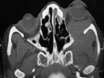
A computed tomography scan revealed a lateral orbital mass pushing the eyeball medially and extending through the inferior oblique fissure into the infratemporal fossa (Figure 1). The paranasal sinuses were within normal limits.
Axial computed tomography scan showing left lateral orbital mass extending into the infratemporal fossa and displacing the eyeball.

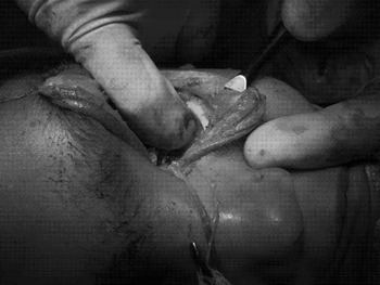
Surgical treatment was undertaken. A horizontal skin incision was made from the lateral canthus along the superior border of the zygomatic process. Orbital exenteration was performed, along with transorbital mobilisation of the mass, in order to visualise the margins of the inferior oblique fissure. After thorough washing, the margins of the inferior oblique fissure were punched out superiorly (involving the zygomatic bone and the greater wing of the sphenoid bone) and laterally (involving the zygomatic bone). Minimal curetting was performed at the inferior border of the inferior orbital fissure, lateral to the infraorbital nerve, which was partially exposed surgically. After sufficient widening of the inferior oblique fissure, the tumour mass extension within the infratemporal fossa was excised in one piece, with the help of sharp dissectors and a bovie. A potential space was thus created, allowing the surgeon's index finger to easily penetrate the defect to a depth of approximately 1 cm (Figure 2). The posterior surface of the maxilla, medial surface of the zygomatic bone, pterygomaxillary fissure and pterygoid muscles could be easily palpated. No major osteotomy was performed, and no injury to any neural structure was encountered. Following the radical tumour excision, there was moderate bleeding (possibly from the pterygoid venous plexus) which was easily controlled with pressure from ribbon gauze packing. The eye was bandaged in the usual fashion.
Intra-operative photograph after total removal of the tumour together with its infratemporal fossa component. The infratemporal fossa is approached via the inferior oblique fissure. The opening thus created is large enough to allow the surgeon to insert a finger to a depth of approximately 1 cm, in order to palpate the bony walls and musculature of the infratemporal fossa.

The post-operative period was uneventful. The packing was removed the day after surgery, revealing a clean, dry, healthy cavity.
Discussion
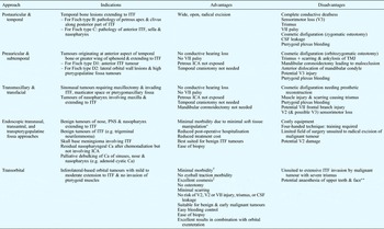
Conventional approaches to the infratemporal fossa require complex surgery which involves significant time, effort, cost and morbidity. Table I summarises the relevant approaches and compares their advantages and disadvantages with those of the novel transorbital approach described above. Inferolateral orbital tumours with infratemporal fossa extension are usually accessed either by a lateral bony orbitotomy approach, exposing the relevant lateral skull base, or by a combination of a preauricular subzygomatic approach together with transorbital exenteration. The incorporation of transorbital approach has been described as complementing the access obtained via classical approaches, thus enhancing exposure of the orbital apex and cavernous sinus. The transorbital approach is usually reserved for benign tumours of the orbital apex and cavernous sinus which are associated with loss of vision, and for low-grade malignancy with minimal involvement of the orbital soft apex and/or optic nerve. Accessing the infratemporal fossa via the inferior oblique fissure has not previously been described.
Approaches to infratemporal fossa

*Compared with conventional open approaches, which risk trismus, V3 and VII injury, scarring, cosmetic disfiguration, and possible cerebrospinal fluid (CSF) leakage. †Comparable with endoscopic approach. ‡Unless prosthetic eye required with orbital exenteration. **Due to injury of posterior-superior dental nerve and infraorbital nerve, respectively. ITF = infratemporal fossa; V3 = third division of Vth cranial nerve; ICA = internal carotid artery; TMJ = temporomandibular joint; V2 = second division of Vth cranial nerve; PNS = paranasal sinus; Ca = carcinoma
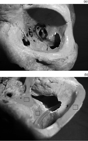
The inferior oblique fissure is formed by the zygomatic bone, the greater wing of the sphenoid bone and the posterosuperior border of the maxilla. The infraorbital nerve lies within its groove in the medial part of the inferior oblique fissure, which is contiguous with the pterygomaxillary fissure. The tendinous ring which constitutes the origin of the intraocular muscles encroaches only on the medial part of the inferior oblique fissure, sparing the lateral portion. Muscle attachments on the anterior bony surfaces of the infratemporal fossa include the crest and infratemporal surface of the sphenoid, the lateral pterygoid plates, and the maxillary tubercle (which constitute the origin of the pterygoid musculature). Hence, muscle attachments are absent from the inner surface of the zygomatic bone, the majority of the posterior surface of the maxilla, and part of the infratemporal fossa surface of the greater wing of the sphenoid bone. Viewed from inside the orbit, this bare bony area predominantly encompasses the lateral two-thirds of the inferior oblique fissure. It is therefore possible to punch out the bone surrounding the lateral part of the inferior oblique fissure, including the zygomatic bone and the greater wing of the sphenoid, in order to widen the inferior oblique fissure superolaterally, thus enlarging the entrance to the infratemporal fossa for better access and visualisation (Figure 3a). Such enlargement does not affect the muscular attachments of this region, nor does it predispose to injury of any vital structure (Figure 3b). Relevant neurovascular structures in this region include the inferior orbital artery, posterior-superior dental artery and nerve, and, rarely, the buccal artery and nerve. No post-operative sensory deficits were encountered in the presented patient; however, any potential sensory morbidity which occurs is likely to be well compensated over time.
(a) Anterior view of the orbit and (b) inferolateral view of the infratemporal fossa seen through the inferior oblique fissure after its margins have been punched out in a superolateral direction. The osteotomy site is seen to be well removed from the tendinous ring attachment of the intraocular muscles (a; circular marking). The osteotomy site also does not involve any muscle attachments (b; shaded areas with overlying squares indicate muscle attachments), and creates a wide communication between the orbit and the infratemporal fossa (indicated by tubing).

If a biopsy is required, the infratemporal fossa can be approached transorbitally with an intact periorbita. A subconjunctival incision is extended deeply onto the periosteum of the infraorbital rim (bony margin), and care is taken to elevate the periosteum off the floor and lateral wall of the orbit until the inferior oblique fissure is delineated. This procedure has been found to be quick and very well tolerated, with virtually no morbidity (Table I). This approach can be further refined by incorporating transorbital endoscopy of the infratemporal fossa via the inferior oblique fissure. This gives a better chance of visualising the anatomy and pathology within the infratemporal fossa, and hence can facilitate a better tumor-free-margin resection.
• Various surgical approaches have been described for tumours of the infratemporal fossa, depending upon their extension, and for space-occupying lesions of the orbit and adjacent extracranial skull base
• Surgery for primary inferolateral orbital tumour extending to the infratemporal fossa usually involves a combination of transorbital and preauricular approaches
• A novel, minimally morbid approach is presented which accesses the infratemporal fossa via the floor of the orbit, after first enlarging the inferior oblique fissure
• This approach is advantageous due to minimal morbidity (including no eyeball traction), excellent cosmesis, no osteotomy, minimal scarring, no trismus, no Vth or VIIth cranial nerve injury, no cerebrospinal fluid leakage, easy haemostasis, ease of biopsy, and excellent results when combined with orbital exenteration
Regarding this procedure, it is important to note that a special periorbital retractor has been designed which causes minimal alteration to the fluid dynamics of the eyeball or optic nerve. This retractor has been named the ‘Torita’ (an abbreviation of ‘trans-orbital retractor for infra-temporal fossa access’). The design has been sent for patenting and will be published elsewhere in due course.
Conclusion
Various surgical approaches have been defined for tumours of the infratemporal fossa, depending upon their extension, including: post-auricular – transtemporal; preauricular – subtemporal; transmaxillary; and transnasal endoscopic and endoscopy-assisted. Infratemporal fossa approaches may also be used as an adjunct to the transcranial-subtemporal, Le Fort 1 and anterior subfrontal approaches.
Surgical approaches to space-occupying lesions of the orbit and adjacent extracranial skull base may include simple osteotomy, transmaxillary orbitotomy, lateral bony orbitotomy and a transfrontoethmoidal approach.
Primary inferolateral orbital tumours extending to the infratemporal fossa are usually accessed via a combination of transorbital and preauricular approaches.
This paper presents a novel, minimally morbid approach for such cases, in which the infratemporal fossa is approached via the floor of the orbit, after enlarging the inferior oblique fissure. Compared with other, previously described approaches, this new route is advantageous due to: minimal general morbidity; no morbidity due to eyeball traction; excellent cosmesis; no need for osteotomy; minimal scarring; no risk of trismus, cerebrospinal fluid leakage, or Vth or VIIth cranial nerve injury; easy haemostasis; ease of biopsy; and excellent results in combination with orbital exenteration.
This new approach is however not suited to cases of extensive invasion of the infratemporal fossa by malignant tumour causing severe trismus, and also carries a risk of potential anaesthesia of the upper teeth (supplied by the posterior-superior dental nerve) and face (supplied by the infraorbital nerve).
Acknowledgements
The author acknowledges his colleagues Drs V Veerendra and B K Ojha, and his team members Drs S Chakraboroty, P Narayan, R Agarwal and R Verma, for their contribution to case management and reference collection.