Introduction
The in utero hormonal milieu is believed to play an important role in fetal programming of later health and disease. Prenatal androgen exposure is of particular interest, given the important role that sex hormones play in the development of many mammalian body systems including the reproductive system and brain. In animal models, manipulation of sex-typical androgen concentrations during critical periods of early development can elicit changes in reproductive functionReference Birch, Padmanabhan, Foster, Unsworth and Robinson 1 – Reference Witham, Meadows, Shojaei, Kauffman and Mellon 3 as well as behavior (both reproductive and social).Reference Goy and McEwen 4 , Reference Hines 5 In humans, evidence from clinical conditions such as congenital adrenal hyperplasia (CAH), in which typical fetal endocrine activity is altered, reinforce the important role that these hormones play during fetal development.Reference Constantinescu and Hines 6 – Reference Mueller, Grissom and Dohanich 9 Much less is known about how the typical range of variation in fetal androgen exposure contributes to postnatal outcomes.
Unfortunately, gaining insight into the fetal hormonal milieu presents a considerable challenge to human research. Indexing fetal hormone concentrations through sampling of fetal blood or amniotic fluid is impossible to do on a population level and is no longer a viable research tool. Assessment of exposure through alternative media (including maternal serum saliva and hair) is feasible, but is still problematic because it: (1) may not be informative about fetus’ level of exposure;Reference Kuijper, Ket, Caanen and Lambalk 10 and (2) requires recruitment during pregnancy, which is impractical for many types of studies, particularly those examining health outcomes presenting much later in life. For these reasons, there has been great interest in establishing biomarkers of prenatal exposure to sex steroids, particularly androgens, that can be reliably measured later in life. Two putative biomarkers of prenatal androgen exposure, anogenital distance (AGD) and 2:4 digit length ratio, have emerged based on animal models and the human literature. Both biomarkers are gaining popularity as research tools and are appealing because their measurement is non-invasive, inexpensive and replicable.Reference Sathyanarayana, Beard, Zhou and Grady 11 , Reference McIntyre 12
AGD, the distance from the anus to the genitals, is 50–100% longer in males than in females in humans and most other species. Androgen-insensitive male rodents show shortened AGD,Reference Yeh, Tsai and Xu 13 and prenatal exposure to anti-androgens is associated with shorter AGD in males in both animal models and humans infants.Reference Swan, Main and Liu 14 – Reference Welsh, Saunders and Fisken 17 AGD may serve as a marker of reproductive health in humans. Infant boys with hypospadias and cryptorchidism tend to have shorter AGD than controlsReference Swan, Main and Liu 14 , Reference Hsieh, Eisenberg and Hittelman 18 and in adult men, short AGD is associated with poorer semen qualityReference Mendiola, Stahlhut, Jorgensen, Liu and Swan 19 , Reference Eisenberg, Shy, Walters and Lipshultz 20 and lower testosterone levels.Reference Eisenberg, Jensen, Walters, Skakkebaek and Lipshultz 21 By contrast, prenatally androgenized female rodents show longer, more masculine AGDReference Dean, Smith, Macpherson and Sharpe 22 and the same is true of female infants with CAH, who experienced elevated androgen levels in utero.Reference Callegari, Everett, Ross and Brasel 23 Little research has examined the extent to which AGD is an indicator of reproductive health in women; however, one study found that women with longer AGD are more likely to have multi-follicular ovaries than women with shorter AGD.Reference Mendiola, Roca and Minguez-Alarcon 24
A second proposed measure of prenatal androgen exposure, the ratio of the lengths of the second and fourth digits (or 2D:4D), is also sexually dimorphic and has been widely, and controversially, studied in relation to psychosocial and physiological endpoints in both sexes.Reference McIntyre 12 , Reference Dean and Sharpe 25 Typically, in males, the fourth digit is longer than the second, while the opposite is frequently true in women, resulting in females having a higher 2D:4D ratio, on average, than males.Reference Peters, Mackenzie and Bryden 26 In rodent models, experimental manipulation of prenatal androgen and estrogen activity alters these sex-typical digit ratios.Reference Auger, Le Denmat and Berges 27 , Reference Zheng and Cohn 28 Evidence from clinical populations provides additional support for a possible relationship between prenatal hormones and digit ratios. Some, but not all, studies have found that women with CAH tend to have a lower, more masculine 2D:4D than female controls,Reference Brown, Finn, Cooke and Breedlove 29 – Reference Okten, Kalyoncu and Yaris 33 whereas males with complete androgen insensitivity (AIS) have a higher, more feminine 2D:4D than controls.Reference Berenbaum, Bryk, Nowak, Quigley and Moffat 34 In cross-sectional studies, 2D:4D ratio has been linked to adult circulating sex steroid hormone concentrations, (most notably, testosterone and estradiol) in both sexes.Reference Manning, Scutt, Wilson and Lewis-Jones 35 , Reference McIntyre, Chapman, Lipson and Ellison 36 Like AGD, variation in 2D:4D ratio has been associated with semen quality and fertility in males.Reference Manning, Scutt, Wilson and Lewis-Jones 35 , Reference Auger and Eustache 37 , Reference Lu, Huo, Liu, Shi and Zhao 38 In females, associations between 2D:4D and reproductive function are less well-characterized; however, digit ratio has been linked to sexual orientation and breast cancer, among other endpoints.Reference Brown, Finn, Cooke and Breedlove 29 , Reference Hong, Zhan-Bing and Zhi-Yun 39 , Reference Muller, Baglietto and Manning 40
One potential complication for the use of these measures as biomarkers of prenatal androgen exposure in reproductive-age women is the possibility that they also covary with the dramatic cyclical changes in reproductive hormone concentrations across the menstrual cycle. For instance, a rodent study found that although AGD measurements were consistent within estrous cycle stages, they varied across stages, and after adjusting for body weight, AGD was longer in diestrus than metestrus.Reference Dusek and Bartos 41 In a study of adult women, use of hormonal contraception predicted shorter AGD, further suggesting that adult circulating hormone levels could affect the measurement.Reference Mendiola, Roca and Minguez-Alarcon 24 We know of no direct assessment of changes in AGD in women across the menstrual cycle, however, other aspects of genital anatomy and physiology in cycling women, such as clitoral volume and vascularization, do show cyclic variation.Reference Morotti, Battaglia and Persico 42 A study of 2D:4D ratio in adult women also found within-cycle variation in finger lengths and digit ratios in naturally cycling women (n=13), whereby the ratio tended to be higher during the pre-ovulatory cycle phases and decline after ovulation; in hormonal contraception users (n=6), patterns differed slightly.Reference Mayhew, Gillam, McDonald and Ebling 43 However, the validity of the digit measurement technique used in that study, which was based on photocopies of the hand, has been questioned.Reference Auger and Eustache 37
Thus, before AGD and 2D:4D can be more widely implemented as a research tools in adult women, it is methodologically important to establish whether there is cyclical variation in AGD and 2D:4D measurements. It is also of additional interest to determine the extent to which these two proposed biomarkers of the prenatal hormonal milieu are correlated. Although two observational studies in rodents have not found correlations between AGD and 2D:4D,Reference Manno 44 , Reference Hurd, Bailey and Gongal 45 in experimental models, male rodents with feminized digit ratios also developed hypospadias,Reference Zheng and Cohn 28 suggesting that digit ratios and genital development may share common developmental pathways. This is further supported by human evidence that certain HOX gene mutations are associated with altered limb and reproductive development in both sexes.Reference Goodman and Scambler 46 However, to our knowledge, to date, no published work has examined the association between digit ratio and AGD in a human population. To this end, in the current study, we employed a longitudinal study design, measuring AGD and 2D:4D digit ratio in 12 naturally cycling women for the duration of three menstrual cycles, taking measurements during the follicular, peri-ovulatory and luteal phases of each cycle.
Methods
Study population and overview of study activities
Subjects were recruited through flyers posted at the University of Rochester Medical Center from 2013 to 2014. Eligibility criteria included age 18–40, nulliparous with no pregnancy lasting >10 weeks, not currently using any form of hormonal contraception, regularly menstruating, body mass index (BMI) of 20–35 kg/m2, no evidence of any hormonal disorder (including polycystic ovary syndrome) and no history of injury to or surgery on the genital region. All study activities were approved by the Research Subjects Review Board at the University of Rochester, and written informed consent was obtained from all subjects. At consent, information was collected regarding subjects’ demographics, lifestyle and gynecological history. Participants were instructed to contact the study team by day 3 of their next menstrual cycle (where day 1 is the first day of bleeding) to schedule physical exams for the early follicular phase (days 5–9), mid-cycle (days 13–15) and the luteal phase (days 19–22). At each visit, the subjects underwent: (1) AGD measurements and (2) digit measurements. This process was repeated for 2 additional months, for a total of 3 months of follow-up with each subject (for a total of nine study visits per subject). Two trained examiners conducted all study activities, including AGD and digit measurements.
AGD measurements
Following procedures described elsewhere,Reference Mendiola, Roca and Minguez-Alarcon 24 AGD measurements were taken using Vernier (dial) calipers. All measurements were made with the subject lying on an examination table in the lithotomy position, with the thighs positioned at a 45° angle (as measured with a protractor) in relation to the table. First, AGD-AC was measured as the distance from the center of the anus to the superior aspect of the clitoris (Fig. 1). Second, AGD-AF was measured as the distance from the anus to the base of the fourchette (the bottom opening of the vagina). Each measurement was repeated three times with the calipers closed in between, to ensure independence of measurements.
Measurement of anogenital distance in women as adapted with permission from Sathayanarayana et al. (2010).Reference Sathyanarayana, Beard, Zhou and Grady 11

Digit measurements
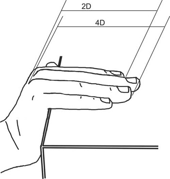
Digit measurements were made based on fixed anatomical landmarks using methods developed and described by Auger and EustacheReference Auger and Eustache 37 . Of several measurement methods tested in their study, this one showed by far the highest correlation with digit measurements made by the gold standard, radiographs. As per their method, the subject’s hand was positioned such that the digits were flat on the edge of the surface of the table with the palm wrapped downward at an angle of 100–120° relative to the fingers (Fig. 2). Using a clean set of Vernier calipers set flat on their side, the second and fourth digits of each hand were measured from the base of the proximal phalanx to the end of the distal phalanx. Each digit was measured three times, for a total of 12 measurements/subject per visit (2 fingers×2 hands×3 measurements/finger). At each visit, 2D:4D ratio was calculated as the mean of the three measurements of the second digit divided by the mean of the three measurements of the fourth digit. Thus, each subject had a total of 18 digit ratio values (one per hand per visit), in contrast to 27 sets of AGD measurements (three per visit each of AGD-AC and AGD-AF).
Direct measurement of digit lengths using calipers as described by Auger and Eustache (2011).Reference Auger and Eustache 37

Statistical analyses
We first calculated summary statistics on the study population for our variables of interest. These included our four outcome variables (AGD-AF, AGD-AC, 2D:4D right hand and 2D:4D left hand) as well as several potential covariates (BMI, height, weight, age at menarche and examiner). We used t-tests and correlations to examine relationships between these variables, and investigate possible multi-collinearity. In particular, we examined the correlation between AGD measures and digit ratios, using the woman, rather than the visit or cycle, as the unit of analysis. To do so, we averaged all measurements taken at all nine visits for each subject and calculated Spearman’s correlations based on that average. We first examined correlations across all subjects, and second, recalculated correlations excluding subjects who reported having ever suffered an injury to the second or fourth digits.
We then selected variables for inclusion in multivariable models. We chose height as our preferred measurement of body size given that digit lengths are more plausibly related to skeletal size than body mass.Reference Barut, Tan and Dogan 47 Our primary predictor, cycle phase, was modelled as two categorical variables (follicular and luteal phases, with mid-cycle as the referent). Thus, our final models included cycle phase, height and age at menarche as independent variables. In analyses predicting digit ratios, we included self-reported history of injury to the relevant digits as an additional covariate. Owing to the nested structure of the data (including multiple measurements, multiple visits and multiple cycles for each subject), to optimize power we fit multi-level linear regression models with a variance components covariance matrix because we had no assumptions as to the relationship between the measurements across cycle phases. Four models were fit, one for each of the four outcomes (AGD-AC, AGD-AF, 2D:4D right and 2D:4D left). We considered the addition of an examiner term in multivariable models, however, there were no significant differences in AGD or digit measurements across examiners, and thus examiner was not retained in final models. In sensitivity analyses, we considered weight and BMI as possible covariates, rather than height. Intra-observer variation was calculated as the coefficient of variation (CV). Across all analyses, standard model assumptions were checked. All analyses were conducted in SAS Version 9.3 (SAS Institute, Cary, NC, USA) and all P-values reported are two-tailed with an α-level of 0.05.
Results
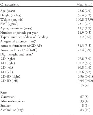
A total of 12 women participated in this longitudinal study of variation across the menstrual cycle, and complete measurements were obtained for all subjects at all study visits (Table 1). The mean age was 25.6 years (minimum–maximum: 19–30) and all women reported cycling regularly (11–13 menstrual periods/year). The average age at menarche was 11.7 (minimum–maximum: 9–16) and the mean BMI was 23.1 kg/m2 (minimum–maximum: 20–26). Eight subjects were Caucasian and four were African-American. All subjects reported being right handed. As the primary outcome measure of the study was AGD, potential subjects were not pre-screened for digit injuries and indeed, three subjects reported a history of injury to the second or fourth digits of the right hand, and three subjects reported injuries to the digits of the left hand. These included digit dislocations, fractures, breaks, all occurring at least several years before participation in the study.
Characteristics of the study population (n=12)

BMI, body mass index; 2D:4D, second to fourth digits of the hand.
a To obtain this value, we took the within-woman mean for each measurement, then took the mean across women.
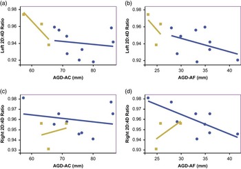
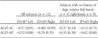
Digit ratios in the right and left hand were highly correlated (r=0.70) and the two AGD measurements (AGD-AC and AGD-AF) were moderately correlated with one another (r=0.53). In bivariate analyses, AGD and digit ratios were generally inversely related (Table 2; Fig. 3). When all subjects were included in the analyses, the left hand digit ratio was significantly, inversely associated with AGD-AF (r=−0.57, P=0.05) and results were similar, albeit weaker, for AGD-AF (r=−0.52, P=0.08). No statistically significant associations were observed with right hand digit ratio and AGD measurement. When subjects with a history of digit injury were excluded from analyses, associations between left hand digit ratio and AGD were attenuated (AGD-AC: r=−0.37, P=0.33; AGD-AF: r=−0.33, P=0.38), however, a moderate correlation was observed between right hand digit ratio and AGD-AF (r=−0.61, P=0.08).
Bivariate relationships between AGD measures (AGD-AC, AGD-AF) and digit measurements (right and left hands). Blue circles indicate subjects with no history of digit injuries. Tan squares indicate subjects with a history of digit injuries. AGD, anogenital distance; AC, anus to clitoris; AF, anus to fourchette.

Spearman’s correlations between anogenital distance and digit length measures within the entire cohort and the subset of women with no history of digit injuries [r (P-value)]Footnote a

2D:4D, second to fourth digits of the hand; AGD, anogenital distance; AC, anus to clitoris; AF, anus to fourchette.
a For each individual, AGD and digit ratio values used in the correlations represent the average of all values overall visits (i.e. average of nine sets of digit ratios and 27 sets of AGD measurements per woman).
In multivariable models, AGD-AF and AGD-AC measurements taken during the follicular and luteal phases did not differ from measurements taken mid-cycle (Table 3). Similarly, there were no significant differences in digit ratio in either hand across the menstrual cycle. Age at menarche was inversely associated with AGD-AC (β=−2.26, 95% CI: −3.38, −1.14), but not AGD-AF. By contrast, age at menarche was positively associated with digit ratio in both the left (β=0.005, 95% CI: 0.002, 0.007) and right (β=0.003, 95% CI: 0.001, 0.004) hands. Height was similarly positively associated with right hand digit ratio (β=0.003, 95% CI: 0.0001, 0.004), but not left hand digit ratio. Finally, self-report of history of injury to the right digits showed a weak association with right hand digit ratio (β=−0.009, 95% CI: −0.017, 0.001); however, self-report of history of injury to the left digits was not associated with left hand digit ratio (β=0.010, 95% CI: −0.002, −0.022). In sensitivity analyses, the inclusion of BMI or weight (rather than height) did not change the estimates for the effects of cycle phase on AGD or digit ratios (not shown). Associations with age at menarche at AGD-AC were attenuated when either BMI (β=−1.20, 95% CI: −2.51, 0.11) or weight (β=−1.20, 95% CI: −2.46, 0.06) was included rather than height. Associations between age at menarche and digit ratios on both hands were slightly attenuated, but still statistically significant when BMI or weight was considered instead of height (not shown).
Multi-level, multivariable linear regression models predicting four proposed biomarkers of prenatal androgen exposure AGD-AC, AGD-AF, 2D:4D (right), 2D:4D (left) (n=12 women, measured at three points in the menstrual cycle, for three cycles)

CI, confidence interval; 2D:4D, second to fourth digits of the hand; AGD, anogenital distance; AC, anus to clitoris; AF, anus to fourchette.
a Referent: mid-cycle (days 13–15).
b For right ratio, this is right finger injuries and for the left ratio, this is left finger injuries.
Inter-examiner variation (as calculated by CV) was low and consistent across all cycle phases for both AGD and digit lengths (Table 4). The CVs were higher for AGD-AF (ranging from 5.0 to 5.6% across phases) than for AGD-AC (ranging from 2.1 to 3.1% across phases). CVs for digit lengths were under 1.0% for all cycle phases.
Mean (s.d.) for the coefficient of variation (CV)Footnote a within examiner by cycle phase

2D:4D, second to fourth digits of the hand; AGD, anogenital distance; AC, anus to clitoris; AF, anus to fourchette.
a CV=(standard deviation/mean)×100.
Discussion
In the current longitudinal study of cycling women, we investigated whether two proposed biomarkers of prenatal androgen exposure, AGD and digit ratio, vary according to menstrual cycle phase. Our results demonstrate that both measures are stable across the menstrual cycle, which is important if they are to be used as research tools in adult women. In addition, despite the small sample size, we found inverse associations between AGD and 2D:4D, as would be predicted based on the literature, and in particular, left hand digit ratios showed stronger associations with AGD measures. We found further evidence of the developmental origins of these biomarkers in that both AGD and digit ratios were associated with age at menarche.
Previous work has suggested that AGD may vary in relation to circulating hormone levels in adult women. A rodent study found that AGD was significantly different during diestrus as compared to metestrus,Reference Dusek and Bartos 41 and in a population of young Spanish women, those using hormonal contraception had significantly shorter AGD than naturally cycling women, indicating that perhaps AGD is responsive to exogenous hormones during adulthood. Unfortunately, in that cross-sectional study, all AGD measurements were taken during the early follicular phase, so cyclical variation could not be examined.Reference Mendiola, Roca and Minguez-Alarcon 24 However, our results suggest that within-woman variation in endogenous ovarian hormones across the menstrual cycle is unlikely to alter AGD. A longitudinal study is needed to determine whether this is true of women using hormonal contraception as well.
One study reported cyclical fluctuation in digit ratios in naturally cycling women, but not hormonal contraception users, across the menstrual cycle.Reference Mayhew, Gillam, McDonald and Ebling 43 One possible explanation for the discrepancy between the current results and that study is differences in measurement techniques. The technique used in the current study was specifically chosen because, of all methods examined, it most closely correlated with digit lengths measured from the gold standard, radiographs, which reflect only bone length and not the fat pad at the distal phalanx.Reference Auger and Eustache 37 Other popular measurement techniques based on photocopying the hand or drawing an outline show typically lower correlation with radiograph-based measurements and capture greater variation in fat deposition.Reference Auger and Eustache 37 , Reference Wallen 48 Fat tissue has estrogen and androgen receptors (ARs),Reference Dieudonne, Leneveu, Giudicelli and Pecquery 49 , Reference Laughlin, Barrett-Connor and May 50 and indeed, there appears to be cyclical variation in other soft-tissue traits in women, including those that are bilaterally symmetrical, such as breast and ear size.Reference Scutt and Manning 51 This may explain the within-cycle variation in digit ratios observed in previous work using photocopy-based measurements.Reference Mayhew, Gillam, McDonald and Ebling 43 Given that variation in bone length, rather than fat mass, is typically of greatest interest with respect to prenatal exposures, the current measurement technique, which is stable across the cycle, may be preferable as a research tool in the future.
If digit ratios and AGD both convey information about prenatal androgen exposure, then it follows that the two anatomical measures may be associated. Even within this small sample, the two biomarkers showed relationships in the expected direction, such that longer (more ‘masculine’) AGD was associated with lower (more ‘masculine’) digit ratio, and vice versa. These correlations were significant only for the left hand ratio and only when we considered the full sample (including women with a history of digit injuries). Unfortunately, for each hand, three (of 12) subjects reported having sustained an injury to either the second or fourth digit at some point in their lifetimes and it is unknown whether those injuries may have altered their digit ratio. Further research in a larger sample of women is needed to confirm this preliminary finding of within-individual associations between AGD and digit ratios, as is analogous work in men and children.
To our knowledge, no prior studies have simultaneously examined AGD and digit ratios in humans, but there are several reports in animal models. In a study of female Rhesus macaques exposed to exogenous androgens during early to mid-gestation, AGD was masculinized (lengthened) compared with controls, whereas 2D:4D was increased, due to elongation of the second digit.Reference Abbott, Colman, Tiefenthaler, Dumesic and Abbott 52 As absolute digit lengths are longer in male macaques than females, the elongation of the second digit was interpreted as masculinization by the authors, however, the overall effect was unexpectedly, a feminization of the 2D:4D ratio. Notably, the elongation was limited to the right hand and was only evident in photocopy-based measurement, not in radiographs, suggesting that soft tissue, but not bone growth, was affected in androgenized animals.Reference Abbott, Colman, Tiefenthaler, Dumesic and Abbott 52 Interestingly, these results contrast with work in rodents, in which manipulation of the androgen to estrogen signaling preferentially altered the growth of the fourth digit.Reference Zheng and Cohn 28 In CD-1 mice, the fourth digit shows increased expression of ARs and estrogen receptor α (ER-α) as compared with the second digit. Inactivation of the AR or the administration of estrogen from gestational day 12.5 to 15.5 resulted in decreased growth of the fourth digit in male mice, whereas inactivation of ER-α or the administration of androgens during the same time period increased growth of the fourth digit in females, leading the authors to conclude that fourth digit bone growth drives sex differences in 2D:4D ratio. Although that study did not specifically report on the correlation between digit ratios and AGD in their models, the authors noted that males with experimentally feminized digit ratios also had hypospadias,Reference Zheng and Cohn 28 a hallmark of the testicular dysgenesis syndrome. That mutations in certain HOX genes are linked to alterations in both limb and reproductive development in humans (males and females) is additional evidence of shared ontogeny.Reference Goodman and Scambler 46 It is worth noting that two observational studies in rodents have not found correlations between AGD and digit ratios, however, in those studies, no sexual dimorphism in digit lengths was observed.Reference Manno 44 , Reference Hurd, Bailey and Gongal 45 Further observational work examining the association between digit ratios and genital development in both control and clinical populations (e.g. CAH, AIS and males with genital anomalies) is needed to inform this discussion.
We also found that age at menarche was a predictor of both digit ratio and AGD. Earlier age at menarche was associated with longer (more masculine) AGD and a lower, more masculine digit ratio. The direction of the relationship is somewhat unexpected given that a later age at menarche (here associated with more feminine digit ratio and AGD) has been associated with subfecundity and lower ovarian hormone concentrations in some, but not all, studies.Reference Apter, Reinila and Vihko 53 – Reference Guldbrandsen, Hakonsen and Ernst 55 In the only study of AGD in adult women, no associations with age at menarche were reported.Reference Mendiola, Roca and Minguez-Alarcon 24 However, our results are in line with recent work on age at menarche and digit ratios. A prospective study that measured digit lengths in pre-menarcheal girls and followed them until the occurrence of menarche found a positive relationship between the two.Reference Oberg and Villamor 56 Similarly, in two large, independent samples (The Avon Longitudinal Study of Parents and Children and the Brisbane Adolescent Twin Study), a single variant in the LIN28B gene, a regulator of developmental timing, was linked to both high digit ratios and delayed menarche.Reference Medland, Zayats and Glaser 57 However, two other studies have found inverse associations between menarcheal timing and 2D:4DReference Manning and Fink 58 , Reference Matchock 59 and another has found no association.Reference Helle 60 Notably, the studies finding inverse associations relied on self-reported age at menarche, and in one study, self-measured digit lengths, raising the possibility of considerable measurement error.Reference Manning and Fink 58 It is plausible that pubertal timing might be associated with AGD and digit ratios through the prenatal hormonal milieu.Reference Yermachenko and Dvornyk 61 It is also possible that there are genetic polymorphisms that influence genital and digit development, as well as postnatal developmental trajectories.Reference Medland, Zayats and Glaser 57 , Reference Sathyanarayana, Swan and Farin 62 For example, polymorphisms of ESR1, encoding ER-α, are associated with both AGD in boys as well as growth and timing of pubertyReference Sathyanarayana, Swan and Farin 62 , Reference Kang, Kim, Park, Yoon and Shim 63 and it is plausible that similar genetic mechanisms may operate in females. LIN28B presents one possibility that merits further research.
As in other studies, relationships across AGD measures and digit ratios across hands were not always consistent. Here, age at menarche was strongly associated with AGD-AC, the longer of the two AGD measures, but more weakly with AGD-AF. Notably, in this and other work,Reference Mendiola, Roca and Minguez-Alarcon 24 AGD-AF shows greater inter-observer variation, suggesting that the fourchette landmark may be more difficult to identify. Similarly, the associations between AGD measures and left hand digit ratio were far stronger than the associations with right hand digit ratio. However, it is also possible that digit injuries altered the digit ratio, particularly on the right hand, thus obscuring our ability to detect relationships, if any. Differences in digit ratios in the right and left hands are frequently observed in studies, and there is conflicting evidence as to which hand may be a better index of prenatal androgen exposure, if any.Reference Oberg and Villamor 56 , Reference Ventura, Gomes, Pita, Neto and Taylor 64 – Reference Putz, Gaulin, Sporter and McBurney 67 Handedness may be important to consider in this respect. In this study, all women were right handed, however, future research including both right and left handed individuals may help to further explain the differences observed when right or left hand 2D:4D is considered.
A notable strength of our study was the longitudinal design, which provided increased power even with a small sample. Each subject had three study visits at pre-defined points in the cycle, for three cycles. At each study visit, three sets of AGD and digit measurements were made. Despite this intensive protocol, all subjects completed all study visits, so our data set is complete. We used multi-level modelling to take advantage of the numerous measurements per subject. Nevertheless, it is possible that the study was insufficiently powered to detect associations and a larger sample size is preferable for future work. This is particularly true for the ‘woman-level’ questions emerging from our data, namely further exploring the relationship between AGD and 2D:4D as well as better understanding how these biomarkers relate to other developmental endpoints, such as age at menarche.
Ultimately, additional research is needed to determine whether either of these anatomic measures is a valid index of prenatal androgen exposure, and the extent to which they predict reproductive outcomes in women. Our results simplify this future work, by demonstrating that using the measurement techniques that we have followed herein, there is little, if any, cyclical variation in the measures in naturally cycling, nulliparous women. Whether these findings can be extrapolated to women using hormonal contraception or parous women (whose AGD may be altered after vaginal childbirth) is unknown. Nevertheless, the results speak to the feasibility of using both measures as research tools that may aid in understanding the downstream sequelae of the prenatal hormonal milieu in women.
Acknowledgements
The authors thank Lynda Kochman, RN and Shaya Greathouse for their work and Dr. Kathy Hoeger for her assistance with study design. In addition, the authors thank Dr. Sarah Thurston for her biostatistical expertise and Rebecca Rowley for her illustration.
Financial Support
Funding for the current study was provided by the Mae Stone Goode foundation with additional support from the National Institutes of Health (K12 ES019851-01; P30 ES001247).
Conflicts of Interest
None.
Ethical Standards
The authors assert that all procedures contributing to this work comply with the ethical standards of the Helsinki Declaration of 1975, as revised in 2008, and it has been approved by the institutional committee at the University of Rochester School of Medicine and Dentistry.