Introduction
In bovine species, the extent of the primordial follicle population is highly variable at birth, ranging from 14,000 to 250,000, and this follicular reserve undergoes a progressive reduction during female reproductive life, and is also correlated with fertility (Ireland et al., Reference Ireland, Zielak-Steciwko, Jimenez-Krassel, Folger, Bettegowda, Scheetz, Walsh, Mossa, Knight, Smith, Lonergan and Evans2009). During the initiation of growth, some primordial follicles leave the reserve pool of quiescent follicles to enter the growing pool (primary, secondary, tertiary, and preovulatory stages). The rate of activation of primordial follicles affects reproductive lifespan and fertility, Monniaux et al. (Reference Monniaux, Clément, Dalbiès-Tran, Estienne, Fabre, Mansanet and Monget2014) discussed the link between the ovarian reserve of primordial follicles and the dynamic reserve of antral growing follicles, and reported that the size of the reserve of primordial follicles formed early in life can determine the ovarian activity of the adult. Cushman et al. (Reference Cushman, de Souza, Hedgpeth and Britt1999) confirmed this positive correlation between the number of primordial follicles and the number of growing follicles. Recently, Favoreto et al. (Reference Favoreto, Loureiro, Ereno, Pupulim, Queiroz, Silva and Barros2019) showed that cows with a higher antral follicular count (AFC) have higher numbers of preantral follicles compared with those with low AFC, reinforcing the relationship between preantral follicle population and ovarian activity. AFC can be classified as high, intermediate or low based on the frequency distribution of visible antral follicles. Cushman et al. (Reference Cushman, de Souza, Hedgpeth and Britt1999) demonstrated that, in any herd, AFC is associated with ovarian follicle reserve and is an indicator of fertility status. The population of antral follicles appears to be highly variable between cows (Burns et al., Reference Burns, Jimenez-Krassel, Ireland, Knight and Ireland2005; Rico et al., Reference Rico, Fabre, Médigue, di Clemente, Clément, Bontoux, Touzé, Dupont, Briant, Rémy, Beckers and Monniaux2009) and, considering that these antral follicles are either the direct targets of gonadotropin treatments during superovulation or are collected by ovum pick-up (OPU) for in vitro fertilization, this variability constitutes the most limiting factor for the success of assisted reproductive technologies and embryo production (Singh et al., Reference Singh, Domínguez, Jaiswal and Adams2004; Mossa et al., Reference Mossa, Duffy, Naitana, Lonergan and Evans2007). Ireland et al. (Reference Ireland, Scheetz, Jimenez-Krassel, Themmen, Ward, Lonergan, Smith, Perez, Evans and Ireland2008) showed that cows with high AFC produce a higher number of mature oocytes and that there is a positive correlation with the total number of morphologically viable oocytes and the number of cumulus–oocyte complexes (COCs) recovered (Silva-Santos et al., Reference Silva-Santos, Santos, Koetz Júnior, Morotti, Siloto, Marcantonio, Urbano, Oliveira, Lima and Seneda2014). Furthermore, the ovarian follicular reserve can also be evaluated through ovarian volume (OV) (Kwee et al., Reference Kwee, Elting, Schats, McDonnell and Lambalk2007), which varies in accordance with the reproductive stage, the oestrous phase and the gestation period, and can be used as a parameter in the clinical and functional evaluation of the organ (Chacur et al., Reference Chacur, Valentim, Martinez, Tostes and Kronka2006). Ovarian volume can be used as a reproductive parameter because it has a direct relationship with hormone production during the oestrous cycle (Penitente-Filho et al., Reference Penitente-Filho, Jimenez, Zolini, Carrascal, Azevedo, Silveira, Oliveira and Torres2015).
The number of oocytes collected by OPU and, consequently, total embryo production by IVF is highly correlated with ovarian AFC (Tamassia et al., Reference Tamassia, Heyman, Lavergne, Richard, Gelin, Renard and Chastant-Maillard2003; Ireland et al., Reference Ireland, Ward, Jimenez-Krassel, Ireland, Smith, Lonergan and Evans2007). Additionally, Mossa et al. (Reference Mossa, Walsh, Butler, Berry, Carter, Lonergan and Evans2012) reported that cows with low AFC have lower pregnancy rates compared with cows with high AFC. However, a recent study showed that cows with lower AFC have larger follicular diameters and a higher conception rate (Morotti et al., Reference Morotti, Moretti, dos Santos, Silva-Santos, Ramos Cerqueira and Seneda2018). Studies have also shown that there is a positive correlation between AFC and the total number of healthy follicles and oocytes present in the ovaries (Cushman et al., Reference Cushman, de Souza, Hedgpeth and Britt1999). In this sense, studying the relationship between AFC and the composition of the ovarian follicular reserve will provide the basis for understanding the mechanisms controlling the maintenance of follicular reserve and oocyte competence that directly affect fertility in cattle.
As the relationship between AFC and levels of degeneration of preantral follicles, as well as with the capacity of oocyte meiotic progression and ultrastructure of COCs matured in vitro have not been investigated, it is hypothesized that there is an association between AFC and OV and that high AFC is related with the meiotic progression of COCs in bovine females. Therefore, the aims of this study were to evaluate in bovine species the relationship among AFC, OV, population and viability of preantral follicles, ability of oocyte meiotic progression and ultrastructure of COCs matured in vitro.
Materials and methods
All procedures and protocols related to animals were approved previously by the Ethics Committee for the use of animals (Standing Committee/PRP – UFLA) of the Federal University Lavras-MG, Minas Gerais, Brazil, with protocol no. 068/14. All chemicals used were purchased from Sigma Chemical Company (St. Louis, MO, USA), unless otherwise indicated in the text.
Experiment 1: Relationship between AFC and population and survival of preantral follicles in bovine ovaries
Procedures
Ovaries were collected from cyclic, multiparous, crossbred cows (Bos taurus indicus, 3–10 years old with mean age of 6 years) in a local slaughterhouse from the south of Minas Gerais State, Brazil. Cows, with predominance of Bos taurus indicus, that had body condition between 5 to 7 were used in this study (Lowman, Scott, Somerville, Reference Lowman, Scott and Somerville1976).
Immediately post mortem, the ovaries were washed in 70% alcohol for 10 s and twice with 0.9% saline solution (NaCl) at 37°C, supplemented with 100 IU/ml penicillin and 100 mg/ml streptomycin (Sigma Chemical Company, St Louis/USA). The ovaries were transported within 1 h in 4% saline solution containing antibiotics to the laboratory (Chaves et al., Reference Chaves, Martins, Saraiva, Celestino, Lopes, Correia, Verde, Matos, Báo, Name, Campello, Silva and Figueiredo2008). The ovaries were classified as high AFC class (≥50 follicles, 3–10 mm); intermediate AFC (30–49 follicles, 3–10 mm) or low AFC (<30 follicles, 3–10 mm), based on the classification standardized by Rodrigues et al. (Reference Rodrigues, Oliveira, Loiola, Ferraz, Chalhoub, Bittencourt, Araujo, Santos, Bittencourt and Filho2015). For each ovarian category (high, intermediate or low AFC), 10 ovaries were fixed for histological analysis.
Histological analysis
For histological studies, from each ovary (n = 10) in each category (high, intermediate or low AFC), 10 fragments (3 mm × 3 mm × 1 mm) from cortical tissues were selected at random. The fragments were fixed in 4% paraformaldehyde in PBS (pH 7.4) directly overnight at room temperature. After fixation, the ovarian fragments were dehydrated in a graduated ethanol series, clarified with xylol and soaked in paraffin wax. After inclusion in paraffin (Synth, São Paulo, Brazil), they were serially sectioned at 7 μm intervals. From each ovarian fragment, 20 sections were performed, mounted on glass microscope slides, stained with the periodic acid method Schiff–haematoxylin and analyzed to count the number of follicles (primordial, primary, secondary) and their morphology (healthy and degenerate). Histological sections were examined under a light microscope (Nikon, Tokyo, Japan).
Stages of follicle development and survival were evaluated in the total area of histological sections. The follicles were classified as primordial when they had an oocyte surrounded by one layer of flattened granulosa cells or with the presence of both flattened and cuboidal cells. Growing follicles were classified as primary when they contained one layer of cuboidal cells, while secondary follicles had two or more layers of cuboidal granulosa cells around the oocyte. The follicles were classified individually as histologically healthy when they had an intact oocyte surrounded by well organized granulosa cells and without the presence of a pyknotic nucleus. Degenerated follicles were defined with a retracted oocyte, pyknotic nucleus, and disorganized granulosa cells detached from the basal membrane (Silva et al., Reference Silva, van Den Hurk, Costa, Andrade, Nunes, Lobo and Figueiredo2004).
Experiment 2: Relationship among AFC, OV, ability of oocytes to resume meiosis and ultrastructure of in vitro matured bovine COCs
Ovaries, follicular classification and oocyte aspiration
Ovaries (n = 210) were collected as described previously in experiment 1. The ovaries were classified as high AFC (≥50 follicles; n = 84); intermediate AFC (30–49 follicles, n = 68) or low AFC (<30 follicles; n = 58), in accordance with Rodrigues et al. (Reference Rodrigues, Oliveira, Loiola, Ferraz, Chalhoub, Bittencourt, Araujo, Santos, Bittencourt and Filho2015). In all three classes, the COCs were aspirated from follicles between 3 and 8 mm in diameter using 10 ml syringes and 40G needles. COCs were counted and classified in accordance with Stringfellow and Givens (Reference Stringfellow and Givens2010), i.e. grade I: oocytes with compact cumulus with more than three cell layers, ooplasm with homogeneous granules, surrounded by the zona pellucida and with brown colour; grade II: oocytes with up three layers of cumulus cells, ooplasm with heterogeneously distributed granules that may be more concentrated in the centre and lighter on the periphery, or condensed in a place appearing as a dark spot, ooplasm filled all spaces within the zona pellucida; grade III: oocytes with expanded cumulus, some contraction in the ooplasm, with space between the cell membrane and the zona pellucida, irregularly filling the perivitelline space. Grades I, II and III oocytes were used for in vitro maturation, as Bakri et al. (Reference Bakri, Ibrahim, Osman, Hasan, Jaffar, Rahman and Osman2016) showed that there are no differences in apoptosis of early embryos after maturation and fertilization of oocytes of these three classes.
Measurement of total OV
In total, 210 ovaries were classified based on total OV (from right and left) as large (>11 mm3, n = 64), intermediate (>8 and ≤11 mm3, n = 62) and small (≤8 mm3, n = 64). Length, width and height of the ovary were measured using a digital caliper (150 mm/0.01 mm Powerfix Nf, Digimess, São Paulo, Brazil). The OV was calculated using the formula: 0.5 (correction factor) × length × width × height (Swanson et al., Reference Swanson, Sauerbrei and Cooperberg1981). The sum of the average volumes of the right and left ovaries was defined as the total OV. The presence or absence of corpus luteum and follicles larger than 10 mm was considered in the study.
In vitro maturation of COCs
COCs (n = 319) collected from ovaries with high AFC (n = 177), intermediate AFC (n = 96) and low AFC (n = 106) classes were maturated in vitro. Viable COCs were placed in 90-μl droplets of maturation medium (maximum 30 oocytes/droplet) consisting of Tissue Culture Medium (TCM-199) supplemented with 22 μg/ml sodium pyruvate, 5 μg/ml bovine follicle stimulating hormone (FSH), 10 IU/ml LH, 20 μg/ml estradiol, 20 mg/ml amikacin and 10% fetal bovine serum, and equilibrated in an incubator for at least 2 h. In vitro maturation of COCs was performed for 24 h at 38.7°C with 5% CO2 in air in an humidified atmosphere.
To evaluate oocyte meiotic progression after IVM, the COCs (n = 319) from ovaries with high AFC (n = 177), intermediate AFC (n = 96) and low AFC (n = 106) were analyzed by fluorescence microscopy. To evaluate chromatin configuration and meiotic progression, cumulus cells were vortexed and oocytes were fixed in 4% paraformaldehyde for 15 min and transferred to 0.5% Triton X-100 solution. Changes in the chromatin configuration were evaluated in 10-μl droplets containing 10 μg/ml of bisbenzimide trichlorohydrate (Hoechst 33342) and analyzed using a fluorescence inverted microscope (Leica, DMI4000B). Fluorescence signals generated by the Hoechst 33342 stain were collected at 568 nm. The oocytes were classified based on stage of nuclear maturation: germinal vesicle (GV), germinal vesicle breakdown (GVBD), metaphase I (MI), anaphase I (AI) and telophase I (TI) and metaphase II (MII) (Bezerra et al., Reference Bezerra, Silva, Rissi, Rosa, Cesaro, Costa, Gonçalves and Silva2016).
Ultrastructural characteristics of in vitro matured COCs from ovaries with high, intermediate or low AFC
In vitro matured COCs (n = 30) from cows with different AFC, i.e., high (n = 10), intermediate (n = 10) and low (n = 10) were examined by transmission electron microscopy. COCs were fixed in Karnovsky’s solution (4% paraformaldehyde and 2.5% glutaraldehyde in 0.1 M sodium cacodylate buffer pH 7.2) for 4 h at room temperature (approximately 25°C). After fixation, cultured COCs were maintained in sodium cacodylate buffer. The specimens were post-fixed in 1% osmium tetroxide, 0.8% potassium ferricyanide and 5 mM calcium chloride in 0.1 M sodium cacodylate buffer for 1 h at room temperature. Then, they were washed in sodium cacodylate buffer and counterstained with 5% uranyl acetate. The samples were dehydrated in solutions with increasing concentrations of acetone and then incorporated in epoxy resin (Epoxy-Embedding Kit, Fluka Chemika–BioChemika). Subsequently, they were cut into semi-thin sections (2 μm), stained with toluidine blue and analyzed by optical microscopy at ×400 magnification. Ultra-thin sections (70 nm) were obtained from bovine COCs classified as morphologically healthy in semi-thin sections. Subsequently, ultra-thin sections were contrasted with uranyl acetate and lead citrate and examined under a Fei Tecnai Spirit electron microscope. Ultrastructure parameters evaluated were the density and integrity of the cytoplasmic organelles of the oocyte and granulosa cells and the degree of cytoplasmic and nuclear vacuolization.
Statistical analysis
Statistical analyzes from both experiments were performed using SAS® statistical software (SAS, Cary, NC, USA). The general data were tested for normality by the Shapiro–Wilk test in accordance with the UNIVARIATE procedure and, when necessary, transformed to achieve normality. The AFC and OV values for cows were submitted and compared with those following the GLIMMIX procedure. All variables were submitted to correlation test using the CORR procedure. Data are presented as mean ± standard error of the mean (SEM). The effect of AFC class on the percentage of oocyte distribution at meiosis stages (GV, GVBD, metaphase I, anaphase I and telophase I and metaphase II) was submitted to the GENMOD procedure as binomial distribution. Differences were considered significant when P-values were < 0.05.
Results
Experiment 1: relationship between AFC and preantral follicle population and survival in bovine ovaries
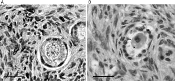
Histological analysis revealed the morphology of healthy (Fig. 1A) and degenerate preantral follicles (Fig. 1B) found in ovaries with high, intermediate or low AFC.
Morphological characterization of healthy and degenerate preantral follicles in ovaries of high, intermediate and low antral follicular count (AFC). (A) Healthy primordial follicle; (B) degenerated primary follicle. GC, granulosa cells; Nu, oocyte nucleus; O, oocyte. Scale bars, 50 µm.

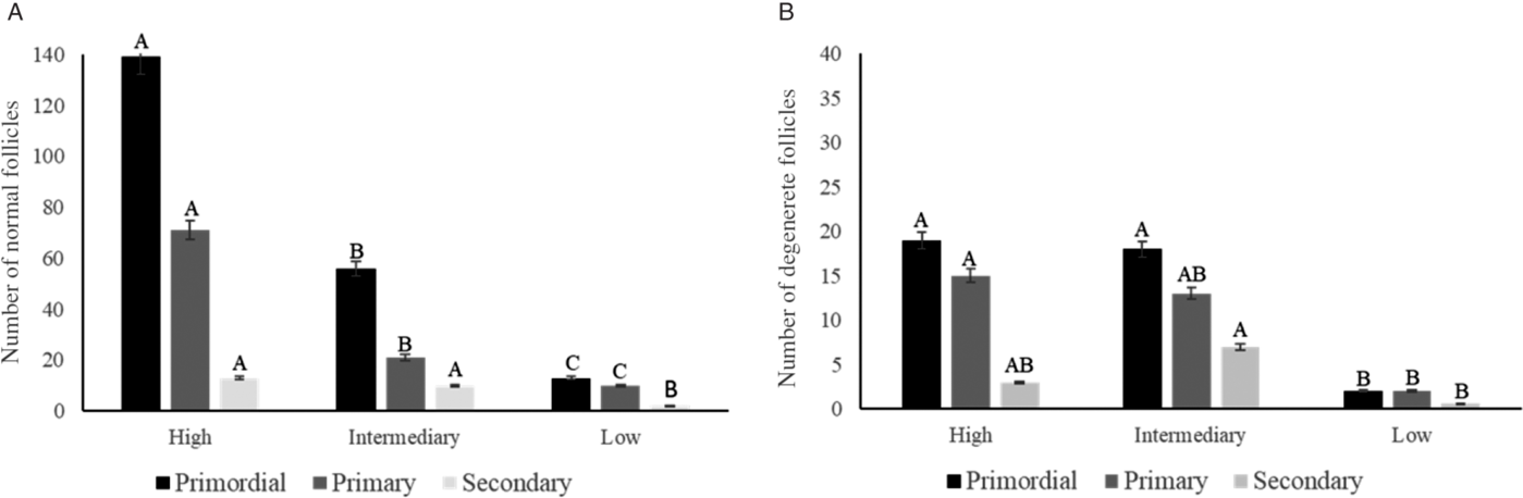
The number of morphologically healthy primordial and primary follicles (Fig. 2A) in ovaries with high AFC was significantly higher (P < 0.001) compared with ovaries with intermediate or lower AFC. In addition, the number of healthy secondary follicles was higher (P < 0.05) in ovaries with high AFC than in low AFC ovaries (Fig. 2A). Conversely, the number of degenerated primordial follicles (Fig. 2B) was higher (P < 0.05) in ovaries with high or intermediate AFC compared with those of low AFC. The number of degenerated primary follicles in high AFC ovaries was higher (P < 0.05) compared with those found in low AFC ovaries (Fig. 2B). The number of degenerated secondary follicles in ovaries of intermediate AFC was higher (P = 0.01) compared with in low AFC ovaries (Fig. 2B).
Mean number of morphologically healthy follicles (A) and degenerated (B) primordial, primary and secondary follicles in ovaries with high, intermediate, and low AFC. A–CCapital letters indicate differences between ovaries with high, intermediate and low AFC (P < 0.05). Numbers are mean ± standard error of the mean (SEM).

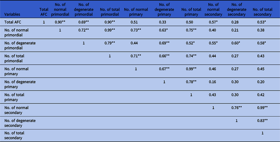
The correlation between AFC and follicular categories is shown in Table 1. A positive correlation between AFC and the number of healthy primordial follicles (r = 0.90, P < 0.01), degenerated primordial follicles (r = 0.69, P < 0.01) and primordial follicles (r = 0.90; P < 0.01) was observed. A positive correlation was also seen between the number of healthy secondary follicles (r = 0.57, P < 0.05) and the total number of secondary follicles (r = 0.53, P < 0.05). The number of healthy primordial follicles was correlated positively with the number of degenerated and total primordial follicles (r = 0.72, r = 0.99, P < 0.01; Table 1) and with healthy, degenerated and total primary follicles (r = 0.73, P < 0.01, r = 0.63, P < 0.05, r = 0.75, P = 0.01; Table 1). The number of degenerated primordial follicles was correlated positively with the number of total primordial follicles (r = 0.79, P < 0.01; Table 1), the number of degenerated and total primary follicles (r = 0.69, P < 0.01, r = 0.52, P < 0.04; Table 1) and the number of healthy, degenerated and total secondary follicles (r = 0.55, r = 0.60, r = 0.58, P < 0.05; Table 1), respectively. There was a positive correlation between the number of total primordial follicles and the number of healthy, degenerated and total primary follicles (r = 0.71, r = 0.66, r = 0.74, P < 0.01, respectively; Table 1).
Coefficient of correlations between antral follicular count (AFC) and categories of healthy, degenerated and total follicles
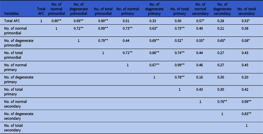
*P < 0.05; **P < 0.01.
Experiment 2: Relationship among AFC, OV, ability of oocyte meiotic resumption and ultrastructure of in vitro matured bovine COCs
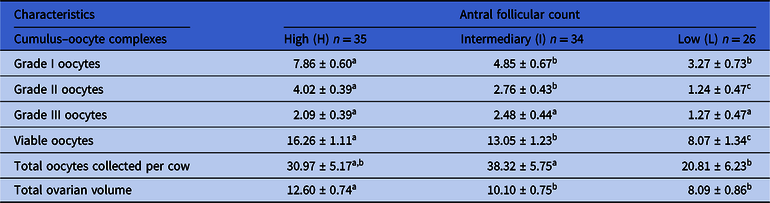
Table 2 shows the mean number of COCs recovered from each ovarian category. The numbers of grade I and grade II COCs were higher (P < 0.05) in ovaries with high AFC when compared with those of intermediate or low AFC. Moreover, the mean number of grade II COCs in intermediate AFC ovaries was higher when compared with low AFC ovaries. A significant reduction in the number of viable oocytes was seen in ovaries of high to intermediate and low AFC, while cows with high AFC had larger ovary volume compared with those with intermediate and low AFC (P < 0.05, Table 2). The total number of oocytes was lower (P < 0.05) in the low AFC ovaries when compared with the intermediate AFC ovaries (Table 2).
Mean number of grades I, II and II cumulus–oocyte complexes recovered from ovaries with high, intermediate and low antral follicular count

a,b,cIn the same line, the superscript letters indicate differences (P < 0.05).
Numbers are mean ± standard error of the mean (SEM).
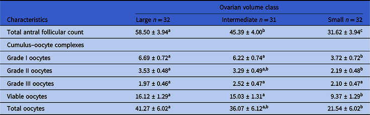
Table 3 shows the relationship between OV and the quality of COCs. Total AFC was higher (P < 0.05) in ovaries with a large volume when compared with those of intermediate or small volumes. Total AFC was higher (P < 0.05) in ovaries of intermediate volume when compared with small volume ovaries (Table 3). The numbers of grade I and viable oocytes were higher (P < 0.05) in ovaries with large or intermediate volumes when compared with those of small volume. The mean numbers of grade II oocytes and total number of oocytes were higher (P < 0.05) in ovaries with large volume compared with those of small volume. There was no difference in grade III oocyte between OV classes (P > 0.05; Table 3).
Relationship of ovarian volume with the quality of collected cumulus–oocyte complexes

a,bOn the same line, the superscript letters indicate differences (P < 0.05).
Numbers are mean ± standard error of the mean (SEM).
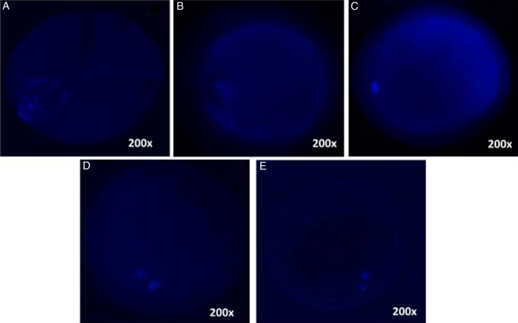
After 24 h of IVM, 62.38% (199/319) of all oocytes (high, intermediate and low AFC) reached metaphase II (Table 4). The proportion of oocytes in MII was higher (P < 0.05) in the ovaries of high AFC when compared with those of intermediate and low AFC. The percentages of oocytes in metaphase I, anaphase I and telophase I were higher (P < 0.05) in oocytes from ovaries with intermediate AFC compared with those of high and low AFC. The percentages of oocytes at the GV stage (P < 0.05) and germinal vesicle breakdown (GVBD) stage (P < 0.0001) in oocytes from low AFC ovaries were higher compared with those observed in oocytes from high or intermediate AFC ovaries (Fig. 3A–E and Table 4).
Percentage (%) of oocytes in the stages of germinal vesicle, germinal vesicle breakdown, metaphase I, anaphase I, telophase I and metaphase II from ovaries with high, intermediate and low antral follicular count

AI, anaphase I; AFC, antral follicular count; GV, germinal vesicle; GVBD, germinal vesicle breakdown; MI, metaphase I; MII, metaphase II; TI, telophase I.
a,b,cIn the same column, the superscript letters indicate differences (P < 0.05).
Photomicrography of bovine oocytes at different stages of nuclear maturation in ovaries with high, intermediate and low antral follicular count (AFC) evaluated under a fluorescence microscope. (A) Germinal vesicle (GV) and (B) germinal vesicle breakdown (GVBD) in ovaries of low AFC. (C) Metaphase I (MI) and (D) telophase I (TI) in ovaries of intermediate AFC. (E) Metaphase II (MII) in ovaries of high AFC. Magnification: ×200.

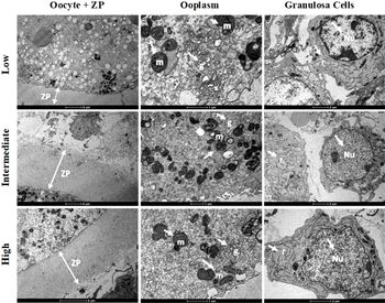
Oocytes from ovaries with different AFC showed large numbers of mitochondria with visible crests, the presence of intracellular vacuoles, uniformly distributed lipid droplets, Golgi complexes and well developed endoplasmic reticulum. It was not possible to observe ultrastructural differences between organelles in oocytes collected from ovaries with high, intermediate or low AFC. The zona pellucida was also intact in oocytes from ovaries with different AFC. Granulosa cells showed well defined nuclei that were usually rounded or with slight indentations, and had well developed endoplasmic reticulum, as well as Golgi complex. Mitochondria had well defined crests and some lipid drops were also seen. All the characteristics observed in the granulosa cells were similar between ovarian categories (Fig. 4).
Representative micrographs of cumulus–oocyte complexes (COCs) from ovaries of low, intermediate and high antral follicular count (AFC). g, Golgi complex; m, mitochondrion; Nu, nucleus; r, rough endoplasmic reticulum; ZP, zona pellucida.

Discussion
This study showed that cow ovaries with high AFC have higher preantral follicle populations and higher numbers of degenerated primordial and primary follicles compared with those with intermediate or low AFC. Previous studies have confirmed the positive correlation between the number of primordial follicles and the number of antral follicles (Cushman et al., Reference Cushman, de Souza, Hedgpeth and Britt1999; Scheffer et al., Reference Scheffer, Broekmans, Looman, Blankenstein and Fauser2003; Ireland et al., Reference Ireland, Scheetz, Jimenez-Krassel, Themmen, Ward, Lonergan, Smith, Perez, Evans and Ireland2008; Favoreto et al., Reference Favoreto, Loureiro, Ereno, Pupulim, Queiroz, Silva and Barros2019). As, in the bovine ovary, the number of primordial follicles is related to the number of antral follicles, there is a clear association between ovarian reserve and reproductive longevity. Therefore, depletion of the ovarian follicular population is associated with reproductive senescence in bovine females (Cushman et al., Reference Cushman, de Souza, Hedgpeth and Britt1999; Modina et al., Reference Modina, Tessaro, Lodde, Franciosi, Corbani and Luciano2013). In accordance with our study, cows with high AFC had higher numbers of degenerated primordial and primary follicles and this finding could also have an effect on reproductive longevity. Previous studies have reported the degeneration rate of preantral follicles in the goat ovaries (Silva et al., Reference Silva, Ferreira, Costa, Santos, Carvalho, Rodrigues, Lucci, Báo and Figueiredo2002) and, more recently, Meng et al. (Reference Meng, Jan, Hamer, van Pelt, van der Stelt, Keijer and Teerds2018) showed that preantral follicle atresia is the result of activation of different cell-death pathways and that, in preantral follicles, this occurs mainly via enhanced granulosa cell autophagy.
In the present study, the numbers of grade I oocytes and OV were higher in high AFC ovaries compared with intermediate and low AFC ovaries. Previous studies have shown that females with a higher oocyte production have a higher number of viable oocytes (Mercé et al., Reference Mercé, Bau, Barco, Troyano, Gay, Sotos and Villa2006; Silva-Santos et al., Reference Silva-Santos, Marinho, Santos, Machado, Gonzalez, Lisboa and Seneda2013). The oocytes derived from high AFC cows have a higher ability to be fertilized compared with those from low AFC cows (Nagai et al., Reference Nagai, Yanagawa, Katagiri and Nagano2015). COCs derived from early antral follicles in ovaries with high AFC have higher maturational ability, and are assumed to have higher fertilizability compared with those from ovaries with low AFC (Nagai et al., Reference Nagai, Yanagawa, Katagiri and Nagano2016). In addition, high numbers of antral follicles are positively associated with in vitro embryo production, but not conception rate (Santos et al., Reference Santos, Silva-Santos, Barreiros, Morotti, Sanches, de Moraes, Blaschi and Seneda2016). More recently, Morotti et al. (Reference Morotti, Moretti, dos Santos, Silva-Santos, Ramos Cerqueira and Seneda2018) showed that cows with low AFC had a greater conception rate compared with those with intermediate or high AFCs, which suggested that the number of antral follicles may influence the reproductive performance. Another study showed a positive correlation between AFC and ovarian reserve, the total population of oocytes recovered by OPU and oocytes candidates for in vitro fertilization (Lonegro et al., Reference Lonegro, Napoli, Pesce and Chacón2016).
In the present study, the mean numbers of grade I and viable oocytes were higher in ovaries with large or medium volumes. Other studies have also shown a positive correlation between OV and the number of grade I oocytes (Mercé et al., Reference Mercé, Bau, Barco, Troyano, Gay, Sotos and Villa2006; Penitente-Filho et al., Reference Penitente-Filho, Jimenez, Zolini, Carrascal, Azevedo, Silveira, Oliveira and Torres2015). Modina et al. (Reference Modina, Tessaro, Lodde, Franciosi, Corbani and Luciano2013) observed a significant reduction in weight, length and height of ovaries in cows with low AFC compared with cows with high AFC. Variation among animals with different AFC during follicular waves was positively correlated with variation in ovarian weight and total number of follicles (Ireland et al., Reference Ireland, Scheetz, Jimenez-Krassel, Themmen, Ward, Lonergan, Smith, Perez, Evans and Ireland2008). Kwee et al. (Reference Kwee, Elting, Schats, McDonnell and Lambalk2007) demonstrated that AFC and total OV were factors in the prediction of ovarian response to IVF. Other studies have correlated AFC positively with ovarian weight and total number of healthy oocytes present in ovaries (Cushman et al., Reference Cushman, de Souza, Hedgpeth and Britt1999; Ireland et al., Reference Ireland, Smith, Scheetz, Jimenez-Krassel, Folger, Ireland, Mossa, Lonergan and Evans2011).
For the percentage of oocytes that reach the MII, the proportion of oocytes in MII was higher in the ovaries of high AFC when compared with those intermediate or low AFC. Nagai et al. (Reference Nagai, Yanagawa, Katagiri and Nagano2016) showed that the improved meiotic development of oocytes from ovaries with high AFC may be related to the high proliferation of granulosa cells. Additionally, Favoreto et al. (Reference Favoreto, Loureiro, Ereno, Pupulim, Queiroz, Silva and Barros2019) recently reported that ovaries of heifers with AFC have increased expression of genes associated with cellular differentiation, follicle development and cellular signalling, especially growth factors. It is also important to consider the differences in gonadotrophin secretion between animals of high and low AFC groups. Lower blood progesterone concentrations were found in animals with low AFC compared with high AFC ovaries (Jimenez-Krassel et al., Reference Jimenez-Krassel, Folger, Ireland, Smith, Hou, Davis, Lonergan, Evans and Ireland2009; Ireland et al., Reference Ireland, Smith, Scheetz, Jimenez-Krassel, Folger, Ireland, Mossa, Lonergan and Evans2011). The secretion of gonadotrophins may be increased chronically in low AFC females during follicular waves (Burns et al., Reference Burns, Jimenez-Krassel, Ireland, Knight and Ireland2005; Ireland et al., Reference Ireland, Ward, Jimenez-Krassel, Ireland, Smith, Lonergan and Evans2007; Jimenez-Krassel et al., Reference Jimenez-Krassel, Folger, Ireland, Smith, Hou, Davis, Lonergan, Evans and Ireland2009). Ovarian function is changed, probably due to desensitization or decoupling of gonadotrophin receptors in granulosa, thecal and luteal cells (Amsterdam et al., Reference Amsterdam, Hanoch, Dantes, Tajima, Strauss and Seger2002).
In conclusion, cow ovaries with high AFC have higher preantral follicle populations, but also have higher numbers of degenerated primordial and primary follicles compared with those with intermediate or low AFC. The number of grade I oocytes and the proportion of oocytes that reached MII after in vitro maturation was also higher in COCs collected from ovaries of high AFC.
Financial support
This research was financed by Fazenda Casa Branca Agropastoril Careaçu-MG in association with the Federal University of Ceará – UFC, Sobral-CE, Federal University of Lavras – UFLA, Lavras-MG and the Laboratory of Ultrastructure of CNPqAM/FIOCRUZ, Recife – PE.
Conflicts of interest
There was no conflict of interest that could be perceived as prejudicial to the impartiality of the research reported.
Ethical standards
The authors declare that all procedures were performed in accordance with national and institutional guides on the care and use of animals. All procedures and protocols related to animals have been approved previously by the Ethics Committee of the use of animals (Standing Committee/PRP – UFLA) of the Federal University Lavras-MG, Minas Gerais, Brazil, with protocol no. 068/14.