Introduction
Even though lichens can be found in most ecosystems of the world (Nash Reference Nash and Nash1996), many lichen-forming fungal symbionts cannot survive without the photobiont, reflecting the ecologically obligate nature of the fungal partner in the symbiosis (Honegger Reference Honegger and Nash1996). Unlike the mycobionts, many photobionts are thought to be free-living except members of Trebouxia De Puymaly (see Ahmadjian Reference Ahmadjian1988; Mukhtar et al. Reference Mukhtar, Garty and Galun1994; Tschermak-Woess Reference Tschermak-Woess1978). Flexibility in the taxonomic specificity and ecological selection of the photobiont must allow for alternative partnerships in different habitats. A highly selective lichen association may be recognized when the uncommon algal species in the pool of all available algae form lichen associations. Selectivity refers to the availability and preferential interactions of the symbionts in nature (Galun & Bubrick Reference Galun, Bubrick, Linskens and Heslop-Harrison1984). A low level of selectivity, reported for the genus Umbilicaria, was considered to be advantageous to the mycobiont where a high diversity of photobionts was available to associate with the fungi (Romeike et al. Reference Romeike, Friedl, Helms and Ott2002). A highly specific lichen association may be recognized if one fungal taxon is always associated with one algal taxon and less specific when a fungal taxon associates with more than one algal taxon (Smith & Douglas Reference Smith and Douglas1987). Specificity refers to the range and taxonomic relatedness of symbiotic partners. For further explanation of selectivity and specificity in lichen associations see Honegger (Reference Honegger and Nash2008).
Since the list of mycobionts and photobionts was published by Tschermak-Woess (Reference Tschermak-Woess and Galun1988), photobiont species are becoming known for a number of taxa of lichen-forming fungi. It has been shown that lichen fungi in the Lecanorales (except Cladoniaceae and Stereocaulaceae) associate with members of the algal genus Trebouxia (Beck Reference Beck1999; Beck et al. Reference Beck, Friedl and Rambold1998; Dahkild et al. Reference Dahkild, Kallersjo, Lohtander and Tehler2001; Helms et al. Reference Helms, Friedl, Rambold and Mayrhofer2001; Kroken & Taylor Reference Kroken and Taylor2000; Ohmura et al. Reference Ohmura, Kawachi, Kasai and Watanabe2006). Fungi in the Cladoniaceae and Stereocaulaceae associate with green algae in the genus Asterochloris (sensu Tschermak-Woess Reference Tschermak-Woess1980) (Nelsen & Gargas Reference Nelsen and Gargas2008; Piercey-Normore & DePriest Reference Piercey-Normore and DePriest2001). Morphological and genetic characteristics of Trebouxia and Asterochloris have been described by Friedl (Reference Friedl1989); Rambold et al. (Reference Rambold, Friedl and Beck1998) and Tschermak-Woess (Reference Tschermak-Woess1980). One species in Trebouxia, T. jamesii, is considered to be highly diverse (Kroken & Taylor Reference Kroken and Taylor2000). Therefore, tentative names have been assigned to several clades within T. jamesii based on sequence variation and the fungal taxon associated with the alga (Hauck et al. Reference Hauck, Helms and Friedl2007; Kroken & Taylor Reference Kroken and Taylor2000).
The identity and variability in photobionts is commonly evaluated through nucleotide sequence polymorphism in the internal transcribed spacer (ITS) of the ribosomal DNA (rDNA). The same region has also been used to examine variation among photobionts across large geographic regions (Ohmura et al. Reference Ohmura, Kawachi, Kasai and Watanabe2006; Kroken & Taylor Reference Ohmura, Kawachi, Kasai and Watanabe2000; Yahr et al. Reference Yahr, Vilgalys and DePriest2004) showing some differentiation. Population studies at smaller geographic scales using RFLPs (Robertson & Piercey-Normore Reference Robertson and Piercey-Normore2007; Piercey-Normore Reference Piercey-Normore2006) showed less differentiation and high levels of gene flow suggesting that a diversity of algae is available to associate with lichen fungi at these smaller geographic scales. Therefore, on continuous substrata with distinct spatial boundaries, algae should become available to freely associate with any lichen fungal propagule that is present on the substratum, suggesting that algae are shared equally among lichen fungi within the same spatially defined habitat. However, if selectivity or specificity by the fungus constrains the photobiont, algae will not be shared equally within a lichen community.
The first objective of this study was to infer the taxonomic identity of the photobiont by comparing sequence similarity of photobionts in a lichen community with those from published taxa in NCBI GenBank from authentic morphological species. It is expected that the algae will be members of the genus Trebouxia. The second objective was to infer gene flow in the photobiont with the hypothesis that the photobiont will show low levels of population subdivision i.e. high levels of gene flow. The third objective of this study relied on the results of the first two objectives. The third objective was to investigate selectivity and specificity by examining the photobiont distribution in a community of lichens growing on continuous substrata with distinct spatial boundaries. Because algal dispersal has been shown to occur at small geographic scales, the hypothesis is that such a community of lichen-forming fungal species will share algal taxa suggesting low levels of selectivity and specificity.
Methods
Field collection
Epiphytic lichens were collected from the trunks of 20 Jack Pine trees (Pinus banksiana Lamb.) in Manitoba. Two transects were established, and each of ten trees within one metre of the transect was sampled. The location of the north end of transect 1 was N 51°7`35.3" W 96°15`21.8" and the location of the north end of transect 2 was N 51°7`54.5" W 96°16`18.3". Transect 1 was 60·1 metres long and transect 2 was 39·6 metres long. Both Jack Pine stands had similar ground vegetation, were both over 60 years old (80–90% Jack Pine and 10–20% trembling aspen), contained similar species of lichen-forming fungi, and were approximately 1 km apart. The lichen community on the tree trunk was confined to 20 cm to the left and 20 cm to the right of due north. and between 100 and 150 cm above ground level. Therefore, lichens were collected from an area of 200 cm2 on each tree.
Two healthy thalli of each species were collected and were at least two centimetres long, in the case of fruticose species, or two centimetres in diameter, for foliose species. Vouchers of lichen thalli were air dried and deposited in the cryptogamic division of the University of Manitoba Herbarium (WIN). The collection numbers for the thalli collected in the present study were those of the first author and ranged from 06001 to 06203.
DNA extraction and amplification
A piece of young thallus, 0·5 to 1 cm, was used in the DNA extractions. The total cellular DNA was extracted with liquid nitrogen in a cetrytrimethylammonium bromide (CTAB) buffer following a protocol modified from Grube et al. (Reference Grube, DePriest, Gargas and Hafellner1995). The DNA pellet was suspended in 50 μl warm, sterile, distilled water and stored at −20°C.
The algal internal transcribed spacer (ITS) of the nuclear ribosomal DNA (rDNA) and part of the small subunit (SSU) were amplified by PCR from 149 lichen specimens. All amplifications were in 20 μl reaction volumes. Ten to 150 ng of DNA were added to each PCR reaction, which contained amplification buffer (50 mM potassium chloride, 10 mM Tris-HCl (pH 8·3)), 200 μM of each dNTP, 2 mM magnesium chloride, 0·5 μM of each primer, and 2 units of Taq DNA polymerase (Invitrogen, Burlington, ON, Canada), The primers used were the algal specific primer AL1700F (5`-CCCACCTAGAGGAAGGAG-3`, Helms et al. Reference Helms, Friedl, Rambold and Mayrhofer2001) located in the SSU rDNA and ITS4 (5`-TCCTCCGCTTATTGATATGC-3`, White et al. Reference White, Bruns, Lee, Taylor, Innis, Gelfand, Sninsky and White1990) located in the large subunit rDNA. Amplifications were performed in a thermal cycler (Biometra T-Gradient; Tampa, FL, USA) with an initial denaturing of the template DNA for 5 minutes at 94°C followed by 34 cycles of denaturing for 1 minute at 94°C, annealing for 1 minute at 54°C, and extension for 2 minutes at 72°C. Samples were then stored at 4°C.
Restriction Fragment length polymorphisms (RFLP) and electrophoresis
Each 20 μl reaction included 2 units of the enzymes HaeIII and HpaII (Invitrogen, Burlington, ON, Canada), manufacturer's buffer, and approximately 200 ng of DNA, The reactions were carried out at 37°C following the manufacturer's directions. The presence or absence of an intron in the PCR product was recorded before the PCR product was digested allowing for similar banding patterns of the ITS-RFLP to be distinguished. Bands less than 100 base pairs were not scored for genotype determination. The RFLP genotype was based only on the ITS region.
PCR product was electrophoresed on a 1% agarose gel while the digested PCR product was electrophoresed on a 2% agarose gel. All gels were stained with ethidium bromide (0·5 μg ml−1) and run at 130 volts until the loading dye (6 × bromophenol blue) was 1 cm from the bottom of the gel. The gels were electrophoresed in 1 × TBE buffer (89 mM Tris-HCl pH 8·0, 89mM boric acid, 20mM EDTA) and visualized with ultraviolet light in an Alphalmager 2200 transilluminator (Fisher Scientific, Nepean, ON, Canada). The fragment lengths of the digested PCR product were estimated by comparison with a 1kb DNA size standard (Invitrogen, Burlington, ON, Canada). A single representative sample of each RFLP banding pattern was selected for sequencing to show DNA variation in the algae from different lichens and to allow sequence comparison with other species of Trebouxia to infer algal identity. Identical RFLP patterns may not have identical DNA sequences but will only have the same four-base restriction enzyme recognition sequence in the same locations resulting in an underestimation of the number of algal taxa. Therefore other algal taxa may be present within each RFLP genotype. RFLP genotypes have previously been used for both symbionts in Evernia mesomorpha (Piercey-Normore Reference Piercey-Normore2006) and for the algal symbiont in Robertson & Piercey-Normore (Reference Piercey-Normore2006). Samples that were selected for sequencing were 06019 (T. impressa B from Parmelia sulcata; EU683442), 06021 (T. jamesii B from Tuckermanopsis americana; EU683441), 06102 (T. impressa A from Usnea lapponica; EU683442), 06106 (T. impressa C from Flavoparmelia caperata; EU683444), and 06117 (T. jamesii A from Evernia mesomorpha: EU6S3440).
DNA sequencing
Eight replicate 50 μl volumes of PCR product of the RFLP representatives were pooled for sequencing. The pooled 400 μl of PCR product was precipitated by adding 0·2 volumes of 5M NaCl and 2·5 volumes of anhydrous ethanol, and centrifugation at 13 000 rpm for 10 min. The DNA pellet was resuspended in 20 μl of sterile, distilled water. The 20 μl of resuspended DNA was gel purified by excising the band from a 1% agarose gel, overnight freezing, crushing the frozen gel block in a folded parafilm strip, and retrieving the liquid containing the DNA by pipette transfer to a tube. DNA precipitation was repeated and the pellet was again resuspended in 20 μl sterile distilled water.
Cycle sequencing reaction volumes were 20 μl and contained 120 ng of purified PCR product, BigDye v. 3.1 (Applied Biosystems, Foster City, CA, USA) according to the manufacturer's directions, and the same PCR primers were used for sequencing. Post reaction cleanup followed the manufacturer's directions for the EDTA and ethanol precipitation, and the samples were dried in a DNA 120 SpeedVac (ThermoSavant, Holbrook, NY, USA). The dried product was resuspended in 20 μl of formamide and, after denaturation at 95°C for 5 minutes, the samples were immediately placed on ice and loaded into a 96-well plate for sequencing (3130 Genetic Analyser, Applied Biosystems, Foster City, CA, USA). Nucleotide sequences were edited in Sequencher v. 4.6 (Gene Codes Corporation, Ann Arbor, ML USA) and all sequences were manually aligned using Se-Al v. 1.0 (Rambaut Reference Rambaut2001). The ITS regions were delimited by comparison with GenBank sequences (accession numbers AJ249571, Z68698) and the ambiguous bases (approximately 100 bp) at each end of the IT S region were removed for the analyses. These ambiguous nucleotides resulted from sequencing ambiguity rather than taxon ambiguity.
Data analyses
Analysis of molecular variance (AMOVA) in GenAlEx version 6 (Peakall & Smouse Reference Peakall and Smouse2005) was used to determine the population structure of the photobionts. The data were analysed by defining a population in terms of the number of trees and in terms of the algal taxonomic groupings inferred from similarity with GenBank sequences. Populations referring to number of trees were defined in four ways: 1) each tree being a population with 10 populations per transect, 2) every two trees being a population to give 5 populations per transect, 3) the first three, middle four, and last three trees being populations to give three populations per transect, and 4) the first five and last five trees being populations to give two populations per transect. In every instance, each transect was defined as a region. The taxonomic groupings were 1) data from all algal samples analysed together and 2) each algal species analysed separately. In the event that a population had a sample size of one associated with it, it was removed from the analysis. For all AMOVA analyses, 9999 permutations were used. The distribution of the algal genotypes was examined at three levels; within populations (ϕPR), among populations (ϕPT), and among transects (regions; ϕRT). ϕPT is a measure of population differentiation analogous to FST (Weir & Cockerham Reference Weir and Cockerham1984). Euclidean pairwise differences between populations were obtained from AMOVA. A P value of 0·05 was used to determine significance for the values of the ϕ statistics and the pairwise differences. Since seven different population definitions were used, the Bonferroni correction for testing multiple hypotheses on the same data was applied by multiplying each of the P-values by seven.
The most likely identity of the photobionts was determined by a NCBI GenBank BLAST search (Altschul et al. Reference Altschul, Madden, Schaffer, Zhang, Zhang, Miller and Lipman1997) of the ITS region using the nucleotide collection, optimizing for highly similar sequences. Phylogenetic analysis using Maximum Parsimony (MP) was performed in PAUP 4.0 (Swofford Reference Swofford2003) using a heuristic search with 1 000 replicates of random addition of taxa, tree-bisection-reconstruction (TBR) branch swapping, MulTrees off, and saving no more than 1 000 trees in each replicate. Bootstrap values were determined by using 500 bootstrap replicates (Felsensten Reference Swofford1985) with simple addition of taxa and TBR branch swapping. Nucleotide sequence similarities were determined using the ‘uncorrected p’ distance in PAUP 4.0. Maximum likelihood/Bayesian analysis, in MrBayes v3.1.2 (Huelsenbeck et al. Reference Huelsenbeck, Ronquist, Nielsen and Boliback2001; Ronquist & Huelsenbeck Reference Ronquist and Huelsenbeck2003), was performed using 1 450 000 generations with random starting trees. A six-parameter GTR (General Time Reversible) model was applied with a gamma shaped parameter and proportion of invariable sites uniformly distributed. This was the best model estimated with Modeltest 3.7 (Posada & Crandall Reference Posada and Crandall1998) that was available in MrBayes. Both runs converged on similar likelihood values and were terminated when the standard deviation of split frequencies fell below 0·01. Posterior probability values greater than 99 are reported on the phylogeny. A neighbour joining tree was also created using the Kimura 2 parameter distance model (Kimura Reference Kimura1980). The Bayesian and NJ Trees are not presented.
Results
Genetic variation in the photobiont
Seven samples of 149 thalli produced multiple bands in PCR where a single band was desired and 19 samples did not produce any PCR product. As a result, algal rDNA was amplified from 123 thalli. Samples with multiple bands will be investigated in further studies. Two length classes of rDNA fragments were amplified from the algal DNA, an intron-plus fragment (1100 bp long) and an intron-minus fragment (700 bp long). Therefore the intron was approximately 400 bp long. The intron was determined to be located after position 1512 and was homologous to the SSU 1512 intron in algae (Friedl et al Reference Friedl, Besendahl, Pfeiffer and Bhattacharya2000).
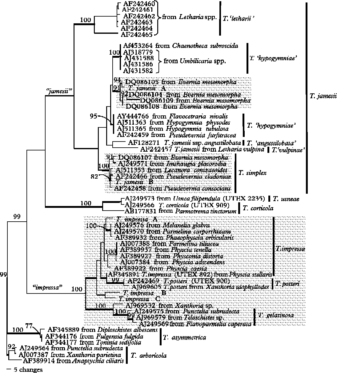
Five algal ITS rDNA genotypes are reported (Fig. 1). Although the forward primer site was located at the 3` end of the SSU, it amplified the 1512 SSU intron with the ITS region producing two length variants, one with and one without the intron. Therefore, sequenced representatives of each different RFLP pattern were used to determine ITS genotypes. The intron was present in T. jamesii A, T. jamesii B, and T. impressa A. It was absent from T. impressa B and T. impressa C. Each of the five ITS genotypes was defined by diagnostic ITS banding patterns: two fragments of lengths 490 and 216 bp (Trebouxia jamesii A); three fragments of lengths 329, 291 and 225 bp (T. jamesii B); four fragments of 405, 154, 149 and 147 bp (T. impressa A); three fragments of 256, 216 and 108 bp (T. impressa B); and two fragments of 517 and 131 bp (T. impressa C). Since fragments less than 100 bp were not used to determine the ITS genotype, the sums of the diagnostic fragment lengths are not consistent. The actual length of the ITS region in all five samples ranged from 489 to 504 bp. Identification of the algal taxa was based on significantly similar matches after BLAST searches were performed in GenBank. Although the names, T. jamesii and T. impressa, were used to refer to the five algal genotypes because of significant BLAST search results, morphology was not examined and the species epithet could not be confirmed.
One of 430 most parsimonious phylograms showing the phylogenetic placement of five algal symbionts in this study relative to species of Trebouxia from the NCBI GenBank database. The tree is based on the nucleotide sequence of the ITS rDNA amplified from the algal partner in epiphytic lichens. Only those bootstrap values greater than 70% are shown above the branches and posterior probabilities greater than 99% are indicated by thickened lines. Trebouxia arboricola is assigned as the outgroup. The tree is 674 steps long, CI=0.6053, RI=O.8782, and 212 characters are informative. Shaded boxes show the clades containing the algae sequenced in this study.
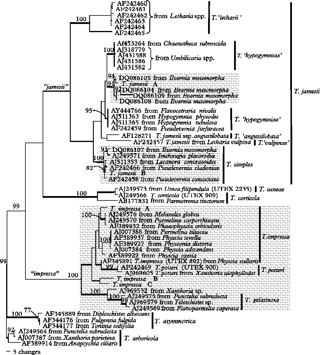
The length of the ITS1 ranged from 267 to 276 bp and the length of the ITS2 ranged from 213 to 233 bp for the five samples sequenced in this study. The length of the 5.8S was constant at 157 base pairs. The length of the entire ITS region sequenced ranged from 637 to 664 base pairs from five representative algal genotypes. After removal of approximately 100 ambiguous bp at each end of the ITS region (see “DNA sequencing” in Methods), the alignment was 537 bp in length. Nucleotide sequence similarity within T. jamesii was 94%, within T. impressa was 86%–90%, and between T. jamesii and T. impressa was 80%–86%.
Photobiont evolution
The MP (Fig. 1), neighbour joining (NJ) and ML/Bayesian trees (data not shown) produced similar topologies with some exceptions. The main difference in topology between the NJ and MP trees was that in the NJ tree, T. impressa (UTEX 891) from Physcia stellaris was nested among members of T. impressa and two samples of T. potteri were sister to T. gelatinosa. In the MP tree two samples of T. potteri were sister to T. impressa (UTEX 891). The difference in topology between the ML/Bayesian and the MP trees was that two clades in the ML/Bayesian tree fell into different positions in the MP tree. The clade containing samples of T. usneae and T. corticola formed a sister clade with the “jamesii” clade in the MP tree, but was nested within the “jamesii” clade in the ML/Bayesian tree. The clade labelled as “jamesii” is paraphyletic with members of T. simplex, and the clade has low bootstrap and posterior probability support. A second clade in the MP tree, labelled as “impressa”, consisted of members of T. impressa, T. gelatinosa and T. potteri, and was supported with 100% bootstrap but not with posterior probabilities (Fig. 1). The outgroup in the MP tree, T. arboricola, formed a sister clade with T. asymmetrica and both clades were sister to the “impressa” clade in the ML/Bayesian tree.
The five sequences from this study fell into either the “jamesii” or the “impressa” clade. Trebouxia jamesii A fell into a highly supported clade containing algae from Evernia mesomorpha. This clade was sister to a clade labelled as T. ‘hypogymniae’ and nested within a second clade labelled as T. ‘hypogymniae’. T. jamesii B fell within a more diverse, but highly supported, clade labelled as T. simplex containing algae from Evernia, Pseudevernia, Imshaugia, and Lecanora. Trebouxia impressa A was nested among other members of T. impressa. Trebouxia impressa B and C fell basal to all the sequences of T. impressa and T. potteri, in the MP trees.
Distribution of photobiont ITS genotypes and mycobiont species
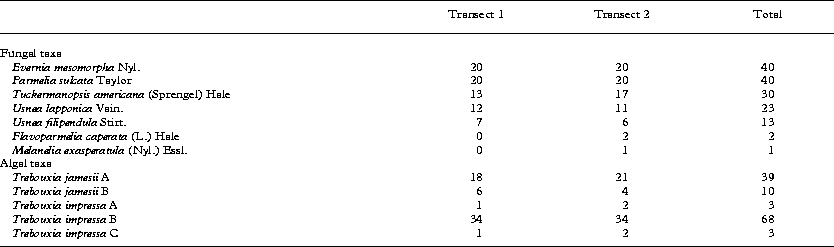
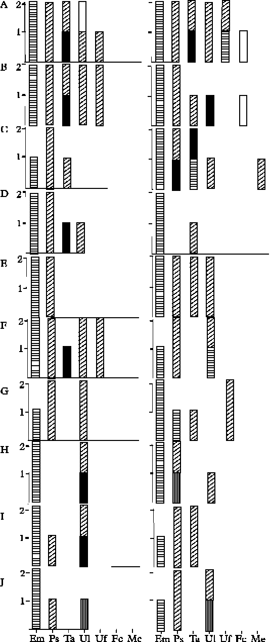
Five algal RFLP genotypes are reported to associate with seven mycobionts. Trebouxia impressa B was the most abundant alga, and T. jamesii A was the second most abundant alga (Table 1). Trebouxia impressa A and T. impressa C were rare, with each observed only three times (Table 1; Fig. 2). Trebouxia jamesii B was more common than T. impressa A and T. impressa C, but less common than T. impressa B and T. jamesii A. The most abundant lichen fungi in both transects were Evernia mesomorpha, Parmelia sulcata, Tuckermanopsis americana and Usnea lapponica (Table 1; Fig 2). Two thalli of Flavoparmelia caperata and one thallus of Melanelia exasperatula were present in transect 2 but were absent from transect 1. One thallus of Ramalina dilacerata was observed in transect 1, but it was not large enough to have allowed for a voucher specimen after DNA extraction and so DNA was not extracted from this lichen.
Distribution of numbers of thalli of the 149 lichen forming-fungal species and 123 algal genotypes among two transects of Jack Pine trees
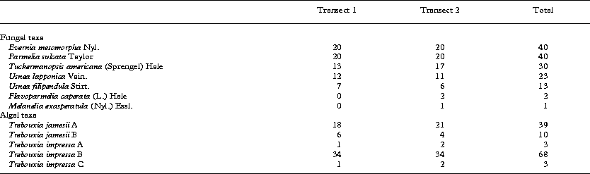
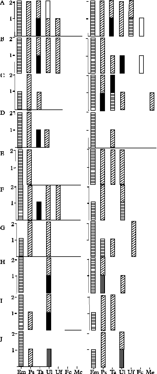
Distribution of the lichen-forming fungal species (x-axis) and the algal partners among ten trees in each of two transects. The graphs in the left column represent the ten trees in transect 1 and those in the right column represent the trees in transect 2. Two graphs in each row, labelled A to J, represent each Jack Pine tree. The y-axis shows the number of samples, x-axis fungal species abbreviated by the first letters for the genus and species names (see Table 1). Horizontal hatching = T. jamesi A; solid black boxes = T. jamesii B; vertical hatching = T. impressa A; diagonal hatching = T. impressa B; solid white boxes = T. impressa C.
Evernia mesomorpha associated exclusively with T. jamesii A in both transects, even though T. jamesii A associated with four additional fungal species (Fig. 2). Parmelia sulcata, Tuckermanopsis americana and Usnea filipendula associated with a larger number of algal genotypes in the second transect than in the first transect. The same number of algal genotypes were associated with U. lapponica in both transects. Trebouxia impressa B, the most common alga, was associated with five of the seven fungi, and T. jamesii B was associated with three fungi.
Population structure
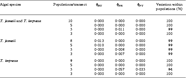
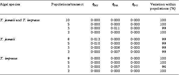
Gene flow was found among the algal partners under all population definitions (Table 2) The majority of the variation present, 99–100%, was within populations in all instances except one. Trebouxia impressa contained less variation within populations (94%) suggesting 6% variation among populations and transects. A pairwise comparison of ϕPT between each of the 20 trees studied did not produce any significant values suggesting gene flow among all populations based on these algal genotypes (data not shown). Even with the Bonferroni correction applied by multiplying the P-values by seven, there were no significant values.
Population structure of lichenized algae using four population definitions for each of three taxonomic analyses based on algal species inferred from GenBank BLAST search. Populations of n=1 were removed from the analysis. No values were significant (P=0·05)
Discussion
Photobiont identity
Photobionts associated with seven species of lichen-forming fungi probably constitute at least five species within the genus Trebouxia (Fig. 1). However, this may be an underestimation of the number of algal taxa present in this lichen community because of the low resolving power of RFLPs. Although the unknown species from this study are closely related to authentic species of T. jamesii and T. impressa in the tree (from UTEX Culture Collection; Hauck et al. Reference Hauck, Helms and Friedl2007) based on ITS rDNA sequence, it does not confirm morphological species.
The clade of T. jamesii has been separated into a number of tentative species including T. ‘letharii’, T. ‘vulpinae’ (Kroken & Taylor Reference Kroken and Taylor2000), T. ‘hypogymniae’, and T. ‘angustilobata’ (Hauck et al. Reference Hauck, Helms and Friedl2007). Based on sequences compared with those reported in Hauck et al. (Reference Hauck, Helms and Friedl2007) and Kroken & Taylor (Reference Kroken and Taylor2000) the photobiont names have been applied to clades in Fig. 1. Since Trebouxia jamesii has been known as a highly variable species (Kroken & Taylor Reference Kroken and Taylor2000), and has been named as T. jamesii or T. simplex in different studies (Hauck et al. Reference Hauck, Helms and Friedl2007; Kroken & Taylor Reference Kroken and Taylor2000; Opanowicz & Grube Reference Nelsen and Gargas2004; Piercey-Normore Reference Opanowicz and Grube2006), T. simplex is included in the T. jamesii clade in this study. Trebouxia jamesii A falls within a highly supported clade with sequences from algae of Evernia mesomorpha that does not contain morphological or accessioned species from other studies suggesting it may be a unique clade. Even though two clades sister to this clade contain sequences labelled as T. ‘hypogymniae’, together the three clades have low support and may represent three different algal lineages. Further sampling of algae representing T. ‘hypogymniae’ would be required to address the question of different lineages. Trebouxia jamesii B falls into another highly supported clade with sequences from T. simplex suggesting that it is T. simplex.
Trebouxia impressa A, B and C form a large group with high bootstrap support but low posterior probability support. This clade labelled as ‘impressa’ represents species of T. impressa, T. potteri and T. gelatinosa as indicated by the morphological species. The authentic species of T. impressa, UTEX892, is not clearly positioned with other morphological species of T. impressa (AJ007388, AJ0073 84; Beck et al. Reference Beck, Friedl and Rambold1998) in the MP tree but they are together in the NJ tree. It is not known whether T. impressa B and C are T. impressa or another species of Trebouxia such as T. flava which is positioned as sister to T. impressa and T. potteri in Hauck et al. (Reference Hauck, Helms and Friedl2007).
Photobiont dispersal
Algal dispersal is supported in this study since no significant population subdivision existed for all definitions of populations (Table 2). This was not surprising because of the small geographic distances between and within transects. Comparison of dispersal distances with other studies show variable dispersal distances. Gene flow was inferred in T. jamesii associated with E. mesomorpha at distances of greater than 50 m (Piercey-Normore Reference Piercey-Normore2006). Wind dispersal of vegetative propagules was suggested to carry algae distances of 30m in Ramalina farinacea and Evernia prunastri (Tapper Reference Tapper1976). Printzen & Ekman (Reference Printzen and Ekman2003) inferred gene flow in the mycobiont of lichen propagules of Cladonia subcervicornis for populations that were 10–15 km apart, and Walser (Reference Walser2004) suggested the maximum dispersal distance for Lobaria pulmonaria propagules was 230 m. Dispersal of vegetative propagules would provide algae with a means of hitchhiking within a vegetative structure that might confer some protection to the alga and carry it longer distances than it might otherwise travel. In addition to vegetative propagules, algal dispersal may also occur as free-living algae. The occurrence of free-living colonies or ‘escaped’ algae of Trebouxia has been disputed (Bubrick et al. Reference Bubrick, Galun and Frensdorff1984; Mukhtar et al. Reference Mukhtar, Garty and Galun1994; Tschermak-Woess Reference Tschermak-Woess1978), but zoospores have been observed (Slocum et al. Reference Slocum, Ahmadjian and Hildreth1980). If zoospores are produced inside the thallus, they can escape from the thallus through cracks in the cortex. These flagellated cells may form colonies close to the thallus in which they were produced (Slocum et al. Reference Slocum, Ahmadjian and Hildreth1980) and become available to associate with germinating fungal spores giving rise to entire populations of the species (Murtagh et al. Reference Murtagh, Dyer and Crittenden2000).
Photobiont selectivity and specificity
Algal taxa were shared among seven lichen-forming fungal species supporting the hypothesis, but the algae were not equally distributed among the fungi. Evernia mesomorpha was associated with a single alga, T. jamesii A, but T. jamesii A also associated with four other species of fungi (Fig. 2). This distribution may be explained if T. jamesii A is the only alga that is genetically compatible with Evernia mesomorpha in this site. However, other algae are known to be compatible with E. mesomorpha (Piercey-Normore Reference Piercey-Normore2006), and one of these, T. jamesii B (T. simplex in Fig. 1), was present in this study. Trebouxia jamesii B was also reported to be associated with other species of Pseudevernia, Imshaugia, and Lecanora (Fig. 1). In this study E. mesomorpha appears to be highly selective of one photobiont and specific to T. jamesii A, but the alga is not specific to E. mesomorpha. Specificity has been shown among members of the Physciaceae (Dahkild et al. Reference Piercey-Normore2001) where four species in the Physciaceae associate only with T. impressa. In this study E. mesomorpha showed high selectivity, but in Piercey-Normore (Reference Piercey-Normore2006) the same mycobiont showed low selectivity using the same molecular marker. Inconsistency in algal selectivity has been reported for three different species of Cladonia (Piercey-Normore Reference Piercey-Normore2004) where C. multiformis and C. rangiferina were highly selective in different geographic regions, but C. gracilis showed low levels of algal selectivity in one geographic area but high levels of selectivity in another geographic area. Habitat features, which were not measured in any of these studies, may be influencing fungal selectivity even within a single species.
Parmelia sulcata associated exclusively with T. impressa B, the most common alga, in transect 1, but it associated with three other algae in addition to T. impressa B in transect 2 (Fig. 2) suggesting low levels of selectivity and specificity. Parmelia sulcata is a pollution tolerant species (Hawksworth and Rose Reference Hawksworth and Rose1970), and is tolerant of many different environmental conditions and habitats. If pollution tolerant lichen-fungi are genetically compatible with many different algae or associate with widespread algae, they may have the capacity to survive in a wide range of habitats. Similarly, three photobiont species were reported to associate with another pollution tolerant lichen-forming fungus, Protoparmeliopsis muralis (Guzow-Krzeminska Reference Guzow-Krzeminska2006) and 21 photobiont genotypes are reported to associate with Parmotrema tinctorum (Ohmura et al. Reference Ohmura, Kawachi, Kasai and Watanabe2006).
The algal distribution observed in this study may be explained by the replacement of locally adapted algae with a common genotype such as T. impressa B if no additional benefit is provided by the locally adapted algal genotype. Alternatively, the widely distributed T. impressa B may be replaced through the effective dispersal of algae that are adapted to particular micro- environmental conditions and confer advantageous properties to the lichen. In both scenarios neither the widely distributed genotype nor the locally adapted genotype may be better adapted in a moderated environment, such as this boreal Jack Pine stand, with low pollution levels. Moderated environments may provide suitable habitat for a higher diversity of algal genotypes by having less selection pressure on the photobiont than more extreme habitats such as iron rich rocks (Beck Reference Beck1999) or polluted urban areas (Ohmura et al. Reference Ohmura, Kawachi, Kasai and Watanabe2006). These more extreme habitats may limit the available pool of algae only to those that can tolerate the more extreme conditions.
Conclusions
The community of algae housed in different lichen thalli within the same strictly defined habitat consisted of divergent algal lineages that were inferred to be at least two species and five genetically differentiated algae. Although these five algal lineages are probably an underestimate of algal diversity in this habitat because of the low resolving power of RFLPs, they show that algal diversity is present within the same confined habitat and that the algal taxa are shared among different mycobionts. This study showed low specificity and variable levels of selectivity with species of lichen-forming fungi. Perhaps in moderated habitats, such as this boreal Jack Pine forest, a large number of algae can survive with many fungi, while in extreme habitats, such as an iron-rich rock, only algae specialized to those conditions can survive. This must be studied further by sampling free-living algae in the same habitat. Even though the algal partners were highly divergent, gene flow was shown to occur under different population definitions in this tightly defined community of lichenized algae. Dispersal by soredia may provide an effective medium for algal dispersal and availability of algae to other fungi in the area. Individual algal and fungal partnerships may be a result of a combination of genetic compatibility between symbionts, algal availability and environmental conditions.
We thank two anonymous reviewers for many helpful suggestions on the manuscript. The study was funded by the Natural Sciences and Engineering Research Council of Canada (NSERC).