Introduction
Brycon orbignyanus is a large tropical species, indigenous to the Paraná and Uruguay River Basins (Carolsfeld et al., Reference Carolsfeld, Harvey, Ross and Baer2004) that is highly dependent on riparian vegetation (Panarari-Antunes et al., Reference Panarari-Antunes, Prioli, Prioli, Galdino, Julio and Prioli2011). In addition to excessive fishing and environmental degradation, the construction of hydroelectric dams in the area has led the species to be classified as close to extinction as the dams impede their reproductive migration (Oliveira et al., Reference Oliveira, Ashikaga, Foresti and Senhorini2017).
Although induced artificial reproduction and incubation are well understood in different teleost species (Faustino et al., Reference Faustino, Nakaghi, Marques, Makino and Senhorini2007), there are few research groups dedicated to studying the basic aspects of the fish embryonic period, which extends from fertilization to hatching. Studies on the early stages of the life cycle are fundamental for knowledge of both taxonomy and species ecology (Sanches et al., Reference Sanches, Nakatani and Bialetzki1999). Reynalte-Tataje et al. (Reference Reynalte-Tataje, Zaniboni-Filho and Esquivel2004) found that embryos and larvae of a species are greatly important and a useful tool to locate spawning areas and study the species growth in the natural environment. Knowledge of the embryonic development stages allows species ontogeny to be studied while also being used in biotechnological processes, as bioindicators of environmental quality to evaluate the effect of toxic substances on aquatic fauna, in repopulation programmes and conservation of rare species (Flores et al., Reference Flores, Araiza and Valle2002; Botero et al., Reference Botero, Fresneda, Montoya and Olivera2004; Ninhaus-Silveira et al., Reference Ninhaus-Silveira, Foresti and Azevedo2006; Paes et al., Reference Paes, Silva, Nascimaneto, Valentin, Senhorini and Nakaghi2014; Santos et al., Reference Santos, Yasui, Xavier, Macedo, Nascimento, Fujimoto, Senhorini and Nakaghi2016).
Fish embryonic development has been studied and divided into stages in several species; among these are Brycon cephalus (Romagosa et al., Reference Romagosa, Narahara and Fenerich-Verani2001), Brycon insignis (Andrade-Talmelli et al., Reference Andrade-Talmelli, Kavamoto, Romagosa and Fenerich-Verani2001), Brycon gouldingi (Faustino et al., Reference Faustino, Makino, Neumann and Nakaghi2015), Brycon amazonicus (Neumann et al., Reference Neumann, Nakaghi, Braga, Faustino and Mendes2013; Silva et al., Reference Silva, Santos, Senhorini, Paes, Valentin, Fujimoto, Nascimento, Yasui and Nakaghi2017); Brycon nattereri (Maria et al., Reference Maria, Ninhaus-Silveira, Órfão and Viveiros2017) and Brycon orthotaenia (Sampaio et al., Reference Sampaio, Prado, Sato, Bazzoli and Rizzo2015). Thus, this research aims at characterizing in depth the external and internal morphological events that occur during oocyte extrusion, fertilization and embryonic development of B. orbignyanus over known time periods. All knowledge regarding its biology is extremely important as it can contribute to studies on repopulation, cryopreservation and reproductive management, aimed at species conservation and survival.
Materials and methods
Collections were carried out during the species reproductive period from October to January. To perform induced reproduction, breeding animals from captivity were selected and were made dry fertilization. Females with an average weight of 4.8 kg and standard length of 46 cm received three doses of common carp pituitary (Cyprinus carpio). The first dose of 0.25 mg/kg body weight (BW) was followed by the second and third doses of 0.5 mg/kg BW each, injected 4 and 16 h after the first dose. Spawning occurred 4 h after the third application. Males with an average weight of 2.1 kg and standard length of 33 cm received a dose of 5.0 mg/kg BW at the same time as the females received the last dose.
After extrusion, the oocytes were placed in a beaker and gently mixed with the added semen. Hydration occurred 1 min later and the eggs were placed in a 1600 litre incubator with water circulation and aeration. The water temperature was kept at 27°C throughout development through heaters with thermostats.
Sampling occurred at the following times: oocyte extrusion, oocyte and semen mixing (time zero), every minute until 10 min were completed, and at 12, 14, 16, 18, 20, 24, 26, 28, 30, 45 and 60 min and after that, every hour until the larvae hatched.
Light microscopy
Samples were fixed in Karnovsky solution (2.5% glutaraldehyde + 2.5% paraformaldehyde) for 24 h, washed in 0.1 M sodium cacodylate buffer, pH 7.4, dehydrated in increasing ethanol series solutions, diaphanized in xylol followed by infiltration and inclusion in paraplast. Subsequently, 4-μm thick histological sections were stained with haematoxylin-eosin and masson trichrome. The analysis and photograph documentation were performed using an Olympus BX 50 photomicroscope.
Scanning electron microscopy
Samples were also fixed in Karnovsky solution for 24 h and then washed in 0.1 M sodium cacodylate buffer, pH 7.2; post-fixed in 1% osmium tetroxide solution for 2 h and washed in the same buffer. Subsequently, the samples were dehydrated in an increasing ethanol series (30, 50, 70, 80, 90, 95, 100%), for 15 min each. Then, they were processed in a liquid crystal (BAL-TEC) Crushed Dryer, mounted on a copper support, metallized with palladium-gold and observed under a scanning electron microscope (JEOL-JSM 5410).
Morphometric analysis
The diameters of the oocytes/eggs were measured under a stereomicroscope using the micrometric ocular lens. Twenty oocytes/eggs were measured from each of the sampling times, and three measurements of diameter were taken for each one. The values were statistically evaluated by Tukey test at 5% (P < 0.05) using ESTAT software.
Results
B. orbignyanus embryonic development lasted 13 h from fertilization to hatching at 27°C. The greenish eggs remained spherical throughout development, and non-adhesive, with a translucent chorion limiting a wide perivitelline space.
The size of the perivitelline space increased up to 30 min after fertilization and after that, diameter is practically maintained. At the time of extrusion oocyte average diameter was 1.31±0.11 mm while, 30 min after fertilization, the egg size, considering the perivitelline space, was 3.32±0.12 mm. At hatching, the larvae measured 3.47±0.15 mm.
Stage 1 (0–20 min)
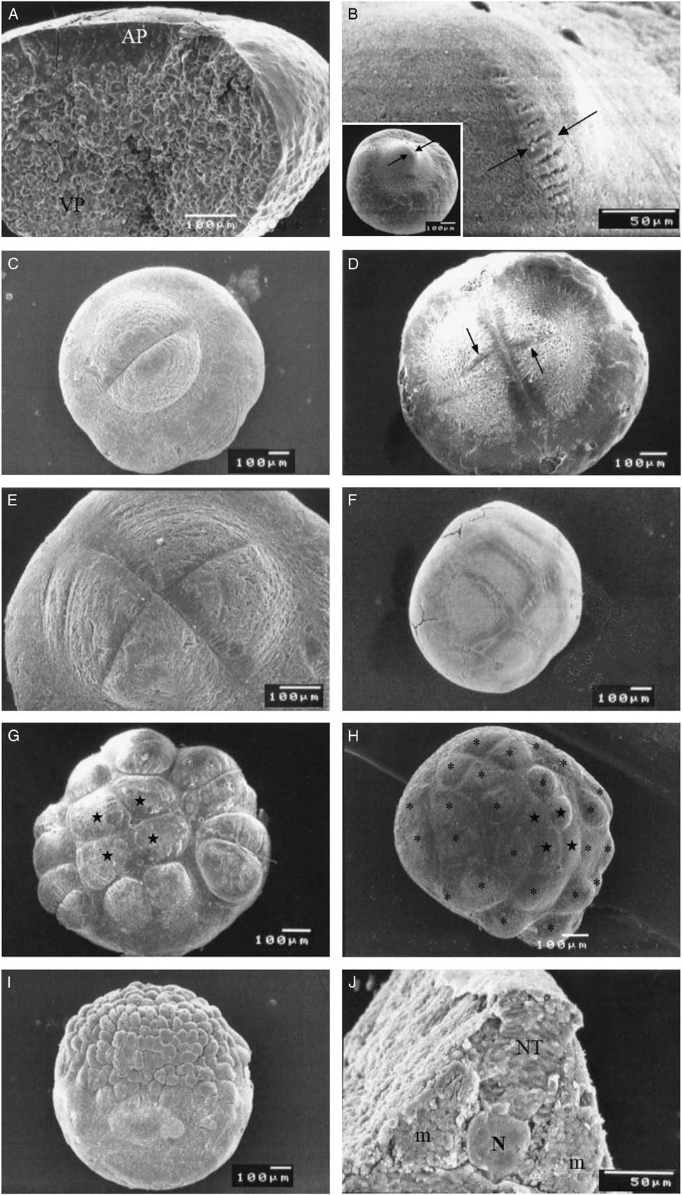
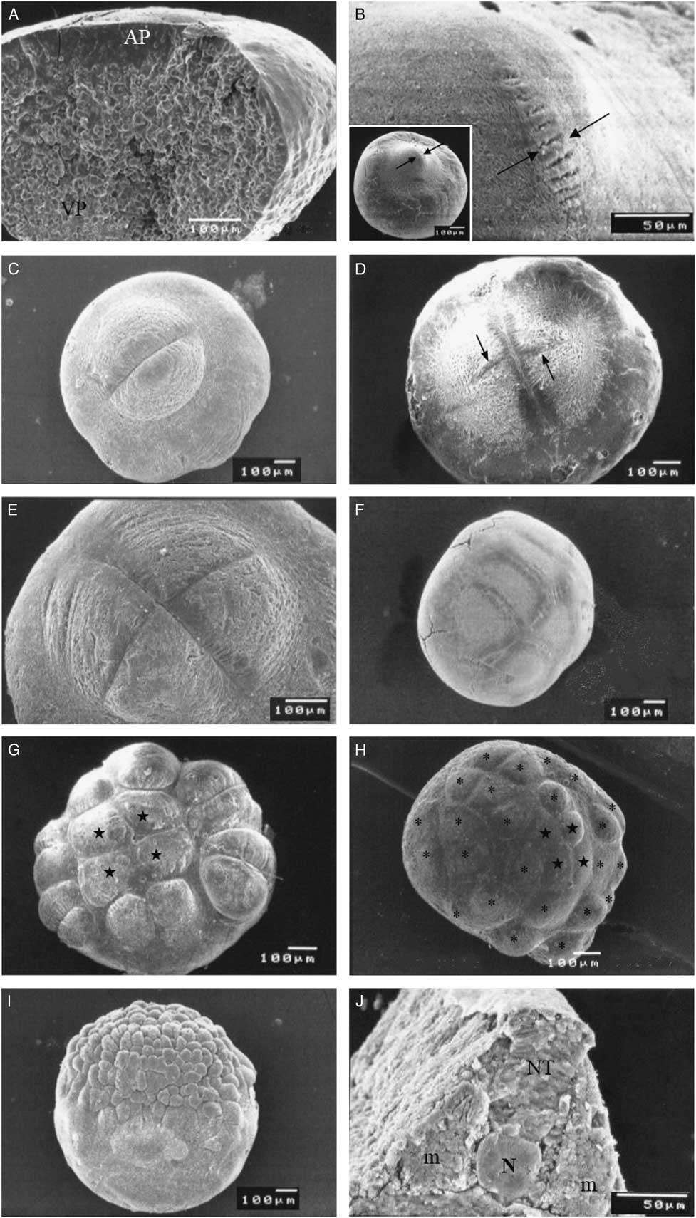
This stage lasted from fertilization to the first cleavage. The female and male pro-nuclei approached for further fusion in the cytoplasm in the animal pole or blastodisc. This region comprising the cortical cytoplasm appeared thick and convex over the yolk sphere, which, at this stage, represented the vegetative pole (Fig. 1A ). It is noteworthy that, at this developmental stage, the cortical cytoplasm was observed to form a thin layer covering the whole yolk.
Scanning electron micrographs (SEM) of B. orbignyanus eggs. (A) Stage 1, time ‘9 min’, showing the formation of the animal and vegetative poles. (B) Stage 2, time ‘20 min’, evidencing the beginning of the first cleavage (arrows). (C) Stage 2, time ‘20 min’, showing egg with 2 blastomeres. (D) Stage 2–3, time ‘26 min’, highlighting the beginning of second cleavage (arrows). (E) Stage 3, time ‘30 min’, egg with four blastomeres. (F) Stage 4, time ‘45 min’, egg with eight blastomeres. (G) Stage 5, time ‘1 h’, egg with 16 blastomeres showing arrangement with four central blastomeres (★) and 12 marginal blastomeres. (H) Stage 6, time ‘1 h’, egg in the 32-cell phase, showing that only 28 blastomeres are observed on the surface, being four central (★) and 24 marginal (*). (I) Stage 7, time ‘2 h’, egg in the morula stage. (J) Stage 10, time ‘8 h’, evidencing the presence of the neural tube (TN), notochord (N) and para-axial mesoderm (m).
Stage 2 (20 to 30 min)
The beginning of the first cleavage occurred in the centre of the blastodisc (Fig. 1B ) and extended to the periphery while simultaneously deepening to the perimeter of the yolk sac, presenting meroblastic or partial type segmentation.
Stage 3 (26–45 min)
The first cleavage furrow, marking the second cleavage developed in two blastomeres, and was vertically oriented (Fig. 1C ). This furrow deepened to divide each blastomere into two of the same size, totalling four blastomeres (Fig. 1D, E ).
Stage 4 (45 min to 1 h)
The third cleavage, which consisted of dividing the four existing blastomeres into eight blastomeres, occurred in a plane parallel to the first cleavage (Fig. 1F ). At this development stage, the resulting eight blastomeres were arranged in two tiers of four blastomeres each, oriented according to the plane of the second cleavage.
Stage 5 (1 h)
Similarly, the fourth cleavage that divided the two rows of four blastomeres into four rows of four blastomeres each, occurred parallel to the second cleavage. The resulting 16 blastomeres were arranged in 12 marginal and four central blastomeres (Fig. 1G ). At this stage, the central projection of the yolk was observed under the blastodisc, raising the four central blastomeres above the cleavage plane.
Stage 6 (1–2 h)
The fifth cleavage was characterized by the formation of 32 cells. The fifth cleavage plane meridionally divided the 12 marginal blastomeres into 24, and horizontally the four central blastomeres into eight, forming one outer and one inner layer, each with four cells in the central region (Fig. 1H ).
Stage 7 (2–3 h)
From the sixth cleavage, the embryo presented cell division plans with diverse orientation, constituting initially 64 to 128 cells, characterizing the morula stage (Fig. 1I ). These cells were arranged in two or more layers forming the dome-shaped blastoderm. The successive cleavages increased the number of cells but not their size.
Stage 8 (2–3 h)
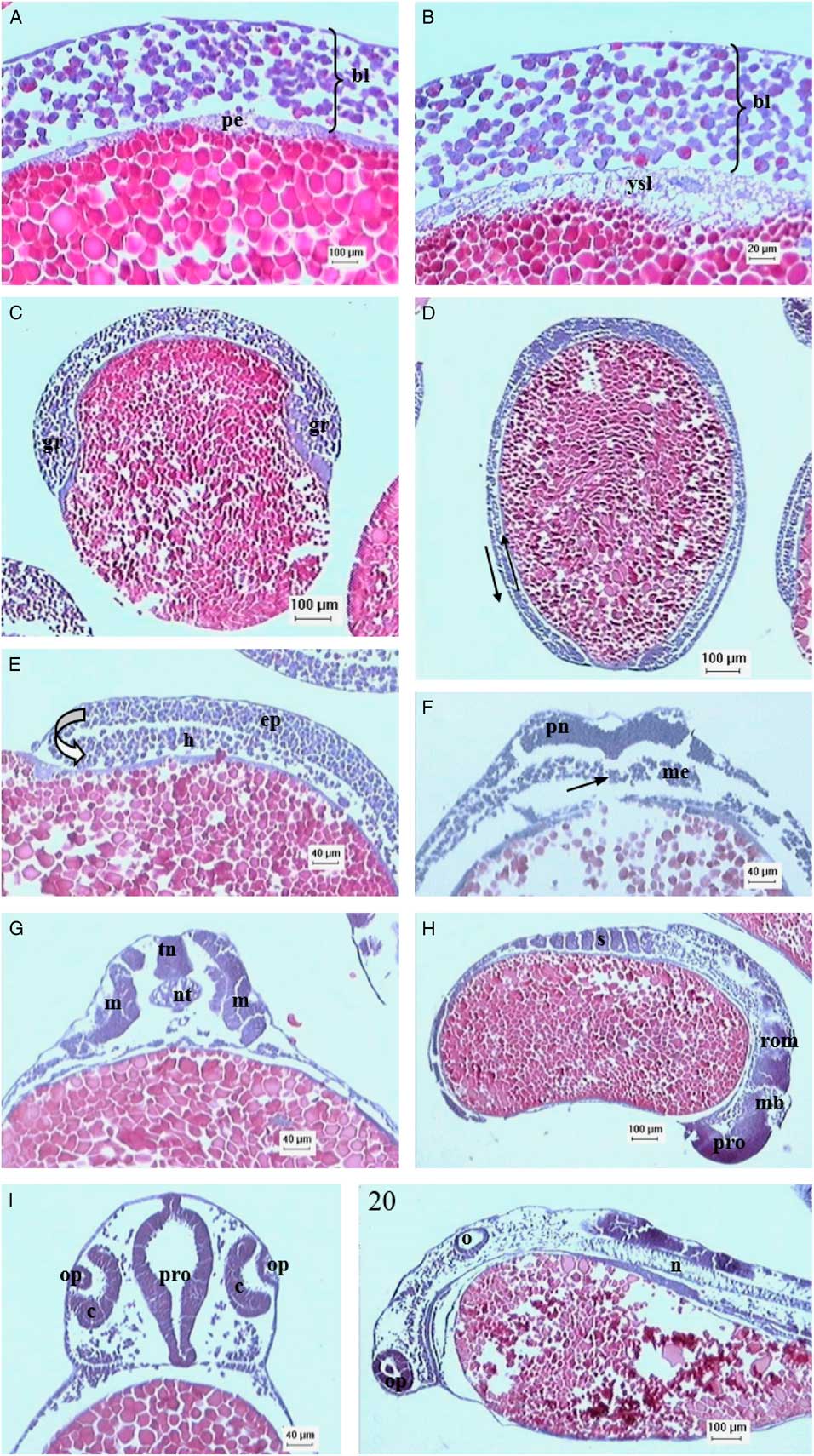
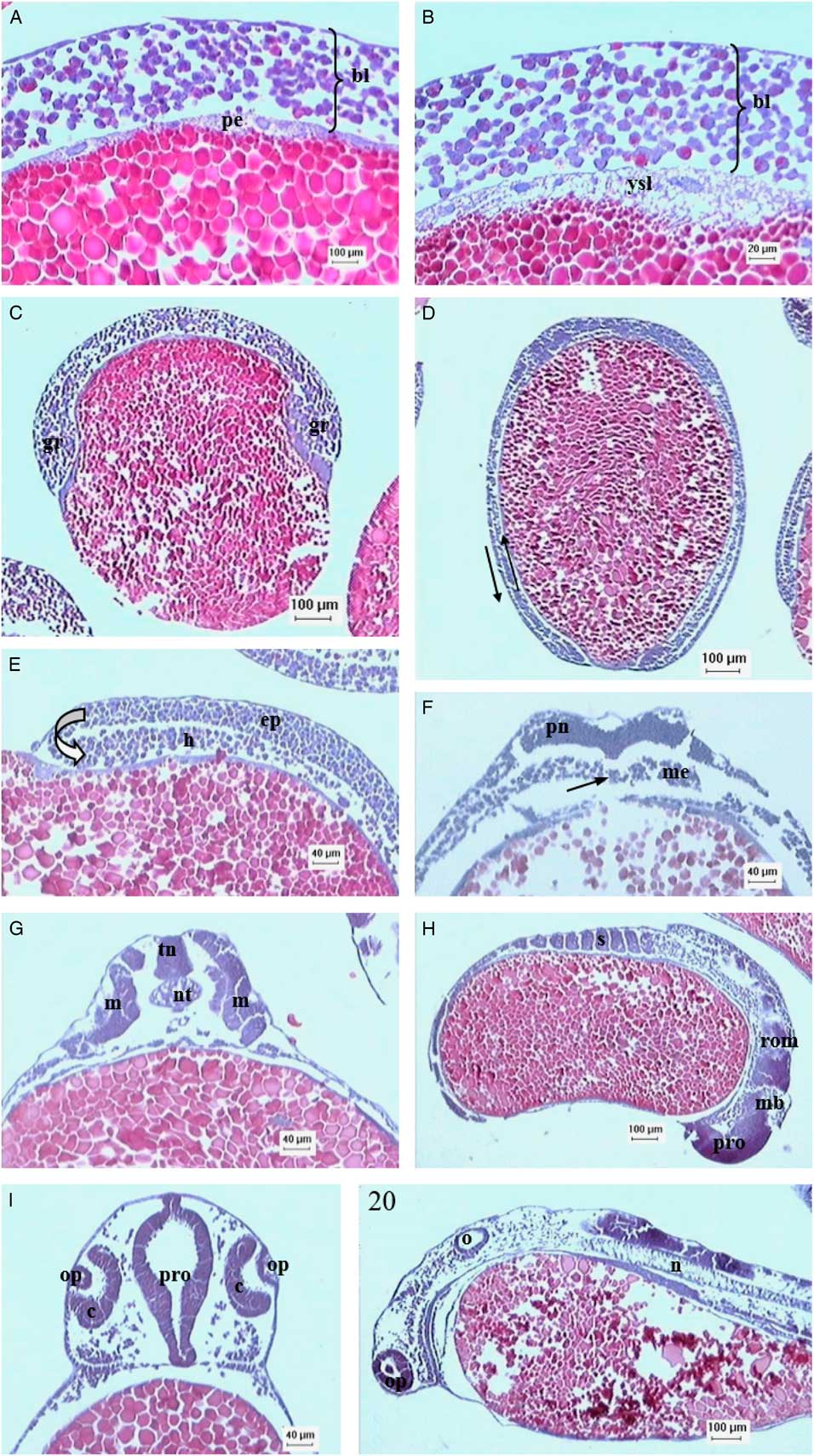
The blastoderm was equally thick in the blastula and at the end of the morula stages. The marginal blastomeres of the blastoderm migrated and positioned themselves in an arranged manner, limiting the vitellus. These cells arranged in a row constitute the periblast, observed around the blastoderm (Fig. 2A ). At this developmental stage, the periblast cells collapse and release their cytoplasm along with their nucleus into the cytoplasm that houses the embryo yolk giving rise to the yolk syncytial layer (YSL) (Fig. 2B ). It was also noted that this stage was characterized by irregular spaces between the cells that constitute the blastoderm (Fig. 2C ). At the end of this stage, epiboly began, that is the blastoderm starts to cover the yolk.
Photomicrographs of eggs/embryo of B. orbignyanus. (A) Stage 9, time ‘4 h’, egg in the blastula phase, evidencing periblast (pe) and blastoderm (bl). (B) Stage 9, time ‘4 h’, showing the formation of the yolk syncytial layer (ysl) and the blastoderm (bl). (C) Stage 9, time ‘4 h’, showing the blastula with irregular spaces between blastoderm cells and formation of the germ ring (gr). (D) Stage 9, time ‘6 h’, showing gastrula at the end of epiboly, highlighting the direction of epiboly (|) (up arrow) and migration (down arrow) (┌) movements. (E) Stage 9, time ‘5 h’, evidencing the migration movement (arrow) performed by the blastoderm cells to form the epiblast (ep) and hypoblast (h). (F) Stage 9, time ‘7 h’, highlighting the formation of a furrow in the neural plate (pn), formed above the notochord (arrow). The mesendoderm (me) is present. (G) Stage 10, time ‘10 h’, showing neural tube (tn), notochord (n) and para-axial mesoderm (m). (H) Stage 10, time ‘8 h’, highlighting the presence of somites (s) and formation of forebrain (pro), midbrain (mes) and rhombencephalon (rom). (I) Stage 11, time ‘11 h’, showing optic or crystalline vesicle (op), optic cup (c) and forebrain (pro). (J) Stage 11, time ‘12 h’, highlighting the optic vesicle (op), optic (o) and notochord (n).
Stage 9 (3–7 h)
The gastrula stage was characterized by the epiboly movement, emergence of the embryonic shield, cell migration with consequent formation of two germ layers. Epiboly was characterized by the continued blastoderm movement to cover the yolk. When 50% of the yolk was covered, the marginal border of the blastoderm thickened forming the germ ring (Fig. 2C ). In this stage, epiboly stops temporarily while cells accumulate perpendicular to the germ ring, forming the embryonic shield. Once the shield is formed, epiboly restarts and the margin of the blastoderm advances along the yolk boundary, completely covering it (Fig. 2D ), forming the yolk sac. At the time epiboly restarted, cells had migrated in the germ ring region forming a fold that projected below and opposite to the movement of blastoderm epiboly (Fig. 2D , E). At this stage, the embryo presented two germ layers, the external or epiblast and the internal or hypoblast (Fig. 2E ).
When the gastrula stage presented more than 50% epiboly, it was noticed that several cell layers started to form between the hypoblast and epiblast. At this stage, this cellular accumulation was considered as the appearance of a possible third embryonic tissue called the mesendoderm (Fig. 2F ). The cells of this new tissue, located below and centrally to the embryonic shield, were considered as axial mesoderm. When epiboly is close to the end, cells start to accumulate in this region, probably forming the second axis of the embryo, known as notochord (Fig. 2F ).
At the end of the gastrula, i.e., when the yolk plug was formed, the onset of the dorsal epiblast thickening was observed immediately above the formed notochord (Fig. 2F ). This region was called the neural plaque, the primordia of the nervous system.
Stage 10 (8 h)
At this stage, thickening of the dorsal epiblast causes a sulcus to form, which later fused its edges into the neural tube (Figs 1J and 2G).
Laterally to the notochord was the para-axial mesoderm (Figs 1J and 2G), which in the anterior portion gave rise to the musculature associated with the organs of this region. Subsequently, the para-axial mesoderm represented the segmentation plate that gave rise to the somites (Fig. 2H ).
Previously, a region representing the pre-chordal plaque was found in the hypoblast. The region of the neural tube above this plate thickened originating the encephalic region, delimiting the forebrain, mesencephalon, and rhombencephalon regions (Fig. 2I ). At this stage, the subcephalic and subcaudal folds appeared caused by the decreasing yolk. The appearance of the folds defined the body shape of the embryo.
Stage 11 (11–13 h)
After 11 h, the optic vesicle started to form by the evagination of the lateral wall of the posterior portion of the forebrain (Fig. 2I ). At this stage, the tail of the embryo was well developed.
The optic vesicle was observed after 12 h (Fig. 2J ) while the tail showed a more intense growth, greatly increasing the length of the embryo. Subsequently, the tail started to move, culminating with the rupture of the chorion and consequent hatching of the larva from 13 h.
At the time of hatching, the digestive system was rudimentarily developed as a massive cord of cells in the mid-posterior region of the embryo, and was characterized by the formation of the posterior intestine.
Discussion
It is known that incubation temperature directly influences the developmental speed of teleosts while possibly leading to anomalies during the embryo and larva formation stages (Santos et al., Reference Santos, Yasui, Xavier, Macedo, Nascimento, Fujimoto, Senhorini and Nakaghi2016). The embryonic development of B. orbignyanus lasted 13 h at 27°C, considered an ideal temperature for the species according to Nakatani et al. (Reference Nakatani, Agostinho, Baumgartner, Bialetzki, Sanches and Cavicchioli1999). Another parameter that should also be considered in the speed of the embryonic development is the size of oocytes and eggs as species with smaller eggs have faster development (Paes et al., Reference Paes, Silva, Nascimaneto, Valentin, Senhorini and Nakaghi2014). The Brycon orbignyanus oocytes had an average diameter of 1.31±0.11 mm at the time of extrusion. It is also known that oocyte and egg diameters vary among species and are associated with both reproductive behaviour and fecundity, presenting inter- and intraspecific alterations (Marques et al., Reference Marques, Faustino, Bertolucci, Paes, Silva and Nakaghi2017). Generally, migratory species that exhibit total spawning, external fertilization and no parental care have a high fertility rate and smaller oocytes/eggs (Vazzoler, Reference Vazzoler1996).
These fish eggs are classified as megalecithal due to the large yolk, and telolecithal because the yolk appears unevenly distributed, concentrated at the vegetative pole and the cytoplasm with the organelles is concentrated in the animal pole (Ribeiro et al., Reference Ribeiro, Leme dos Santos and Bolzan1995). The eggs of Brycon orbignyanus, similar to those of other Brycon species, can also be classified as such (Romagosa et al., Reference Romagosa, Narahara and Fenerich-Verani2001; Andrade-Talmelli et al., Reference Andrade-Talmelli, Kavamoto, Romagosa and Fenerich-Verani2001; Faustino et al., Reference Faustino, Makino, Neumann and Nakaghi2015; Sampaio et al., Reference Sampaio, Prado, Sato, Bazzoli and Rizzo2015).
The B. orbignyanus eggs had a wide perivitelline space, which was also observed in other species of teleost fish (Sampaio et al., Reference Sampaio, Prado, Sato, Bazzoli and Rizzo2015; Marques et al., Reference Marques, Faustino, Bertolucci, Paes, Silva and Nakaghi2017). However, Valentin et al. (Reference Valentin, Nascimento, Silva, Fernandes, Giannecchini and Nakaghi2015) and Santos et al. (Reference Santos, Yasui, Xavier, Macedo, Nascimento, Fujimoto, Senhorini and Nakaghi2016) reported that Betta splendens and Astyanax altiparanae eggs hydrate very little, while presenting a small perivitelline space. The size of perivitelline space depends on the reproductive characteristics of each species (Paes et al., Reference Paes, Silva, Nascimaneto, Valentin, Senhorini and Nakaghi2014), the environment in which the eggs develop and the functions attributed to it such as mechanical protection, osmotic regulation, nutrition and prevention to polyspermia (Laale, Reference Laale1980).
Embryogenesis extends from fertilization to hatching and, for teaching purposes, is usually divided into stages such as cleavage, blastula, gastrula and segmentation (Valentin et al., Reference Valentin, Nascimento, Silva, Fernandes, Giannecchini and Nakaghi2015; Santos et al., Reference Santos, Yasui, Xavier, Macedo, Nascimento, Fujimoto, Senhorini and Nakaghi2016; Marques et al., Reference Marques, Faustino, Bertolucci, Paes, Silva and Nakaghi2017). In B. orbignyanus, the first cleavage divided the egg cell into two same size blastomeres that appeared indistinguishable, similar to Danio rerio (Kimmel et al., Reference Kimmel, Ballard, Kimmel and Ullmann1995) and Zungaru jahu (Marques et al., Reference Marques, Faustino, Bertolucci, Paes, Silva and Nakaghi2017). According to Castellani et al. (Reference Castellani, Tse, Leme dos Santos, Faria and Santos1994), the number of blastomeres increased as their size decreased during cleavage, a fact also observed in B. orbignyanus. The first cleavage in B. orbignyanus started at the centre of the blastodisc, extending laterally and deepened to divide the cell completely, similar to Alosa sapidissima (Shardo, Reference Shardo1995).
In the blastoderm stage, irregular spaces formed between the blastoderm cells, a fact also observed in zebrafish by Kimmel et al. (Reference Kimmel, Ballard, Kimmel and Ullmann1995). These authors suggest that this stage is better defined as ‘stereoblastula’ as it does not present a typical blastocoele.
In the blastula stage, formation of the periblast and the YSL was observed. The YSL is a cytoplasmic continuous layer, located between the blastoderm disc and the yolk, resulting from the incomplete division of the blastomeres (Ninhaus-Silveira et al., Reference Ninhaus-Silveira, Foresti, Azevedo, Agostinho and Veríssimo-Silveira2007). It is noteworthy that the appearance of periblast in teleosts can occur at the morula stage, as reported for Z. jahu by Marques et al. (Reference Marques, Faustino, Bertolucci, Paes, Silva and Nakaghi2017) or at the blastula stage (Kimmel et al., Reference Kimmel, Ballard, Kimmel and Ullmann1995).
At the end of blastula and beginning of gastrula stages, epiboly movement was evidenced in B. orbignyanus eggs. This movement was accompanied by the appearance of a germ ring at the marginal border of the blastoderm, and consequent formation of the embryonic shield. Similar events have been observed in Oryzias latipes (Iwamatsu, Reference Iwamatsu1994), Danio rerio (Kimmel et al., Reference Kimmel, Ballard, Kimmel and Ullmann1995) and Alosa sapidissima (Shardo, Reference Shardo1995). As previously mentioned, in D. rerio, O. latipes and A. sapidissima, formation of the embryonic shield has a beginning delimited by the dorsal lip of the blastoderm, whereas in B. orbignyanus it was not possible to observe beginning of formation of the germ shield through a region delimited such as the dorsal lip.
In B. orbignyanus, when epiboly reached 50% coverage, cells migrated forming a fold that projected below and opposite the epiboly movement of the blastoderm, therefore forming the epiblast and the hypoblast. The epiblast corresponds to the definitive ectoderm, which gives rise to the epidermis, central nervous system, neural crest and sensory placodes (Kimmel et al., Reference Kimmel, Ballard, Kimmel and Ullmann1995; Shardo, Reference Shardo1995). The hypoblast gives rise to a joint layer described as mesoderm and endoderm (Kimmel et al., Reference Kimmel, Ballard, Kimmel and Ullmann1995; Shardo, Reference Shardo1995). Thisse et al. (Reference Thisse, Thisse, Schiling and Postlethwait1993) named this tissue the mesendoderm, emphasizing that only two embryonic layers are present in the gastrula; how this leaflet is subdivided into mesoderm and endoderm remains unknown (Kimmel et al., Reference Kimmel, Ballard, Kimmel and Ullmann1995).
The central nervous system in B. orbignyanus rising from the ectoderm coincides with the formation of this organ system in the other vertebrates (Iwamatsu, Reference Iwamatsu1994; Kimmel et al., Reference Kimmel, Ballard, Kimmel and Ullmann1995; Shardo, Reference Shardo1995). The mesodermal tissues that appeared on both sides of the notochord during the gastrula were organized into segments called somites, similar to those described in Danio rerio (Kimmel et al., Reference Kimmel, Ballard, Kimmel and Ullmann1995); Oreochromis niloticus (Morrison et al., Reference Morrison, Miyake and Wright2001) and Oryzias latipes (Iwamatsu, Reference Iwamatsu1994).
In B. orbignyanus, the anterior region of the neural tube expanded to form the forebrain, mesencephalon, and rhombencephalon regions, similar to that described for Z. jahu (Marques et al., Reference Marques, Faustino, Bertolucci, Paes, Silva and Nakaghi2017) and P. lineatus (Ninhaus-Silveira et al., Reference Ninhaus-Silveira, Foresti and Azevedo2006), emphasizing that in these species the optic primordia developed at the beginning of the prosencephalon formation.
Larvae began to hatching after 13 h, when intense tail movement was observed, culminating with rupture of the chorion. According to Zhang et al. (Reference Zhang, Bhattacharya and Li2009), hatching of many bonefish occurs when glands start to secrete proteolytic enzymes that are associated with tail and body movements.
Chardon et al. (Reference Chardon, Parmentier and Vandewalle2003) stated that rapid embryonic development is typical of migratory species with seasonal reproductive strategies, high fertility and no parental care. Embryonic development in B. orbignyanus consisted of events that have already been described for other tropical teleosts, but differed regarding duration and some species-specific characteristics.
Vandewalle et al . (Reference Vandewalle, Germeau, Besancenet, Parmentier and Baras2005) stated that rapid embryonic development is typical of migratory species with seasonal reproductive strategies, high fertility and no parental care. Embryonic development of B. orbignyanus was divided into 11 stages that consisted of events already described for other tropical teleosts, but differ regarding duration and some species-specific characteristics.
Acknowledgements
The authors thank Buriti Fisheries (Nova Mutum, MT, Brazil) for providing the eggs and fish and to Orandi Mateus, histo-technician from the Histology and Embryology Laboratory of FCAV/UNESP, Jaboticabal-SP, Brazil, for his help in material processing.
Financial support
The authors would like to thank the National Council for Scientific and Technological Development – CNPq for their financial support.
Conflicts of interest
There are no conflicts of interest.