Introduction
Angarotipula Savchenko, Reference Savchenko1961 (Diptera: Tipulidae) has been recognised at the genus level since the publication of the Palaearctic Catalogue of Diptera, Volume 1 (Oosterbroek and Theowald Reference Oosterbroek and Theowald1992), but this change in rank from a subgenus of Tipula Linnaeus, 1758 to genus was never formally proposed (P. Oosterbroek, personal communication) and has been questioned or ignored by the author and other tipulid workers (e.g., Young Reference Young2004; Gelhaus Reference Gelhaus2009). A more detailed investigation was therefore needed to see whether elevating Angarotipula to full generic rank is warranted, and if so, to make this taxonomic change in accordance with the International Code on Zoological Nomenclature (1999).
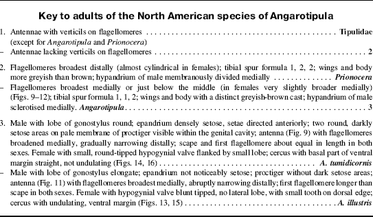
Specimens identified as Angarotipula parrioides (Alexander, Reference Alexander1919) from Alaska, United States of America and northern Canada seemed very similar to northern European specimens identified as Angarotipula tumidicornis (Lundsröm, 1907). I suspected that these may be synonyms, necessitating a review of relevant specimens of both species.
Similarities among the species of Angarotipula and Prionocera Loew, 1844, resulted in several species being erroneously placed in Prionocera rather than in the taxon now recognised as Angarotipula. Therefore, diagnostic characters and similarities between these two genera also needed to be explored.
Material and methods
Specimens from the following collections were used for this study: Albert J. Cook Arthropod Research Collection, Michigan State University, East Lansing, Michigan, United States of America (ARC); Biodiversity Institute of Ontario, Guelph, Ontario, Canada (BIOUG); Canadian National Collection of Insects, Ottawa, Ontario, Canada (CNC); Brodo Collection, Ottawa, Ontario, Canada (FB); Museum of Zoology, Helsinki, Finland (MZH); Royal Ontario Museum, Toronto, Ontario, Canada (ROM); University of Alaska Museum, Fairbanks, Alaska, United States of America (UAM); University of Michigan Museum of Zoology, Ann Arbor, Michigan, United States of America (UMMZ); University of Minnesota Insect Collection, St. Paul, Minnesota, United States of America (UMSP); United States National Museum of Natural History, Washington, District of Columbia, United States of America (USNM); and Zoological Museum, University of Turku, Turku, Finland (ZMUT).
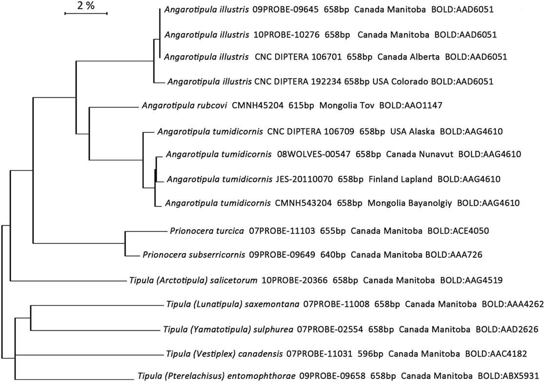
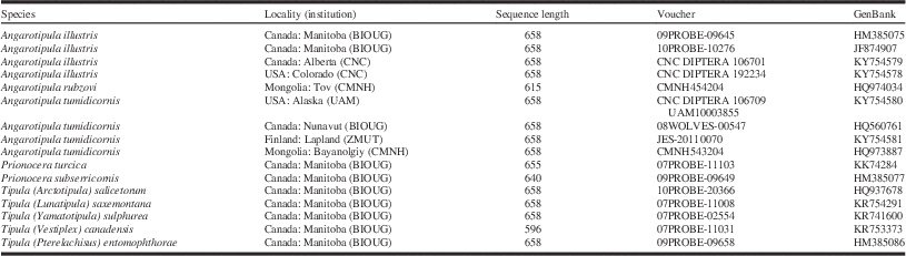
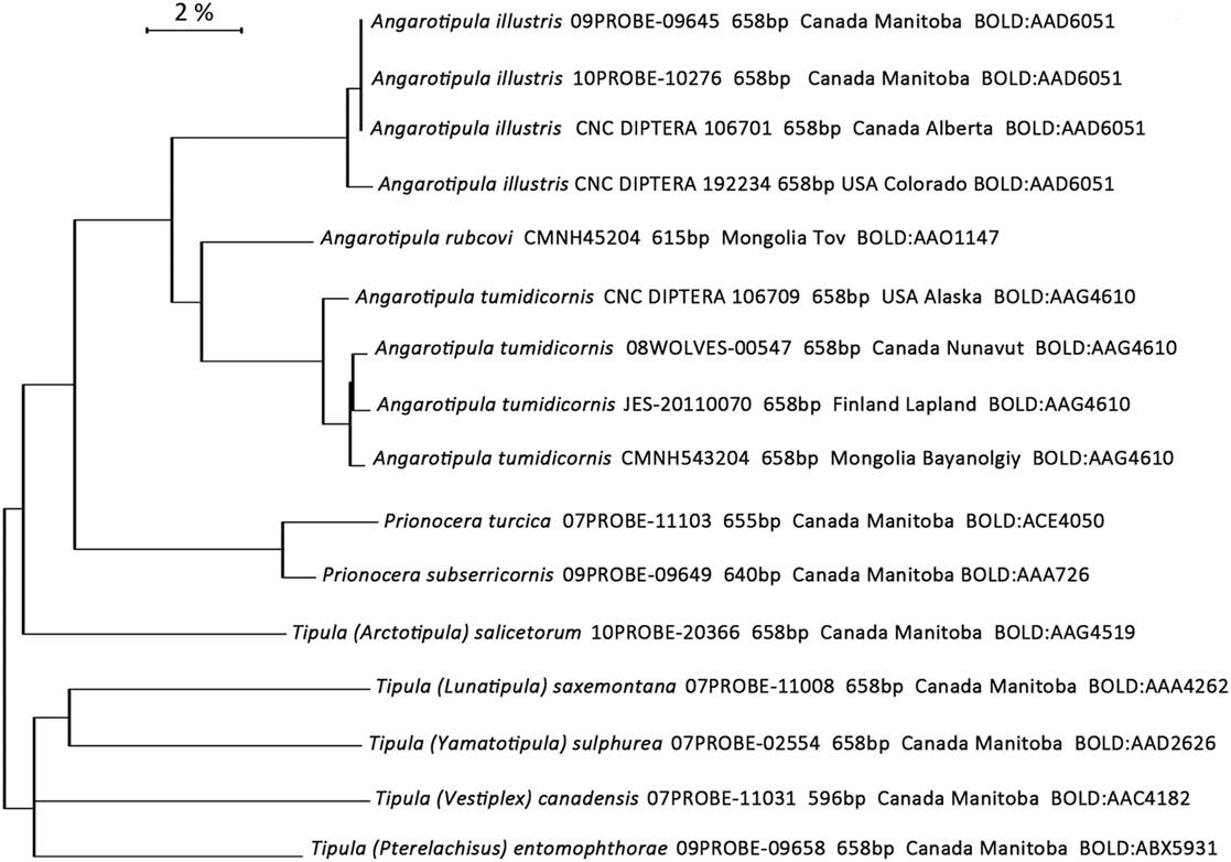
Specimens of A. tumidicornis, A. illustris (Doane, Reference Doane1901), A. rubzovi (Savchenko, Reference Savchenko1961), and various species of Prionocera and Tipula had been submitted for barcoding to the Biodiversity Institute of Ontario in Guelph, Ontario, Canada. A single leg was removed from each specimen to obtain mitochondrial DNA barcodes ((658 base pairs from the 5' end of cytochrome c oxidase subunit I (COI)). The DNA was extracted, amplified, purified, and sequenced following the protocols given by Hajibabaei et al. (Reference Hajibabaei, deWaard, Ivanova, Ratnasingham, Dooh and Kirk2005). Successful COI sequences are deposited in GenBank (Table 1). Cytochrome c oxidase subunit I sequences of >596 base pairs were analysed using neighbour joining (Fig. 1) created in the Barcode of Life Data Systems (BOLD) using the BOLD Aligner (Amino Acid based HMM) (Ratnasingham and Hebert Reference Ratnasingham and Hebert2007).
Neighbour-joining tree of four specimens of Angarotipula illustris, one specimen of Angarotipula rubzovi, four specimens of Angarotipula tumidicornis, two specimens of Prionocera, and five specimens of Tipula with cytochrome c oxidase subunit I (COI) sequences of >596 base pairs (bp).
Voucher information for Angarotipula and other specimens that were submitted for barcoding, including geographic locality, institution housing the specimen (in parentheses), sequence length in base pairs, unique voucher number, and GenBank (www.ncbi.nlm.nih.gov/genbank/) number.

Pupae, larvae, and larval exuviae are best kept in alcohol. Occasionally the alcohol evaporates from a vial leaving shrivelled specimens. These can be retrieved by soaking them in 10% potassium or sodium hydroxide for 24 hours or longer, washing in water, further clearing in lactic acid if warranted, and then storing in alcohol. This is essentially the same procedure used for preparing adult genitalia, except that the genitalia are stored in glycerin.
The terminology describing the male genitalia follows Gelhaus (Reference Gelhaus2009) who strove to provide consistency of morphological terminology in Tipulidae with that across the Diptera (following Cumming and Wood Reference Cumming and Wood2009). The dorsum of the genital capsule is, therefore, referred to as the epandrium rather than the ninth tergite and the ventral part as the hypandrium instead of ninth sternite. This change in terminology is particularly appropriate for the Tipulidae (and the Cylindrotomidae) because the gonocoxites are fused to, and so part of, the hypandrium. Similarly, for consistency, I am adopting the terminology of Ribeiro (Reference Ribeiro2006) and use the terms clasper and lobe of gonostyli instead of inner and outer gonostyli, respectively.
The terminology for describing the larvae follows Gelhaus (Reference Gelhaus1986), Podeniene et al. (Reference Podeniene, Gelhaus and Oyunchuluun2006), and Neugart et al. (Reference Neugart, Schneeberg and Beutel2009) and the terminology for describing the pupae follows Podeniene et al. (Reference Podeniene, Gelhaus and Oyunchuluun2006) and Borkent (Reference Borkent2012).
For citing label data on specimens, a slashed line (/) separates the lines of information on a label and a semi-colon (;) is used to separate the different labels on the specimen. Square brackets ([ ]) are used to indicate additional information not on the original label.
The genus Angarotipula Savchenko, Reference Savchenko1961
Savchenko (Reference Savchenko1961) erected the subgenus Angarotipula to accommodate what he recognised to be a closely related and systematically isolated group (Angara refers to eastern Siberia, the presumed origin of this taxon that Savchenko [Reference Savchenko1961] suggested is part of an ancient Angarian fauna). He selected Tipula tumidicornis Lundström, Reference Lundström1907 as the type species and included T. altivolans (Alexander, 1935), T. laetipennis (Alexander, 1935), T. rubzovi Savchenko, Reference Savchenko1961, and T. indica Edwards, 1926 (with reservations). Here, 14 species are recognised in Angarotipula: the five mentioned above plus A. aspina Liu and Yang, Reference Liu and Yang2010, A. biprocessa Liu and Yang, Reference Liu and Yang2010, A. frommeri (Alexander, 1966), A. heilongjiangana Yang and Yang, 1995, A. illustris (Doane, Reference Doane1901), A. qinghaiensis Yang and Yang, 1996, A. snodgrassiana (Alexander, 1966), A. tokunagana (Alexander, 1964), and A. xuthoptera (Alexander, 1966). Six of these species are known only from China, five are from India, and one of these (A. indica) is also known from Laos. Angarotipula tumidicornis is herein synonymised with A. parrioides, and is recognised as having a Holarctic distribution that includes Mongolia and northeastern China; A. illustris is restricted to the Nearctic and A. rubzovi to the Palaearctic.
Savchenko (Reference Savchenko1961) noted that Lundström (Lundström and Frey Reference Lundström and Frey1916) had reconsidered the taxonomic position of A. tumidicornis, and moved it from Tipula to Prionocera. Four other species now considered to belong to Angarotipula were originally described in Prionocera: A. altivolans, A. laetipennis, A. indica, and A. illustris (as Stygeropis fuscipennis Loew, Reference Loew1866 , Stygeropis being a synonym of Prionocera). The commonality was the lack of verticils on the flagellomeres.
Savchenko (Reference Savchenko1961) adequately distinguished both the males and females of Tipula (Angarotipula) from the other subgenera of Tipula, but for clarity and because the original description is in Russian, a redescription is presented below.
Description of Angarotipula adults
Diagnosis. Flagellomeres lacking verticils; 1, 1, 2 tibial spur formula; small genitalia in both sexes; medium-sized tipulids with brownish-yellow abdomen and wings tinted yellowish brown without other markings.
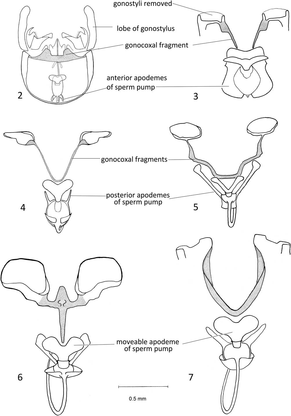
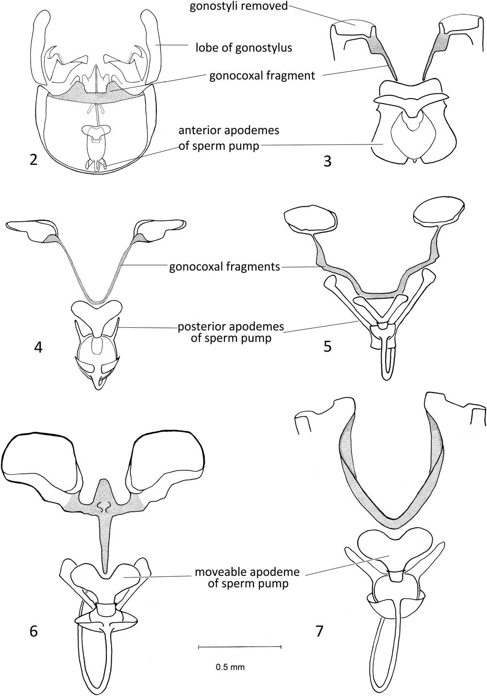
Description. Male: Body size 10–17 mm; wing length 12–17 mm; head and thorax generally greyish brown, abdomen yellowish brown; wings tinted yellowish brown. Head with moderately long rostrum with prominent nasus; antenna with 13 antennomeres covered in short, dense setae; flagellomeres lacking verticils, broadened ventrally near middle of flagellomere. Thorax somewhat flattened dorsally with short, inconspicuous setae; longer setae on anatergite and metakatepisternum. Wings pale yellowish brown; stigma barely discernible; veins brown, media, cubital and anal veins generally bare. Legs of moderate length for family; tibial spur formula 1, 1, 2, (outer spur on mesothoracic leg absent). Genitalia short, prominent sutures separating epandrium from hypandrium; hypandrium sclerotised ventrally, without membranous medial separation; 8th sternite simple. Internal structures (Fig. 2): gonocoxal fragment comprising distal horizontal connection across hypandrium, extending medially around base of gonostyli; sperm pump with narrow, elongate anterior apodeme and shallow posterior apodeme; aedeagal guide (adminiculum) small, tripartite structure, attached to hypandrium distal to sperm pump; central section with overlapping edges dorsally forming tube for aedeagus. Aedeagus emerging anteriorly from sperm pump, curving ventrally, continuing through central portion of aedeagal guide.
Male hypandrium, dorsal view with epandrium removed, gonocoxal fragments marked by stippling. 2. Angarotipula illustris; 3. Prionocera subserricornis (Zetterstedt, 1851) with epandrium, gonostyli, aedeagus, and aedeagal guide removed; 4. Tipula (Arctotipula) salicetorum Siebke, 1870 with epandrium, gonostyli, aedeagus, and aedeagal guide removed; 5. Tipula (Pterelachisus) entomophthorae Alexander, 1918 with epandrium, gonostyli, aedeagus, and aedeagal guide removed; 6. Tipula (Vestiplex) bergrothiana Alexander, 1918 with epandrium, gonostyli, aedeagus, and aedeagal guide removed; 7. Tipula (Yamatotipula) furca Walker, 1848 with epandrium, gonostyli, aedeagus, and aedeagal guide removed.
Female: Similar to male but generally slightly larger, body size: 11.0–17.5 mm, wing length: 15–17 mm. Flagellomeres almost cylindrical. Genitalia short, lightly sclerotised, narrower than preceding abdominal segments; 10th tergite about as long as wide; cercus broad, oriented laterally, with blunt rounded tip; 8th sternite bears short, triangular hypogynial valves barely reaching base of cerci.
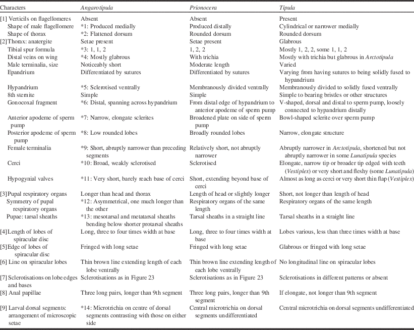
Remarks. Fourteen shared characters of the genus Angrotipula and nine shared characters of the genera Angarotipula and Prionocera are discussed below and listed in Table 2.
Morphological comparisons of the genera Angarotipula, Prionocera, and Tipula.
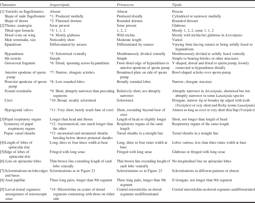
Note: Numbers in square brackets [ ] refer to shared characters of Angarotipula and Prionocera. An asterisk (*), colon with a number indicates a diagnostic character of Angarotipula.
The gonocoxal fragment in the male is so named because it appears derived and modified from the dorsal surface of the gonocoxites that have become reduced internal structures covered by the epandrium. A character of Angarotipula is the horizontal nature of the gonocoxal fragment with no anterior extension towards the sperm pump. In adult male Tipula species, by contrast, the gonocoxal fragment is a V-shaped sclerite connecting the inner edges of the hypandrium, just anterior to the insertion of the gonostyli and extending anteriorly to the sperm pump (Figs. 4–7). In Prionocera, a gonocoxal fragment extends from the distal medial edge of the hypandrium to the posterior apodeme of the sperm pump (Fig. 3).
Dobrotworsky (Reference Dobrotworsky1968) described and illustrated similar sclerites under the epandrium in the Australian genus Ptilogyna Westwood, 1835, and applied the term “genital bridge” for just that section in the centre that connects the two sides and lies just posterior and dorsal to the sperm pump (vesica). He noted that “In Australian species of Dolichopeza and Nephrotoma the sclerotisations arising from the coxites are short and no genital bridge is formed”. This suggests that Dobrotworsky also considered these sclerites, at least the lateral parts arising from the coxites, to be gonocoxal in origin.
Neumann (Reference Neumann1958), however, considered these sclerites to be derived from the genital chamber and he referred to the lateral sclerite as sp2 and the medial sclerite as sp1 (an abbreviation of Skleritespangen). Oosterbroek (Reference Oosterbroek1980) and Tangelder (Reference Tangelder1985), in describing Nephrotoma Meigen, 1803, followed Neumann’s terminology but they considered the bridge to be homologous with the Fp-Fp of Trichocera Meigen, 1803, the lateral plates of the parameres. Cumming and Wood (Reference Cumming and Wood2009) define parameres as a pair of posteriorly directed processes each attached to a gonocoxal apodeme and they may be fused medially. The gonocoxal fragment, however, is attached distally, just anterior to the attachment of the gonostyli, not anteriorly where the gonocoxal apodeme would be. The term gonocoxal fragments includes the previously mentioned sclerites (sp2) as well as the “genital bridge” (sp1) if there is one.
The more-or-less spherical sperm pump in Tipulidae has immoveable sclerites attached to it, quite separate and distinct from the moveable, or ejaculatory apodeme. These immoveable sclerites were referred to as apodemes (Brodo Reference Brodo1987). Sinclair et al. (Reference Sinclair, Cumming and Brooks2013) studying the male terminalia of Diptera, based on muscle attachments between the fixed apodeme and the ejaculatory apodeme, concluded that the fixed sclerites are parameres. This is an intriguing idea because as Ribeiro (Reference Ribeiro2008) shows in the Limnophilinae (Diptera: Limoniidae), parameres are structures closely associated with the sperm pump that is at the base of the aedeagus. The parameres, however, also have a lateral connection to a gonocoxal apodeme. In Tipulidae, the apodemes are part of the sperm pump and lack any connection to the hypandrium (or gonocoxites). More study is needed to establish that these are parameres and for now I agree with Wood (Reference Wood1991) that the Tipulidae lack parameres.
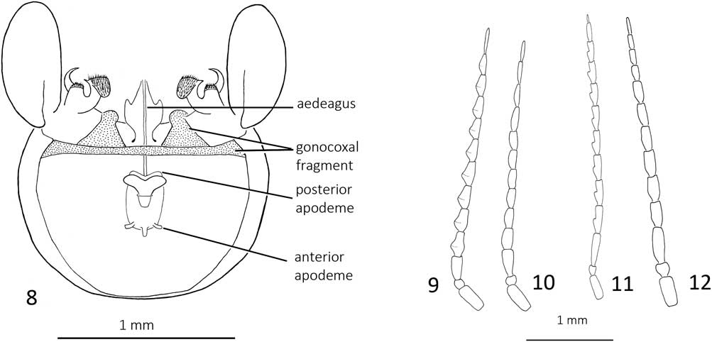
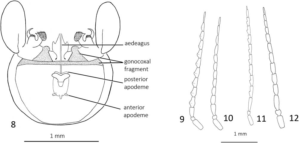
Characters of Angarotipula are the narrow, elongate anterior apodeme and shallow posterior apodeme on the sperm pump (Figs. 2, 8), whereas in Tipula the posterior apodeme is elongate and the anterior apodeme is generally cup shaped, surrounding the anterior end of the sperm pump (Figs. 4–7). In Prionocera the sperm pump is flanked by a broad apodeme that extends slightly anterior to the sperm pump (Fig. 3).
8. Angarotipula tumidicornis, hypandrium, dorsal view with epandrium removed; 9. Male Angarotipula tumidicornis, antenna; 10. female Angarotipula tumidicornis, antenna; 11. male Angarotipula illustris, antenna; 12. female Angarotipula illustris, antenna.
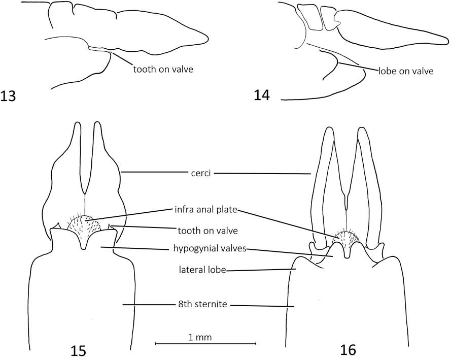
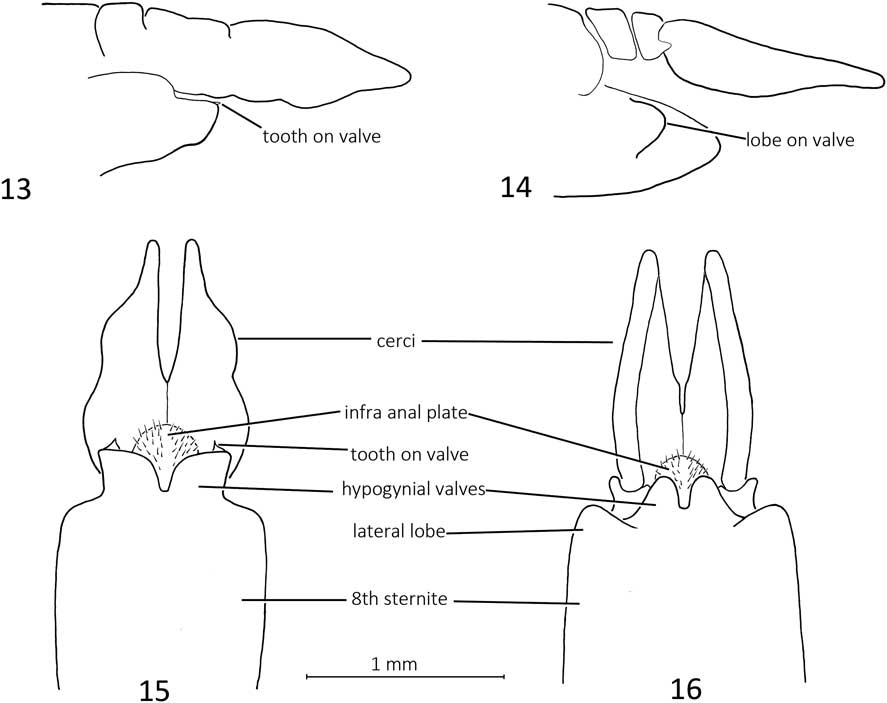
Female terminalia. 13. Angarotipula illustris, left lateral aspect; 14. Angarotipula tumidicornis, left lateral aspect; 15. Angarotipula illustris, ventral aspect; 16. Angarotipula tumidicornis, ventral aspect.
Angarotipula is recognised by having a flatter thoracic dorsum; in other Tipulidae the dorsum is more rounded. Setae on the metakatepisternum are not ubiquitous in the Tipulidae but they do occur in Prionocera and in other genera. Setae on the anatergite, however, are restricted to Prionocera, Brachypremna Osten Sacken, 1887, and Megistocera Wiedemann, 1828 in the Tipulidae. Setae on the anatergite also occur in Ptychopteridae (Ptychoptera Meigen, 1803, Bittacomorphella Alexander, 1916, and Bittacomorpha Westwood, 1835), therefore setae on the anatergite may be part of the ground plan of the Tipulidae rather than a shared character of Angarotipula, Prionocera, Brachypremna, and Megistocera. A lack of setae on the distal branches of R, M, and Cu was noted by Savchenko (Reference Savchenko1961) and is probably a synapomorphy of Angarotipula. The latter state, however, also occurs in the subgenus Tipula (Arctotipula), suggesting independent loss of these setae. A tibial spur formula of 1, 1, 2 is not unique to Angarotipula; the loss of the outer spur on the mesothoracic leg also occurs in some species of Tipula [e.g., T. (Lunatipula) mainensis Alexander, 1915 and T. (Yamatotipula) concava Alexander, 1926]. Short female genitalia are found in some females of T. (Lunatipula) Edwards, 1931 (e.g., T. (L.) fascipennis (Meigen, 1818), T. (L.) mainensis (Alexander, 1915), and T. (L.) megaura (Doane, Reference Doane1901)), but in these species the genitalia are not abruptly narrower than the preceding abdominal segments and the tips of the hypogynial valves generally extend as far as the tips of the cerci.
Diagnosis of Angarotipula pupae
The unusually long and flexible respiratory organ, one being considerably longer than the other, as well as the tips of mesotarsal and metatarsal sheaths curving beneath the shorter sheaths of the protarsi distinguish these pupae from others in the Tipulidae (Fig. 17).
Diagnosis of Angarotipula final instar
The later instars of the two North American species of this genus are distinguished by a pale, longitudinal line on the dorsal abdominal segments, flanked by a darker line, quite evident in A. tumidicornis but fainter in A. illustris. This central line is made up of microtrichia that are different, or differently arranged, from the lateral microtrichia. The spiracular disc has six very long lobes, two to three times their width at base, fringed with long setae and with a thin brown line extending the length of the lobe; three pairs of long anal papillae are present.
Remarks. Angarotipula and Prionocera larvae have similar spiracular discs and similarly long anal papillae and so these genera are difficult to distinguish. The longitudinal dorsal line formed by the different character of the central microtrichia differentiates Angarotipula from Prionocera, at least in North America.
Angarotipula tumidicornis (Lundström, Reference Lundström1907)
Tipula tumidecornis Lundström, Reference Lundström1907: 23–24.
Tipula tumidicornis; Lundström Reference Lundström1907: erratum (spelling corrected)
Prionocera tumidicornis; Lundström and Frey Reference Lundström and Frey1916: 18–19.
Tipula tumidicornis; Lackshewitz Reference Lackshewitz1936: 298.
Tipula (Vestiplex) tumidicornis; Mannheims Reference Mannheims1953: 136; Mannheims Reference Mannheims1954: 45.
Tipula (Angarotipula) tumidicornis; Savchenko Reference Savchenko1961: 348; Mannheims Reference Mannheims1965: 3; Mannheims Reference Mannheims1967: 156; Alexander Reference Alexander1965a: 337; Lantsov and Chernov Reference Lantsov and Chernov1987: 27.
Stygeropis parrioides Alexander, Reference Alexander1919: 9–10c new synonymy.
Tipula (Arctotipula) parrioides; Alexander Reference Alexander1944: 95.
Tipula (Angarotipula) parrioides; Alexander Reference Alexander1965a: 337; Alexander Reference Alexander1965b: 30; Brodo Reference Brodo1990: 473.
Notes on synonymy. Tipula tumidecornis was described by Lundström (Reference Lundström1907) based on 3 males collected in northern Finland (Li. Utsjoki, 25.vi.1894, J. Sahlberg; Lkem. Muonio, 8.viii.1867, J. Sahlberg; Ob. Uleåborg, W. Nylander [no date]). The name was emended to T. tumidicornis by Lundström in an erratum included with the same paper and according to the International Commission on Zoological Nomenclature (1999, Article 32.5.1.1), T. tumidicornis should be accepted as the correct spelling. Two male specimens from ZMH were examined and neither one had the complete data as mentioned in the original description. The male specimen collected by J. Sahlberg from Lapponia is designated as lectotype.
Alexander (Reference Alexander1919) described Stygeropis parrioides from Alaska (west of Konganevik, Camden Bay) based on seven males. Stygeropis is a synonym of Prionocera and S. parrioides was originally placed in Stygeropis because of the lack of verticils on the antennae.
Type material examined. Tipula tumidicornis: LECTOTYPE male (here designated in order to fix identity of the species), labelled: [white, printed] Lapponia; [white, printed] J. Sahlbg.; [white, ink] 1006.; [white, red ink] Tipula / tumide / cornis; [white, printed and ink] Mus. Zool. H:fors / Spec. typ. No. 4088/Tipula tumi-/decornis Lundstr.; [red, ink]LECTOTYPE / Tipula/tumidicornis / Lundström / designated by / Brodo 2016 (ZMH). This specimen has the entire abdomen, both antennae, and both wings intact but lacks all legs.
PARALECTOTYPE male, labelled: [white, printed] Uleåborg; [white, printed] W. Nylander; [white, red ink] Tipula / tumide / cornis; [white printed and ink] Mus. Zool. H:fors / Spec. typ. No. 4089 / Tipula tumide / cornis Lundstr. (ZMH). This specimen is now double mounted on a piece of foam (the original pin was bent), it lacks the abdomen, has only seven flagellomeres on the left antenna and six on the right; left wing tip and entire right wing are mounted on a point.
A male specimen borrowed from USNM had the following four labels: [white, printed] Kantalaks; [white, printed] R. Frey; [blue, printed and ink] PARATYPE / Prionocera tumidecornis Lundst.; [pink, printed and ink] Spec. typ. No…… / Tip. (Prionocera) / tumidecornis Lundstr. This specimen was not mentioned in the original description therefore it should not be considered a paralectotype.
Stygeropis parrioides: HOLOTYPE male, labelled: [white, ink] W of KANGENGEVIK / Camden Bay / Alaska June; [white, printed] Canadian / Arctic / Expedition/ F.J. 1914; [red, printed and ink] HOLOTYPE / Stygeropis / parrioides / C.P. Alexander (CNC). The holotype has both antennae; right wing is missing, left wing tip missing; legs: left prothoracic leg, left mesothoracic leg, and right mesothoracic leg remain; abdomen is intact. PARATYPES: four males, same data as holotype (CNC). One male paratype has the genitalia in a vial pinned with the specimen, one has genitalia intact and two paratypes are missing their abdomens.
Remarks. Alexander (Reference Alexander1919) corrected the spelling of the type locality to Kongangevik. I have compared the types of A. tumidicornis and A. parrioides and conclude that these are synonyms. The neighbour-joining tree (Fig. 1) indicates that the specimens from Canada, Mongolia, Finland, and Alaska are conspecific. There is less than 1% genetic divergence between the Alaskan specimen and the other three specimens of A. tumidicornis. All four specimens were assigned the same barcode index number (BIN) AAG4610.
In their key to species of Angarotipula (Liu and Yang Reference Liu and Yang2010), A. parrioides was differentiated from A. tumidicornis (and from the rest of this genus) by cell m1 in the wing being sessile. This is true of the holotype that was described and illustrated by Alexander (Reference Alexander1919). There is considerable variability in the presence or absence of a stem for cell m1 and for the length of that stem. The wings of several specimens from North America have cell m1 petiolate and some northern European specimens that I have seen have cell m1 stemless. This distinction, therefore, does not hold.
Additional material examined. Canada. British Columbia. Chilkot Pass, 17.vii.1948, Mason and Hughes (1♀, CNC). Manitoba. Churchill, 25.vi.1952, Chillcott (1♂, CNC). Northwest Territories. Aklavik, 16–20.vi.1953, Bird (1♂, CNC); Ford Lake, 63°11'N, 107°19'W, 26.vi.1966, Shewell (1♀, CNC); Reindeer Depot, 2.vii.1948, Vockeroth (2♂, CNC); Tuktoyaktuk, 21 mi E, 10–25.vi.1971, Wood (2♂, CNC); Tuktoyaktuk, 21 mi E, 2–5.vii.1971, Wood (4♂, CNC). Nunavut. Baker Lake [Qaman’tuaq], 26.vii.1947, Freeman (2♀, CNC); Bathurst Inlet, 23.vi.1951, Campbell (2♂, CNC); Boothia Peninsula, Lady Melville Lake, 69°23'N, 93°15'W, 3.vii.1951, Chillcott (3♀, CNC); Spence Bay [Taloyoak], 1–8.vii.1951, Chillcott (6♂, 2♀, CNC); Bylot Island, west, Oikoqtaaluk, 73.1572°N, 79.9495°W, 12 m, Malaise pitfall trap, 3–4.vii.2008, Bolduc and Patenaud, 08WOLVES-00524 (1♀, barcode associated, BIOUG); Bylot Island, west, Oikoqtaaluk, 73.1572°N, 79.9495°W, 12 m, Malaise pitfall trap, 3–4.vii.2008, Bolduc and Patenaud, 08WOLVES-00527 (1♀, barcode associated, BIOUG); Bylot Island, west, Oikoqtaaluk, 73.1572°N, 79.9495°W, 12 m, Malaise pitfall trap, 3–4.vii.2008, Bolduc and Patenaud, 08WOLVES-00549 (1♀, barcode associated, BIOUG); Chesterfield, 9–25.vii.1950, Chillcott (18♂, 10♀, CNC); Chesterfield, 5–29.vii.1950, Vockeroth (6♂, 2♀, CNC); Chesterfield, 27.vi–10.vii.1951, Downe (1♂, 1♀, CNC); Coppermine [Kugluktuk], 14.vii.1951, Hicks (2♀, CNC); Eskimo Point [Arviat], 1.vii.1950, Roberts (9♂, 3♀, CNC); Padlei, 22.vii.1950, Hennigar (1♂, 2♀, CNC); Southampton Island [Shugliaq], Coral Harbour, 30.vi.1952, Ehrlich (60♂, 6♀, CNC); Southampton Island [Shugliaq], Coral Harbour, 5.vii.1948, Shewell (8♂, 4♀, CNC); Victoria Island, 71°17'N, 114°W, 1–10.vii.1975, Wood (1♂, 1♀, CNC); Victoria Island, 71°17'N, 114°W, 1–10.vii.1975, Wood, CNC DIPTERA 106718 (1♂, CNC); Cambridge Bay, 12.vii.1950, Smith (1♀, CNC); Cambridge Bay, 11.vii.1950, Sweatman (10♂, 1♀, CNC).
Finland. Lapland. Muonio [67°57'N, 23°41'E], Wegelius (1♀, ZMH); Pallastunturi [68°05'N], 14.vii.1951, Wegelius (1♀, ZMH); Pallastunturi [68°05'N], 20–22.vi.1953, Wegelius (2♀, ZMH); Pallastunturi [68°05'N], 30.vi.57, Wegelius (3♀, ZMH); Pallastunturi [68°05'N], 26.vii.1951, Kaisila (1♀, ZMH); Rovaniemi [66°39'N 24°32'E], Sahlberg (1♀, ZMH).
Russia. Chukotski Peninsula, Shmidt [Cape], 18.vii.1963, Gorodkov, CNC DIPTERA 106727 (1♂, CNC).
Sweden. Abisko, 8.vii.1951, Vockeroth (1♂, CNC).
United States of America. Alaska. Arctic National Wildlife Refuge (ANWR), Sheenjek River near Last Lake, 690–700 m, 68.61181°N, 143.72828°W±2 km, on aufeis, 5.vii.2000, Johnsen, CNC DIPTERA 192230, UAM100038354 (1♂, barcode associated, UAM); Arctic National Wildlife Refuge (ANWR), Sheenjek River near Last Lake, 690–700 m, 68.61181°N, 143.72828°W±2 km, on aufeis, 5.vii.2000, Omtzigt, CNC DIPTERA 192231, UAM100038388 (1♂, barcode associated, UAM); Sheenjek River Camp 2000, on aufeis, 28.vi.2000, Omtzigt, CNC DIPTERA 106709, UAM10003855 (1♀, UAM); Sheenjek River Camp 2000, on aufeis, 28.vi.2000, Omtzigt, CNC DIPTERA 192232, UAM100038361 (1♀, barcode associated, UAM); Meade River, vii.1953, W. Maher (1♂, 6♀, UMMZ); Point Barrow, vii.1953, Maher (1♂, 1♀, UMMZ); Umiat, 3.vii.1959, Martin (1♀, CNC); Utukok River, 20.vii.1953, Maher (1♂, UMMZ).
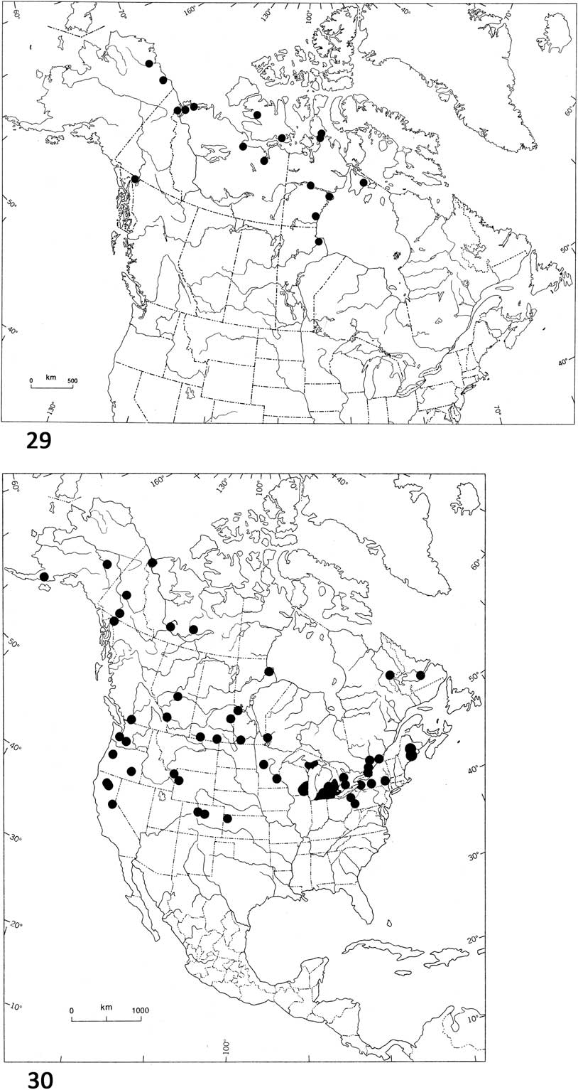
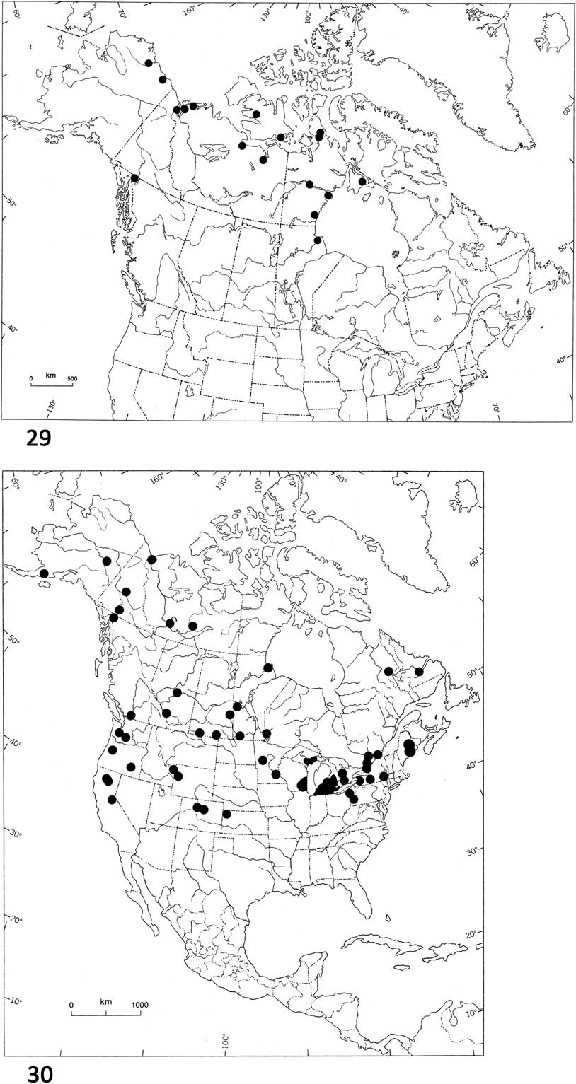
Distribution. Angarotipula tumidicornis is a species of the wetlands in the low Arctic, prevalent on the mainland and on the more southern Canadian Arctic Islands and it has been recorded from Alaska, United States of America (Fig. 29). Its distribution overlaps that of A. illustris at the Mackenzie Delta, Northwest Territories, Chilkot Pass – Atlin area of northern British Columbia, and Churchill, Manitoba. It has been found in the Arctic regions of Scandinavia, Russia, and Mongolia. Lantsov and Chernov (Reference Lantsov and Chernov1987) classified this species as hypoarctic; it was abundant in the more southern tundras of Russia and an important food source for fishes and birds.
Distribution of Angarotipula tumidicornis in North America. Fig. 30. Distribution of Angarotipula illustris in North America.
Remarks. The only record I have from Churchill, Manitoba is a single male collected in 1952. Despite intense collecting in the Churchill area in 1977 and 1978 and again from 2007 to 2010 (the latter for barcoding all species in this area), A. tumidicornis was never found again and probably no longer occurs in the Churchill area.
Redescription of Angarotipula tumidicornis adults
For illustrations see Alexander (Reference Alexander1919: 26c, fig. 13 (male antenna), fig. 22 (lobe of gonostylus), fig. 25 (clasper of gonostylus), fig. 27 (epandrium)); Oosterbroek (Reference Oosterbroek2017, male and female terminalia)); Savchenko (Reference Savchenko1961, fig. 207 (female terminalia), fig. 211 (male terminalia)).
Diagnosis. Flagellomeres without verticils; brown abdomen with contrasting darker longitudinal stripe; males with round claspers of the gonostyli; females with short, triangular hypogynial valves flanked by a small lateral lobe.
Description. Male: (Figs. 8–9) Body length 10.3–12.6 mm; wing length 12.6–14.4 mm. Head with rostrum short, light brown, covered with short dark setae extending across prominent nasus; vertex dark grey medially, lighter brown laterally, with short dark setae; post gena with longer setae. Antenna dark brown, scape and 1st flagellomere subequal, cylindrical, densely covered with short setae and with scattered longer setae; pedicel glabrous; flagellomeres lacking verticils, densely covered with short setae; flagellomeres 2–9 widest in middle, smoothly narrowing distally; 10th and 11th narrower, cylindrical, elongate. Thorax somewhat flattened, yellowish grey with darker grey narrow median stripe, setae short, pale yellow; transverse suture shiny dark brown; scutellum and mediotergite dusky yellowish brown with short setae; pleura yellowish grey, bare except for pale setae on katepisternum, anatergite and metakatepisternum. Wing tinted yellowish brown, slightly darker on costal edge and stigmal region; veins brown; short dark trichia on costa, radius and scattered trichia on R4+5, other veins bare. Halter dusky brown, lighter at tip. Legs: coxae silvery grey, with pale setae; trochanters and femora yellowish brown with short adpressed dark brown setae; tibia and tarsi gradually darker brown, tibial spur formula 1, 1, 2, outer spur on mesotibia missing; claws narrow, simple. Abdomen brown to yellowish brown with darker brown medial dorsal stripe, short dark brown setae on dorsal sclerites directed anteriorly, setae on ventral segments directed posteriorly. Terminalia short, bent dorsally, 8th sternite longer than 8th tergite, hypandrium longer than epandrium, sutures separating latter sclerites; epandrium thickly setose, setae directed anteriorly, shallowly emarginate distally with edge narrowly bare, shiny brown. Gonostylus with clasper short, elongate horizontally, with lateral lobe, covered with short setae except for glabrous anterior tip of triangular beak and short sharply curved posterior hook; lobe of gonostylus round, with setae on both sides, setae directed anteriorly. Proctiger with two black setose areas on membrane visible in dried specimens. Gonocoxal fragment extending across hypandrium distally in straight line, with triangular medial section extending posteriorly to ventral medial edge of hypandrium, lateral to tripartite aedeagal guide.
Female: (Figs. 10, 14, 16) Similar to male except as follows. Body length 11.0–17.5 mm, wing length 15–17 mm. Antenna with 1st flagellomere subequal to scape, slightly broader distally; flagellomeres 2–8 shorter than 1st, very slightly broader ventrally, just below middle; flagellomeres 9–11 narrower, cylindrical and slightly longer than preceding flagellomeres. Abdomen dorsally, yellowish brown with dark brown contrasting central stripe, short setae on segments directed posteriorly. Genitalia short, narrower than preceding abdominal segments; 9th tergite about half length of 8th tergite; cercus narrow dorsally, broad laterally with ventral edge, smoothly curved, narrowing to rounded distal tip, small setose lobe at base of cercus; hypogynial valve short, extending to base of cercus, flanked by short, rounded lobe.
Description of Angarotipula tumidicornis pupae
Material examined. All in alcohol, CNC. Canada. Nunavut. Chesterfield Inlet, 9.vii.1950, Chillcott (1♀ pupal exuviae, adult); 5.vii.1950, Vockeroth (1♂ pupal exuviae, pinned adult); Coppermine [Kugluktuk], 27.vi.1951, Hicks (4♀ pupae; 3♂, 1♀ pupal exuviae); Coral Harbour, 1.vii.1948, Shewell (2♂, 1♀ pupal exuviae); Eskimo Point [Arviat], 24.vi.1950, Roberts (2♀ pupae); Spence Bay, 27.vi.1951, Downe (1♀ pupal exuviae, pinned adult).
Sweden. Abisko, 10.vii.1951, Vockeroth (1♂, 2♀, pupal exuviae retrieved from Carex Linnaeus (Cyperaceae) pond).
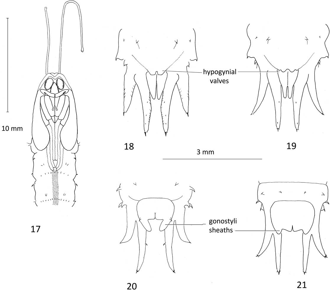
Diagnosis. Very long, unequal, respiratory organs, 9–14 mm for the longer respiratory organs and 5–7 mm for the shorter; tips of mesotarsal and metatarsal sheaths curving beneath shorter sheaths of protarsi. Darker longitudinal line both dorsally and ventrally (Fig. 17).
Angarotipula pupae. 17. Angarotipula tumidicornis, anterior segments, ventral aspect; 18. Angarotipula illustris, female terminalia, ventral aspect; 19. Angarotipula tumidicornis, female terminalia, ventral aspect; 20. Angarotipula illustris, male terminalia, ventral aspect; 21. Angarotipula tumidicornis, male terminalia, ventral aspect.
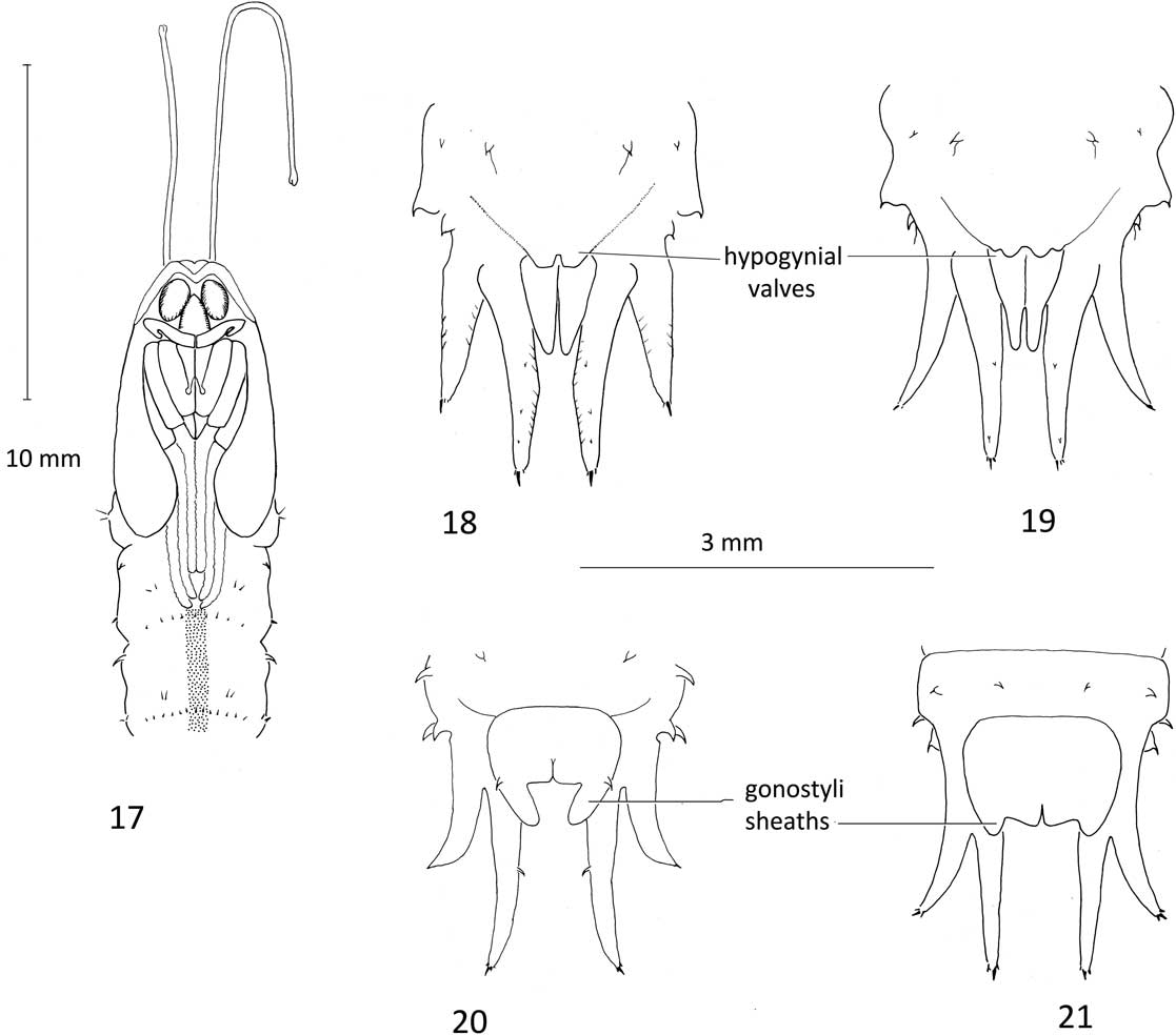
Description. Male length 20–25 mm; female length 22–27 mm. Pupae glabrous, dull pale greyish brown with distinct, darker brown dorsal and lateral stripes on abdomen. Cephalic crest small; maxillary palpus with tip recurved (similar to most other tipulines). Mesonotal respiratory organs long, unequal, tips split into two flaps; shorter respiratory organ (5–7 mm) extending to tip of wing sheaths, longer almost double in length (9–14 mm), about half length of pupa; mesothorax with fine transverse lines arranged in three columns positioned over thoracic stripes of imago, with 4 or 5 widely scattered setae below; sheaths of halter covered by wing sheath. Abdominal segments secondarily divided into anterior and posterior sections, segments 2–7 with similar arrangement of spines: dorsally with fringe of 16–20 tiny spines on posterior section, one or two spines with fine seta attached; laterally with spine on anterior section and two or three smaller spines on posterior section, spines with fine seta attached; ventrally on posterior section, pair of tiny spines on either side of midline, above posterior row of 16–20 slightly larger spines, some with seta attached. Segments 8 and 9 narrower; 9th bearing long sheaths over three pairs of larval spiracular lobes, dorsal sheath shortest, erect, flattened, parallel-sided, terminating in two spines; lateral and ventral sheaths considerably longer, cylindrical, curved, each terminating in three spines, central spine longest. Ventrally, row of four spines anterior to sheath of genitalia, posterior to these, two larger spines laterally, on each side. Male posteriorly with rounded lateral edge of genital sheath over round lobe of developing adult gonostylus (Fig. 21). Female posteriorly with sheath of hypogynial valves indicating rounded tip flanked by smaller lobe (Fig. 19); cercal sheath relatively short, extending half length of ventral spiracular sheath.
Description of Angarotipula tumidicornis last instar
Material examined. All in alcohol, CNC. Canada. Northwest Territories. Tuktoyaktuk, 21 miles E, 2–5.vii.1971, Wood (six larvae). Nunavut. Coppermine [Kugluktuk], 27.vi.1951, Hicks (two larvae); Victoria Island, 71°17'N, 114°W, 1–5.vii.1975, G. Wood and M. Wood (three larvae).
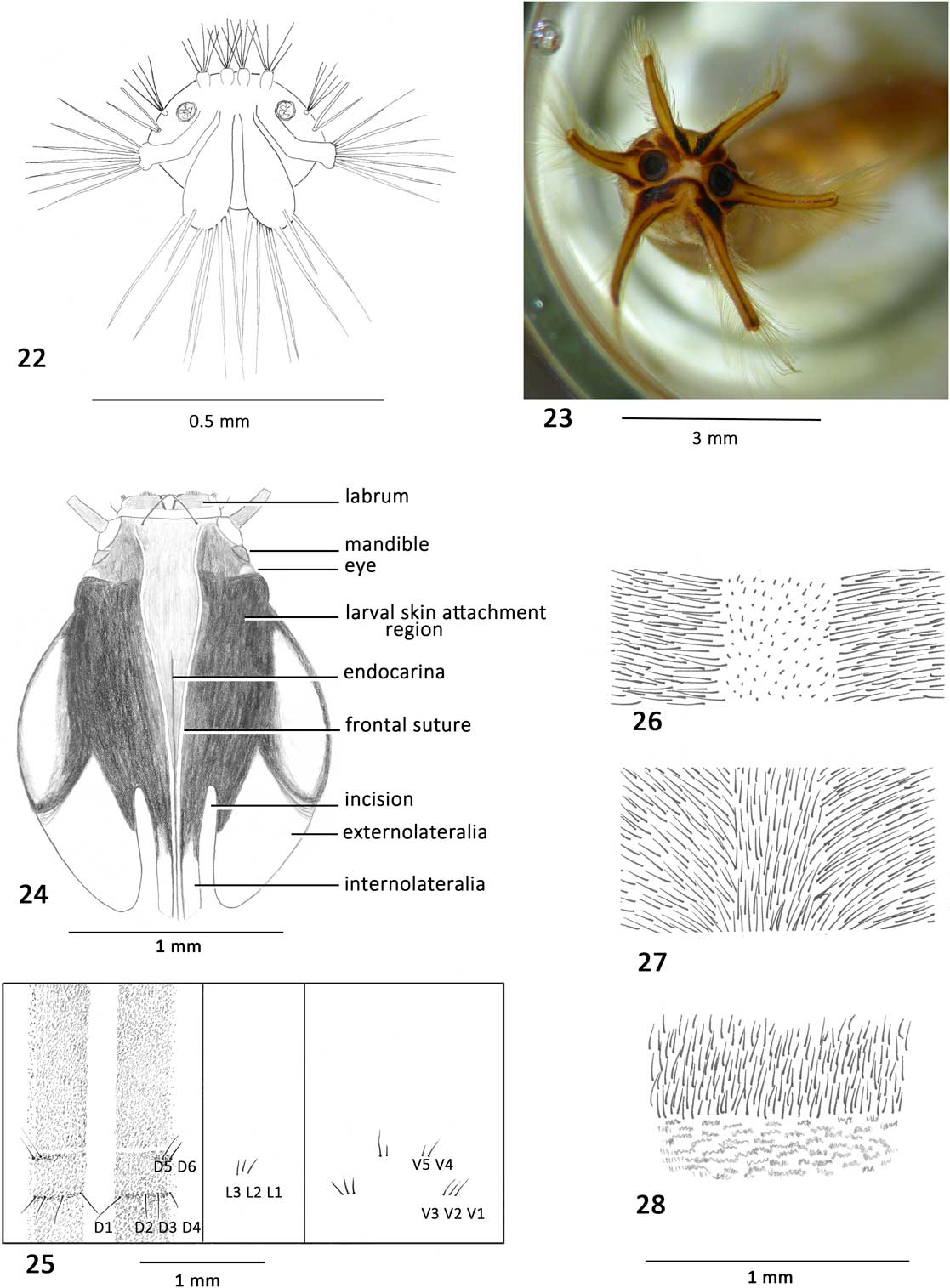
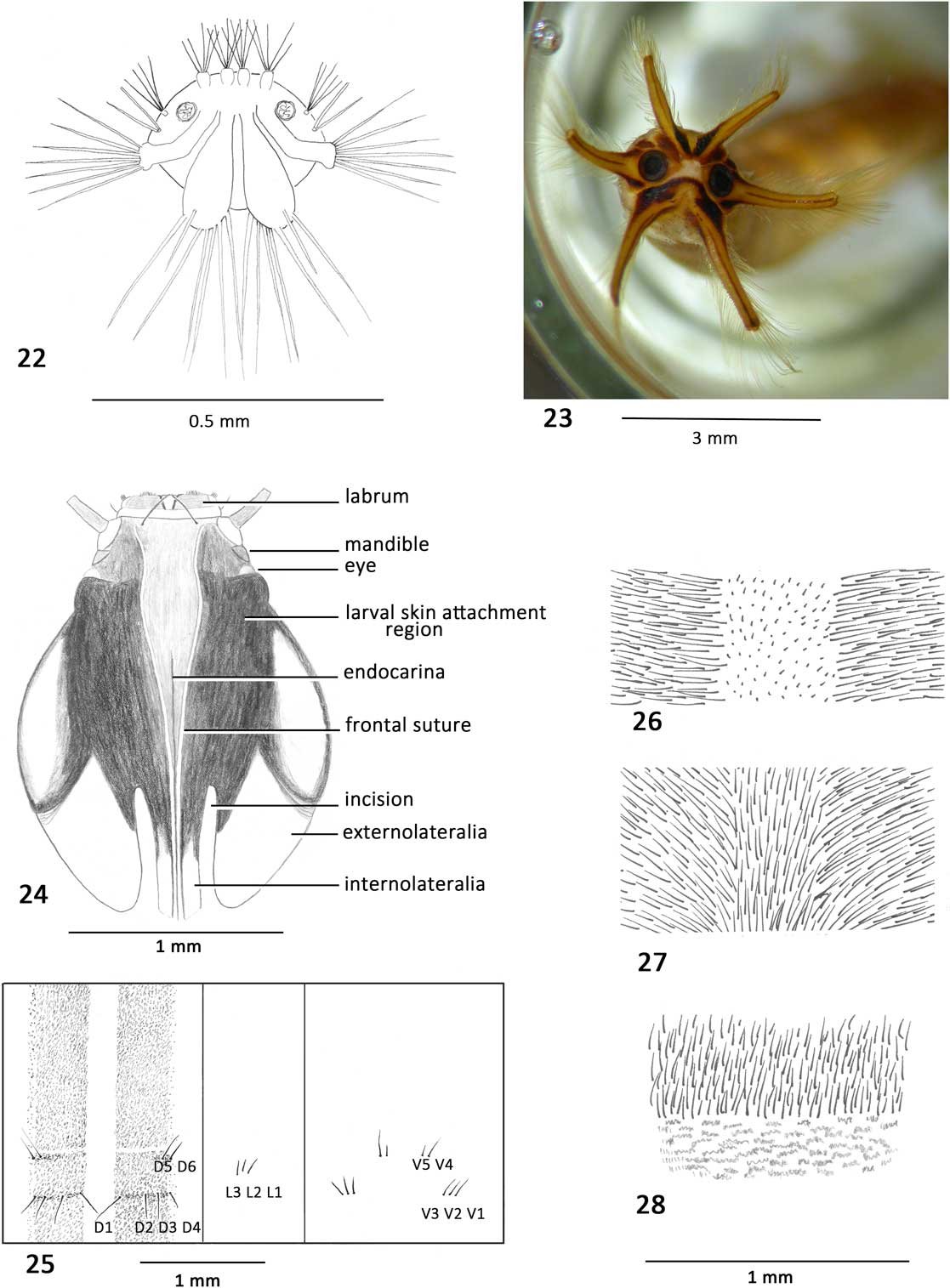
Diagnosis. Pale longitudinal line on dorsum flanked by much darker line, darker line composed of brown microtrichia orientated perpendicular to larval body, pale central area composed of minute microtrichia (Figs. 25–26); six long lobes on spiracular disc fringed with long setae (Fig. 23); three pairs of long anal papillae.
Angarotipula larvae. 22. Angarotipula illustris, 1st instar, spiracular disc; 23. Angarotipula tumidicornis, last instar, spiracular disc; 24. Angarotipula tumidicornis, last instar, head capsule, anterior aspect; 25. Angarotipula tumidicornis, last instar, abdominal segment showing arrangement of setae dorsally, laterally, and ventrally; 26. Angarotipula tumidicornis, last instar, showing arrangement of minute and longer microtrichia on middle section of dorsal abdominal segment; 27. Angarotipula illustris, last instar, showing arrangement of microtrichia on middle section of dorsal abdominal segment; 28. Prionocera dimidiata, last instar, showing arrangement of microtrichia and short microtrichia on middle section of dorsal abdominal segment. Scale line applies to Figures 26–28.
Description. Length 28–33 mm, width 3.2–4.0 mm. Body pale yellowish white with two longitudinal brown stripes dorsally.
Head capsule (Fig. 24). Length 2.2–2.8 mm, width 1.2–1.8 mm. Oval, slightly depressed dorsoventrally, colour varying from black to pale yellow indicating degree of sclerotisation; very heavily sclerotised medially, flanking notal plate, extending half way down distal incision and bordering anterolateral oval area of externolateralia; internolateralia heavily sclerotised anteriorly, fading out distally with tip almost invisible; incision extending about 1/3 length of head; notal plate long, triangular, bounded by frontal sutures, transparent seta on anterolateral corner; endocarina visible on narrowed distal half of notal plate, continuing as narrow, vertical ridge between, and extending as far back as internolateralia. Labrum lightly sclerotised with brush of short setae anteriorly, smaller brush of setae laterally; clypeus narrow, unpigmented, with short, inconspicuous seta laterally; antenna conical, lightly pigmented, on membranous base. Ventral wall of head capsule reduced, foramen occipital widened anteriorly, heavily sclerotised; hypostoma with nine strong, anteriorly directed teeth; mandible heavily sclerotised, with five teeth, lacinia mobilis articulated medially; maxilla with dense microtrichia medially, wedge-shaped cardo bearing two long setae distally, a single seta in membranous area near base of maxilla.
Abdominal segments secondarily divided horizontally, division indicated by pale area, anterior twice length of posterior section (Fig. 25); posterior sections of segments 2–7 with thin brown macrosetae arranged as follows (labelling according to Gelhaus Reference Gelhaus1986; Podeniene et al. Reference Podeniene, Gelhaus and Oyunchuluun2006). Dorsal setae D1, D2, D3, and D4 in line across brown stripe, D1 longest; D5 and D6 shorter, above D3 and D4. Pleura with L1, L2, and L3 close together, at level between two rows of dorsal macrosetae, no L4. Ventrally: V1, V2, and V3 close together and positioned laterally, V4 and V5 medial and anterior to latter. Segment 8 with four long, dark macrosetae ventrally, anterior to anal papillae. Brown microtrichia form darker stripe; minute, pale microtrichia mark pale central stripe (Figs. 25–26). Anterior and posterior edges of dorsal segments as well as horizontal secondary divisions marked by minute microtrichia forming network (rather than distinct rows).
Spiracular disc (Fig. 23) with six long spiracular lobes, three to four times as long as width at base, ventral lobes longest; lobes fringed with long setae; thin brown longitudinal line extending length of each lobe and sclerotised triangular areas at base. Spiracles further apart than width of spiracle. Three pairs of long anal papillae, longer than spiracular lobes.
Angarotipula illustris (Doane, Reference Doane1901)
Stygeropis fuscipennis Loew, Reference Loew1866: 129; Osten Sacken Reference Osten Sacken1878: 40; Alexander Reference Alexander1919: 817, 933; Alexander Reference Alexander1920a: 199.
Prionocera fuscipennis (Loew); Kertész Reference Kertész1902: 278; Alexander Reference Alexander1920b: 978, 996; Alexander Reference Alexander1927: 215; Alexander Reference Alexander1942: 220; Proctor Reference Proctor1946: 330.
Tipula illustris Doane, Reference Doane1901: 97; Alexander Reference Alexander1948: 23 (senior synonym preoccupied by T. fuscipennis Curtis, 1834); Alexander Reference Alexander1961: 81; Alexander Reference Alexander1962: 10; Alexander Reference Alexander1965a: 337; Alexander Reference Alexander1967: 21.
Pachyrhina hybrida Dietz, Reference Dietz1918: 113.
Prionocera hybrida (Dietz); Alexander Reference Alexander1965b: 19.
Tipula (Angarotipula) illustris Doane; Brodo Reference Brodo1987: 72.
Material examined. Over 400 specimens from the following provinces, territories, and states. (ARC, CNC, FB, ROM, UMSP). Canada. Alberta. Banff, Calgary, Kananaskis. British Columbia. Agassiz, Atlin, Mission City. Manitoba. Aweme, Brandon, Churchill, Mile 505, Ninette, Shilo, The Pas. Newfoundland and Labrador. Goose Bay. Northwest Territories. Fort Simpson, Mackenzie District, Yellowknife. Ontario. Bobcaygeon, Ottawa, Marmora, Point Pelee National Park, Saint Lawrence Islands National Park, Southampton. Québec. Gatineau, Kazubazua, Lac Mondor, Port Hope, Schefferville. Saskatchewan. Hyas, Val Marie. Yukon Territory. Swim Lakes, Whitehorse.
United States of America. Alaska. Fairbanks, Savonoski, Naknek Lake. California, S. Lake Tahoe. Colorado. Boulder County, Denver. Michigan. Alger County, Barry County, Berrien County, East Lansing, Genesee County, Gull Lake, Lapeer County, Oakland County, Sandusky. Minnesota. Crockston, Detroit Lakes, Itaska State Park, Ramsey County, Crockster, Erskine, Saint Paul. Wyoming. Teton County.
Distribution. Angarotipula illustris is only found in North America where it has a broad distribution across the boreal and north temperate part of the continent from western Alaska (United States of America) to Goose Bay Labrador (Canada) in the east (Fig. 30). It has been collected as far south as the Lake Tahoe area, California (United States of America) to the Mackenzie Delta (Canada) in the north and is recorded from western Kansas (Gelhaus Reference Gelhaus1982). Its distribution is generally south of that of A. tumidicornis, but these two species occur together in the Northwest Territories, Yukon, and northern British Columbia, regions where the northern boreal forest meets the tundra. Both species are found in the same sort of habitats, marshes, swamps, fens, and along weedy stream beds.
Redescription of Angarotipula illustris adults
The male has been described several times: Loew (Reference Loew1866, as Stygeropis fuscipennis); Doane (Reference Doane1901, as Tipula illustris); Dietz (Reference Dietz1918, as Pachyrhina hybrida); Alexander (Reference Alexander1942, as Prionocera fuscipennis), and the male genitalia have been illustrated by Alexander (Reference Alexander1919, as Stygeropis fuscipennis; 1965a, 1967), but the female has never been described nor illustrated.
Diagnosis. Male and female: antenna without verticils; flagellomeres of male with distinctive shape, broadest in the middle, abruptly narrower distally (Fig. 11). Tibial spur formula 1, 1, 2. Abdomen orange-brown without strongly contrasting darker brown stripe; wings and legs orange-brown; thorax dusky grey. Female genitalia short, abruptly narrower than preceding abdominal segments; hypogynial valves blunt tipped, without lateral lobes (Fig. 13).
Description. Female. Body length: 10.3–14.8 mm; wing length: 12.8–16.2 mm. Head dark brown dorsally with short, dark, stiff setae; pale yellowish white behind eye and on gena; rostrum slightly shorter than vertex, dark dorsally with short stiff, black setae interspersed with long, pale, procumbent setae, pale yellow laterally with long pale setae; nasus dark, about half length of rostrum with dark black setae; palpus brown, palpomeres heavily setose. Antenna 2.5–3.0 mm., brown, scape with very short setae and long scattered setae; 1st flagellomere, cylindrical, slightly longer than scape; succeeding flagellomeres shorter, more slender; 11th almost as long as 10th but narrower with one or two short, dark setae at tip; flagellomeres covered with very short dark setae. Thorax dusky grey dorsally with thin darker stripe, pale golden setae between stripes; pleurae mottled grey and pale yellowish white with setae on katepisternum, anatergite, and metakatepisternum. Abdomen orange-brown; segments covered with short, pale setae; vague darker dorsal stripe sometimes discernible. Wing tinted orange-brown with stigma barely evident. Legs: coxae pale white to blue-grey, with long white setae; trochanters, femora, tibia, and tarsi medium brown; legs paler at base and gradually darkening towards tips. Genitalia (Figs. 13, 15): 9th tergite abruptly narrower than 8th; 10th tergite abruptly narrower and shorter than 9th; cerci fleshy, broader at base with undulating ventral margin, narrowing to rounded tips distally; hypogynial valves narrower than 8th sternite, short, fleshy, blunt tipped with small, sharp tooth on dorso-distal edge.
Description of Angarotipula illustris pupae
Material examined. All in alcohol except for some associated pinned adults, larvae with head capsules exposed are indicated by [ ]. Canada. Manitoba. Churchill, 29.vi.1978, Brodo (1♀ pupal exuviae, 1♀ adult, FB #642); Churchill, 7.vii.1978, Brodo (1♀ pupa, FB #686). Ontario. Lanark County, Silver Lake Provincial Park, 21.v.1984, Brodo (1 larval exuviae, 1♂ pupal exuviae, 1♂ adult, FB #19); Ottawa, Bells Corners, 2.vi.1974, Brodo (1 larval exuvia [head capsule], FB #111); Ottawa, Bells Corners, 4.iv.1981, Brodo (1♂ pupal exuviae, 1♂ adult, FB #349); W. Richmond, 29.iv.1973, Teskey (1 larval exuviae [head capsule], 1♀ pupal exuviae, 1♀ pinned adult, CNC); South March, 17.viii.1970, Teskey (1♀ pupal exuviae, 1♀ pinned adult, CNC); South March, 26.v.1972, Teskey (1 larval exuviae, 21.vi.1972; 1♂ pupal exuviae, 23.vi.1972, 1♂ pinned adult, CNC); Waterloo, Galt, 14.iv.1961, Teskey (four larvae, 1♂, 1♀ pupal exuviae, CNC).
Diagnosis. Very long respiratory organs, one being about 2/3 the length of the other, the longer respiratory organ about 2/3 length of pupa; tips of mesotarsal and metatarsal sheaths curving beneath shorter sheaths of protarsi. A faint longitudinal line dorsally and ventrally.
Description. Male: length 18–20 mm, width 2.3–3.0 mm. Pupa glabrous, yellowish brown to dark brown with faint darker brown longitudinal line dorsally; lateral margins somewhat flattened. Mesonotal respiratory organs long, slender, unequal, tips split into two flaps; shorter respiratory organ 4.5–6.0 mm, extending about 2/3 length of wing sheath; longer respiratory organ 6–9 mm, extending to tip of wing sheath or beyond; mesothorax with fine transverse lines arranged in three columns positioned over thoracic stripes of imago, with single seta laterally near base of wing sheath; sheath of halter covered by wing sheath. Tarsal sheaths of mesothoracic and metathoracic legs curving around shorter prothoracic leg sheaths. Abdominal segments divided into anterior and posterior sections, segments 2–7 with following arrangement of spines: dorsally along posterior edge, 13–15 tiny spines, several with short seta attached near base; laterally with large spine anteriorly, three smaller spines posteriorly; ventrally, posterior section, two small spines on either side of midline and row of 10–13 small spines near posterior edge, many spines with fine seta attached near base. Segments 8 and 9 narrower; segment 9 with three pairs of sheaths over larval spiracular lobes; dorsal sheath shortest, erect, somewhat flattened, parallel-sided, terminating in two straight spines; lateral and ventral sheaths longer, cylindrical, curved, terminating in sharp curved spine with two shorter spines subapically; row of four widely spaced spines ventrally, anterior to genital sheath and two spines laterally, level with base of genital sheath, none of these spines with seta attached; small spine laterally at base of elongate, developing clasper of gonostylus (Fig. 20).
Female: (Fig. 18) Length 15–25 mm, width 2.0–3.6 mm. Similar to male except for terminal segments. Ventral surface of segment 9 with pair of spines on each side, medial spine larger with seta near base; distolateral edge of segment with large spine; three smaller spines, more dorsally positioned, at base of lateral spiracular sheath. Three pairs of sheaths over larval spiracular lobes posteriorly on segment 9; shorter dorsal sheath and longer lateral sheath similar to that of male; ventral sheath with scattered spines on dorsal surface. Cercal sheath short, extending nearly half the length of ventral spiracular sheath; hypogynial sheath short, shallowly lobed, blunt tipped, without small lateral lobe.
Remarks. Alexander (Reference Alexander1920b) described the pupa of A. illustris (as Prionocera fuscipennis). Pupae of A. illustris differ from those of A. tumidicornis in being more uniformly coloured (without darker lines dorsally and ventrally). The arrangement of spines is the same as in A. tumidicornis and, similarly, some of the spines have a fine seta attached near the base. Perhaps more of these spines originally had a fine seta attached but these setae may have fallen off subsequently. The spines on the pupae of A. illustris are more pronounced. In males, the longer lobe of the adult gonostylus is indicated by a longer posterolateral extension of the hypandrial sheath (Fig. 20). In females, the blunt-tipped hypogynial valves lacking lateral lobes are revealed by the shape of the cercal sheath (Fig. 18).
Description of Angarotipula illustris 1st instar and eggs
Material examined. All in alcohol, FB. Canada. Manitoba. Churchill, 9.vii.1978, Brodo (2, 1st instars, FB #85). Ontario. Ottawa, Bells Corners, 45°17'N, 75°48'W, 14.iv.1982, Brodo (1♀, 15 eggs, 10, 1st instars, FB #70). Québec. Schefferville, 26 km SE of, Astray Bog, 1.vii.1981, Brodo (12 eggs, 12, 1st instars, hatched 6.vii.1981, FB #664); Schefferville, 26 km SE of, Astray Bog, 1.vii.1981, Brodo (1♀, 3, 1st instars, FB #679).
Description of eggs of A. illustris. Length 0.72–0.80 mm, width 0.20–0.24 mm. Medium brown, cylindrical, no markings on chorion except micropyle (indentation on ventral surface near anterior end). Terminal filament in form of tightly coiled cap on posterior end on some eggs, unfolding to many times length of egg on other specimens.
Remarks. Savchenko (Reference Savchenko1961) had guessed that the eggs of Angarotipula would possess a terminal filament, a common feature of tipuline eggs laid in an aquatic environment. The terminal filament serves as an anchoring device and tends to make the eggs clump together. In Churchill, Manitoba, a female was observed floating on the water surface and dropping eggs singly, one by one into the water. In the laboratory, eggs were observed being laid during the day. Byers (Reference Byers1961) reported that oviposition by Dolichopeza only occurred at night in the laboratory and that he had never observed Dolichopeza to lay eggs during the day in nature. The longer daylight hours at Churchill may have affected egg-laying time.
Description of 1st instar of A. illustris . Length 1.3–1.6 mm, width 0.3–0.5 mm; spiracular disc width 0.8–1.0 mm. Body white and partially translucent, head capsule pale brown. Spiracular disc (Fig. 22) white with two pairs of pale yellowish-brown sclerites on lateral and ventral spiracular lobes; spiracle small, with scalloped edge; spiracles separated by four times width of spiracle. Disc dorsally with four similar cylindrical structures each bearing five moderately long bristles; sclerite of lateral lobe narrow, elbowed beneath spiracle, slightly broadened tip bearing seven stiff, long, tapering bristles; ventral lobe paddle-shaped with one shorter bristle and six longer, stiff, bristles distally. Two clusters of setae dorsal to lateral lobe: upper cluster with five moderately long bristles arising from small round base; lower cluster with two broad bristles on smaller base. Two long anal papillae ventrally.
Remarks. First instars are quite different from later instars and may hold clues to phylogeny. Byers (Reference Byers1961) described and illustrated the 1st instars of Dolichopeza obscura (Johnson, 1909) and Podeniene et al. (Reference Podeniene, Naseviciene and Podenas2014) did the same for Ctenophora guttata Meigen, 1818 and Nephrotoma flavescens (Linnaeus, 1758). All 1st instars have long bristles of varying lengths. The spiracles are farthest apart in A. illustris.
Discussion
Angarotipula and Prionocera share many similarities in morphological characters in all life stages, and this has been the cause of taxonomic confusion in the past, as discussed above. These morphological characters are described below and compared with those in the genus Tipula and other genera in the Tipulidae. Shared characters of Angarotipula and Prionocera that may be indicative of a sister group relationship between these two genera are numbered in square brackets [ ] and listed in Table 2. Characters defining the genus Angarotipula are numbered, marked with an asterisk (*), and also listed in Table 2.
[1] The adults of both genera lack verticils on the antennae. Verticils on flagellomeres are otherwise ubiquitous in the Tipulidae and are found in the other Tipuloidea families. [2] The presence of setae on the anatergite of the thorax. Setae on the anatergite also occur in the tipuline genera Brachypremna and Megistocera as well as in the Ptychopteridae. This region is bare in all the other genera of Tipulidae and in the other Tipuloidea families. Setae on the anatergite may have evolved more than once, or, this may be a plesiomorphic character of the Diptera.
[3] The pupae of Angarotipula and Prionocera have exceptionally long, slender, mesonotal respiratory organs, longer than the head length of the pupa. Mesonotal respiratory organs in the other genera in the Tipuloidea, if elongate, are barely as long as the pupal head. The respiratory organs in Prionocera are equal in length and range from 2–3 mm in P. subserricornis (Zetterstedt, 1851) (n=6) to 5 mm in P. chosenicola Alexander, 1945 (n=1).
Gelhaus (Reference Gelhaus1986) noted the striking similarity between the larvae of Angarotipula and Prionocera and that these two genera are very difficult to separate. The larvae of both have functional spiracles that are surrounded by six [4] long spiracular lobes, [5] fringed with long setae, [6] with a brown longitudinal stripe from the tip of each lobe to near the base, ventrally, [7] and with a similar pattern of sclerotisation on the spiracular disc. [8] Both Prionocera and Angarotipula have six elongate anal papillae whereas other tipuloids have much shorter anal papillae, some being just four broad lobes. Six long spiracular lobes fringed with long setae are also found in Megistocera, but without the longitudinal brown lines running down the centre of each lobe, and lacking the sclerites at the lobe bases, and with only two long anal papillae (Young Reference Young2004). [9] Dorsum of larvae with longitudinal central stripe formed by microtrichia different from that on either side.
The above nine shared characters suggest that Angarotipula is more closely related to Prionocera than to the several subgenera of the genus Tipula, the genus to which it had been previously assigned.


Acknowledgements
For the loan of types, and other specimens from the Museum of Zoology, Helsinki (MZH), the author is grateful to Pekka Vilkamaa. She thanks Derek Sikes (UAM), Mark O’Brien (UMMZ), Wayne Mathis and the late Holly Williams (USNM), and the late Roland Fischer (ARC), for loans of specimens in their care. Thanks are due to Vladimir I. Lantsov, Russian Academy of Science (Nalchik), for the gift of an important specimen and for translating Russian texts and Russian labels on specimens. Her sincere thanks go to Scott Brooks (CNC) for teaching how to use Adobe Photoshop, Jeffrey Skevington and Jeffrey Cumming for helpful discussions relating to this manuscript and other entomological matters, and especially for allowing free access to the Canadian National Collection of Insects. Help from Shannon Henderson (CNC) and Donna Naughton is also acknowledged. Suggestions and comments from two reviewers and the subject editor greatly improved this manuscript. The author is very fortunate to have the support and good advice of her husband Irwin Brodo.