Introduction: Alzheimer's disease and other dementias
Alzheimer's disease (AD), the most common cause of dementia in the elderly, is an irreversible, progressive neurodegenerative disorder clinically characterized by memory loss, and cognitive and functional decline that leads invariably to death, usually within 7–10 years after diagnosis. The progressive nature of neurodegeneration suggests an age-dependent process that ultimately leads to synaptic failure and neuronal damage in cortical areas of the brain critical for memory and higher mental functions. The increasing age of the population in developed countries suggests that, if unchecked, these disorders will become increasingly prevalent (Masters et al., Reference Masters2006).
In the absence of specific biological markers, direct pathological examination of brain tissue is still the only definitive method for establishing a diagnosis of AD. The typical macroscopic picture is gross cortical atrophy, whilst microscopically there is widespread cellular degeneration and diffuse synaptic and neuronal loss, accompanied by reactive gliosis and the presence of the pathological hallmarks of the disease: intracellular neurofibrillary tangles (NFTs) and extracellular amyloid plaques (Masters et al., Reference Masters2006).
Whilst NFTs are intraneuronal bundles of paired helical filaments mainly composed of the aggregates of an abnormally phosphorylated form of tau protein, neuritic plaques consist of dense extracellular aggregates of β-amyloid (Aβ), surrounded by reactive gliosis and dystrophic neurites (Masters et al., Reference Masters2006). Aβ is a 4-kDa 39–43 amino acid metalloprotein derived from the proteolytic cleavage of the amyloid precursor protein (APP) by β- and γ-secretases. To date, all available evidence strongly supports the notion that an imbalance between the production and removal of Aβ leading to its progressive accumulation is central to the pathogenesis of AD (Villemagne et al., Reference Villemagne, Barrow and Small2006).
Post-mortem studies have shown that the distribution and density of Aβ plaques do not correlate consistently with the degree of cognitive impairment in AD. Conversely, the best physiological correlation has been observed with NFTs and soluble levels of Aβ (Masters et al., Reference Masters2006). These soluble forms of Aβ, in equilibrium with the insoluble Aβ in plaques, are potentially neurotoxic through a number of possible mechanisms, including oxidative stress, excitotoxicity, energy depletion, toxic oxidative interaction with various metal species, inflammatory response and apoptosis (Villemagne et al., Reference Villemagne, Barrow and Small2006). Nevertheless, the exact toxic mechanism by which Aβ effects synaptic loss and neuronal death is still uncertain (Masters et al., Reference Masters2006).
Currently, the clinical diagnosis of AD is based on progressive impairment of memory, decline in at least one other cognitive domain affecting the activities of daily living, and the exclusion of other diseases (McKhann et al., Reference McKhann1984). A period of up to five years of prodromal decline in cognition, known as mild cognitive impairment (MCI), usually precedes the formal diagnosis of AD (Petersen, Reference Petersen2007). About 40–60% of carefully characterized subjects with MCI will subsequently progress to meet criteria for AD over a 3–4-year period (Petersen, Reference Petersen2007).
Frontotemporal lobe degeneration (FTLD) and dementia with Lewy bodies (DLB) account for about 20% of dementia cases in post-mortem studies. While there is no Aβ accumulation in FTLD, the majority of DLB cases show extensive cortical Aβ deposition along the characteristic α-synuclein-containing Lewy bodies. Cortical Aβ is not present in cognitively intact Parkinson's disease (PD) patients. It may be present, however, when PD patients develop dementia along with Lewy bodies in the neocortex. These data suggest a central role for Aβ in several types of dementia (Villemagne et al., Reference Villemagne, Barrow and Small2006).
There is currently no cure for AD, but a growing understanding of the molecular mechanisms of Aβ formation, degradation and neurotoxicity is being translated into new imaging and therapeutic approaches (Masters et al., Reference Masters2006).
Need for disease-specific biomarkers
The clinical diagnosis of AD is typically based on progressive cognitive impairments, whilst excluding other diseases. Clinical diagnosis of sporadic disease is challenging; presenting symptoms are often mild and non-specific and can be attributed to a diverse and overlapping pathology presenting similar phenotypes. Confirmation of an AD diagnosis still relies on autopsy. While clinical criteria, together with current structural neuroimaging techniques (computed tomography (CT) or magnetic resonance imaging (MRI)), are sufficiently sensitive and specific for the diagnosis of AD at the mid- to late stages of the disease, they focus on relatively non-specific features, such as memory and functional activity decline and brain atrophy, which are late features in the course of the disease. The neurodegenerative process usually begins decades before symptoms are evident, making early identification based on structural neuroimaging difficult. This in turn precludes early intervention with disease-modifying medications during the presymptomatic period, which by arresting neuronal loss would presumably achieve the maximum benefits of such therapies (Villemagne et al., Reference Villemagne2008a).
Therefore, a change in the diagnostic paradigm is needed, where diagnosis moves away from identification of signs and symptoms of neuronal failure – indicating that central compensatory mechanisms have been exhausted and extensive synaptic and neuronal damage is present – to the non-invasive detection of specific biomarkers for particular traits underlying the pathological process (Clark et al., Reference Clark2008). Given the complexity and sometimes overlapping characteristics of these disorders, and despite recent advances in molecular neurosciences, it is unlikely that a single biomarker will be able to provide the diagnostic certainty required for the early detection of neurodegenerative diseases like AD; especially for the identification of at-risk individuals before the development of the typical phenotype. Therefore, a multimodality approach combining biochemical and neuroimaging methods is warranted (Shaw et al., Reference Shaw2007).
Valid biomarkers are important as they can be used to identify the risk of developing a disease (antecedent biomarkers), aid in identifying disease (diagnostic biomarkers), assess disease evolution (progression biomarkers) or predict future disease course, including response to therapy (prognostic biomarkers).
While neuropsychological evaluation, cerebrospinal fluid (CSF) assessment of Aβ and tau, and structural and molecular neuroimaging (both 18F-fluorodeoxyglucose (FDG) and amyloid imaging) are all good diagnostic biomarkers for AD, evaluation of the data available to date suggests that amyloid imaging and CSF assessments of Aβ are good antecedent biomarkers at the preclinical and prodromal stages of the disease (Jack et al., Reference Jack2010). Conversely, CSF assessments of tau, structural neuroimaging, FDG and neuropsychological evaluation seem to be excellent biomarkers of disease progression (Jack et al., Reference Jack2010). A review of the literature shows that quantification of Aβ1–42, total tau, and p-tau in CSF, FDG and a long list of assessments of global or regional brain atrophy as well as white matter hyperintensities with MRI, have all been proposed as predictors of conversion to AD (Chang and Silverman, Reference Chang and Silverman2004; Smith et al., Reference Smith2008; Jack et al., Reference Jack2010). While longitudinal studies will further clarify the role of amyloid imaging in predicting conversion from MCI to AD, one of the most important conclusions to be drawn from these studies evaluating Aβ deposition in the brain is that the likelihood of developing AD in a cognitively unimpaired subject with a negative amyloid imaging scan is extremely low (Villemagne et al., Reference Villemagne2011).
The other aspect to be considered is prediction of therapeutic response. Amyloid imaging is an appropriate tool to assess eligibility of adequate candidates for anti-amyloid therapeutic trials, while also monitoring – and potentially predicting – treatment response, thus aiding in reducing sample size and minimizing cost while maximizing outcomes. The results from longitudinal studies support the growing consensus that anti-amyloid therapy may need to be given early in the course of the disease, perhaps even before symptoms appear, to be effective, and that downstream mechanisms may also need to be addressed to successfully prevent the development of AD. Furthermore, the slow rate of Aβ deposition indicates that the time window for altering Aβ accumulation prior to the full manifestation of the clinical phenotype may be very wide (Villemagne et al., Reference Villemagne2011).
Amyloid ligands
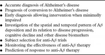
Aβ plaques and NFTs are the hallmark brain lesions of AD. These microscopic aggregates are still well beyond the resolution of conventional neuroimaging techniques used for the clinical evaluation of patients with AD. While selective tau imaging is starting to show some promising results (Fodero-Tavoletti et al., Reference Fodero-Tavoletti2011), the development of molecular imaging methods for non-invasively assessing Aβ burden in AD is already allowing early diagnosis at presymptomatic stages and more accurate differential diagnosis, as well as evaluation of disease-modifying therapy (Villemagne et al., Reference Villemagne2008b). Table 1 indicates the potential clinical roles for Aβ imaging.
Potential clinical roles for Aβ imaging

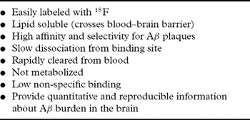
Since Aβ is at the center of AD pathogenesis, and given that several pharmacological agents aimed at reducing Aβ levels in the brain are being developed and tested, much effort is focused on generating radiotracers or agents that allow Aβ imaging in vivo (Villemagne et al., Reference Villemagne2008b). For a radiotracer to be useful as a neuroimaging Aβ probe, a number of key general properties are desirable: it should be a lipophilic molecule that crosses the blood–brain barrier (BBB) and is preferably not metabolized, whilst reversibly binding to Aβ in a specific and selective fashion (Villemagne et al., Reference Villemagne2008b). Furthermore, low non-specific binding to white matter is desirable (Table 2). Therapies, especially those targeting irreversible neurodegenerative processes, have a better chance of succeeding if applied early, making early detection of the underlying pathological process so important.
Ideal Aβ PET radioligand

Several compounds have been evaluated as potential Aβ probes. Mathis and colleagues at the University of Pittsburgh developed numerous Congo red and Chrysamine-G derivatives for potential use as in vivo Aβ probes, but the ability of these relatively large and acidic compounds to cross the BBB was found to be marginal at best. Eventually, it was a thioflavin T derivative, {N-methyl-[C-11]}2-(4′-methylamino-phenyl)-6-hydroxy-benzothiazole ([C-11]6-OH-BTA-1, also known as “Pittsburgh Compound-B” or 11C-PIB, that showed more favorable binding characteristics to fibrillar Aβ both in vitro and in vivo (Mathis et al., Reference Mathis2002).
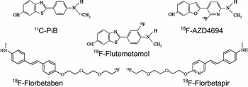
While most PET amyloid imaging agents have been shown to display nanomolar affinity for Aβ fibrils, only a few of these agents have found their way into human clinical trials, with studies conducted in AD patients, healthy age-matched controls (HCs) and patients with other dementias using 11C-PiB (Klunk et al., Reference Klunk2004). 18F-Florbetaben (Rowe et al., Reference Rowe2008), 18F-Flutemetamol (Vandenberghe et al., Reference Vandenberghe2010), 18F-Florbetapir (Wong et al., Reference Wong2010) and 18F-AZD4694 (Jureus et al., Reference Jureus2010) (Figure 1). The different pharmacokinetic and pharmacodynamic characteristics of these radiotracers are discussed in detail elsewhere (Villemagne and Rowe, Reference Rowe2010).
Amyloid ligands. Chemical structure of selected PET amyloid radiotracers that have been used in clinical studies.

To date, 11C-PiB (Figure 1) has been the most widely used of the currently available amyloid tracers. Unfortunately, the 20-minute radioactive decay half-life of 11C limits its use to centers with an on-site cyclotron and 11C radiochemistry expertise. Consequently, access to 11C-labeled radiotracers is restricted and the high cost of studies is prohibitive for routine clinical use. To overcome these limitations, an Aβ imaging tracer labeled with 18F was required. The 110-minute radioactive decay half-life of 18F permits centralized production and regional distribution as currently practiced worldwide in the supply of FDG for clinical use. There are now several amyloid ligands labeled with 18F under clinical development.
The first reported amyloid imaging tracer, 18F-FDDNP, developed at the University of California, Los Angeles (UCLA) by Barrio and colleagues has been shown to bind both the extracellular Aβ plaques and the intracellular NFTs in AD (Shoghi-Jadid et al., Reference Shoghi-Jadid2002). Direct comparison of 18F-FDDNP with 11C-PIB in human subjects showed a very limited dynamic range of 18F-FDDNP retention between HCs and AD patients (Tolboom et al., Reference Tolboom2009).
18F-Florbetaben (also known as 18F-BAY94–9172) (Figure 1), developed by Bayer-Schering Pharma, has been shown to bind avidly to neuritic and diffuse amyloid plaques and to cerebral amyloid angiopathy (CAA). In human studies, 18F-Florbetaben neocortical retention was higher at 90 minutes post injection in AD subjects compared to HCs and FTLD patients (Rowe et al., Reference Rowe2008). Recently completed phase II clinical studies further confirmed these results (Sabri et al., Reference Sabri2010), while a preliminary study showed no pharmacokinetic differences in HCs of different ethnic background (Senda et al., Reference Senda2010). An ongoing longitudinal study in MCI subjects is helping to establish the usefulness of 18F-Florbetaben PET in predicting progression to AD (Ong et al., Reference Ong2010). Matching the reported post-mortem distribution of Aβ plaques, 18F-Florbetaben allowed detection of the presence or absence of AD pathology in the brain in subjects from a wide spectrum of neurodegenerative diseases (Villemagne et al., Reference Villemagne2010) (Figure 2).
Amyloid imaging with 18F-Florbetaben. Representative transaxial PET images obtained 90–120 minutes post injection of 300 MBq 18F-Florbetaben in a healthy control (HC), a participant classified as mild cognitive impaired (MCI), one subject with Alzheimer's disease (AD) and one with frontotemporal lobar degeneration (FTLD). There is cortical retention of 18F-Florbetaben in the frontal, posterior cingulate/precuneus and lateral temporal areas with relative sparing of occipital and sensori-motor cortex in the MCI and AD subjects. In contrast, there is no cortical 18F-Florbetaben retention in the HC or FTLD subjects. All images are scaled to the same SUVR maximum.

Another fluorinated tracer, 18F-Flutemetamol (also known as 18F-GE067; Figure 1), developed by GE Healthcare, has just completed Phase 2 studies, in which it was demonstrated to distinguish between AD patients and HCs, displaying a similar regional distribution to 11C-PIB (Vandenberghe et al., Reference Vandenberghe2010).
Developed by Hank Kung for Avid Radiopharmaceuticals, 18F-Florbetapir (also known as 18F-AV-45; Figure 1) is an amyloid tracer with rapid reversible binding characteristics (Wong et al., Reference Wong2010). Several studies have used 18F-Florbetapir. A multicenter Phase 2 study in AD, MCI and HCs showed the ability of 18F-Florbetapir to discriminate between AD and HC (Sperling et al., Reference Sperling2009). 18F-Florbetapir has been adopted as the amyloid tracer for ADNI 2, and is being used in a large number of therapeutic clinical trials around the world. Recent completion of a Phase 3 study demonstrated a 96% agreement with neuropathology in detecting brain Aβ in vivo (Clark et al., Reference Clark2011). 18F-Florbetapir is about to become the first 18F-labeled radiotracer approved by the Food and Drug Administration (FDA) since FDG.
Still in the early phase of development by Astra-Zeneca, human studies with 18F-AZD4694 (Figure 1) has shown low non-specific binding to white matter, suggesting it might be helpful for the detection of small Aβ cortical deposits at very early stages of the disease process (Jureus et al., Reference Jureus2010).
Amyloid imaging in dementia
Clinical imaging studies with amyloid radiotracers are providing quantitative information on Aβ burden in vivo, leading to new insights into Aβ deposition in the brain and facilitating research of neurodegenerative diseases where Aβ may play a role (Masters et al., Reference Masters2006). Most of the amyloid imaging studies worldwide had been conducted using 11C-PiB.
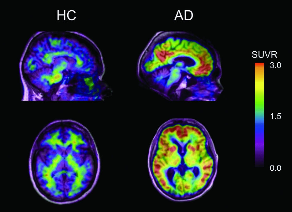
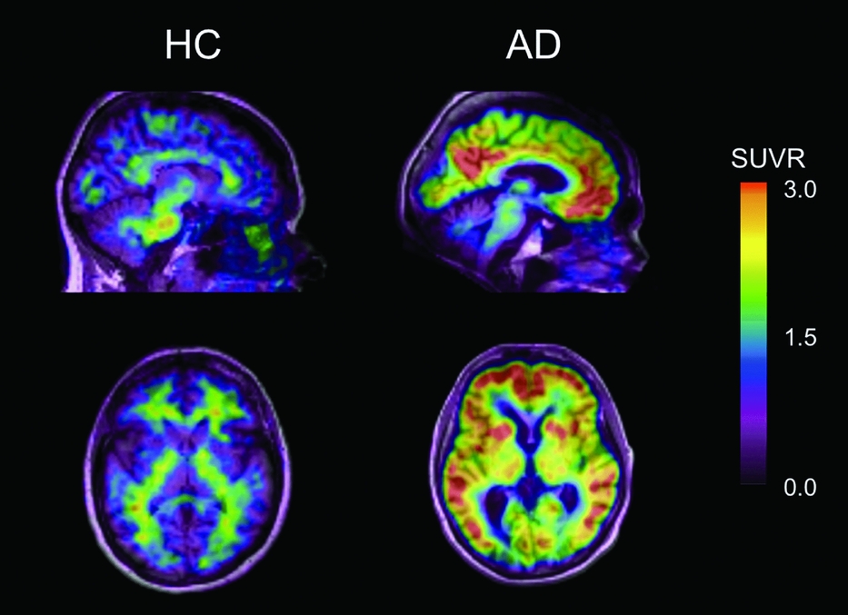
On visual inspection, cortical retention of 11C-PiB, regardless of disease severity, is markedly elevated in AD (Klunk et al., Reference Klunk2004; Rowe et al., Reference Rowe2007), whilst being generally lower and more variable in DLB, probably reflecting a larger spectrum of Aβ deposition (Rowe et al., Reference Rowe2007). In AD, 11C-PiB retention is highest in the frontal, cingulate, precuneus, striatum, parietal and lateral temporal cortex (Figure 3). The occipital cortex, sensori-motor cortex and mesial temporal cortex are usually less affected (Figure 3). With the exception of 18F-FDDNP, which shows high retention in the mesial temporal cortex, a similar pattern of retention in AD is observed with most 11C and 18F amyloid ligands. 11C-PiB binds reversibly, with fastest clearance from the cerebellar cortex and slowest from white matter (Klunk et al., Reference Klunk2004; Rowe et al., Reference Rowe2007). With the exception of familial AD cases (Klunk et al., Reference Klunk2007; Villemagne et al., Reference Villemagne2009a) or prion diseases like Creutzfeldt-Jakob disease (Villemagne et al., Reference Villemagne2009b), absence of neuritic Aβ plaques in the cerebellar cortex of AD patients is reflected in a similar clearance from this region in all groups (Rowe et al., Reference Rowe2007; Ikonomovic et al., Reference Ikonomovic2008). No significant 11C-PiB cortical retention was observed in a preliminary report on PD patients (Johansson et al., Reference Johansson2008), nor when it was used to discriminate between AD, PD, DLB and PD with dementia (Edison et al., Reference Edison2008). 11C-PiB-PET has also facilitated differential diagnosis of dementia in patients with atypical onset of dementia who presented with provisional diagnosis of either progressive non-fluent aphasia – considered by many to be a variant of FTLD – or posterior cortical atrophy, respectively (Ng et al., Reference Ng2007b).
Amyloid imaging with 11C-PiB. Representative 40–70-minute post-injection sagittal and transaxial 11C-PiB PET images overlayed on their respective MRI for a healthy control (HC) subject and an Alzheimer's disease (AD) patient. 11C-PiB PET images show clear differences when comparing cortical 11C-PiB retention in either HC or AD patients. Only non-specific PiB binding in white matter is observed in the HC subject, compared to PiB retention in cortical areas of the AD patient. All images are scaled to the same SUVR maximum.
11C-PiB retention is also elevated in subjects diagnosed with CAA (Johnson et al., Reference Johnson2007), and a similar retention pattern is observed in AD and DLB cases (Rowe et al., Reference Rowe2007). There is usually no cortical 11C-PiB retention in patients with clinical diagnosis of FTLD (Rabinovici et al., Reference Rabinovici2007; Rowe et al., Reference Rowe2007), while analysis of 11C-PiB binding in subjects diagnosed with MCI, a condition that progresses to AD at a rate of approximately 15% per year (Petersen, Reference Petersen2007), has revealed two distinct 11C-PiB retention patterns. About 60% of MCI cases show cortical 11C-PiB binding similar in distribution and sometimes in degree to AD, while the rest exhibit little or no cortical 11C-PiB retention, similar to HC (Pike et al., Reference Pike2007).
Interestingly, about 25–35% of asymptomatic age-matched HCs present with cortical 11C-PiB retention, predominantly in the prefrontal and posterior cingulate/precuneus regions (Rowe et al., Reference Rowe2007; Aizenstein et al., Reference Aizenstein2008; Villemagne et al., Reference Villemagne2008b). These findings are in agreement with post-mortem reports that about 30% of older individuals without dementia over the age of 75 years have Aβ plaques, with deposits occurring well before the onset of dementia (Morris and Price, Reference Morris and Price2001). The retention of 11C-PiB in HCs correlates with subtle memory impairment and a greater risk of cognitive decline (Pike et al., Reference Pike2007; Villemagne et al., Reference Villemagne2008b; Reference Villemagne2011), likely to represent what has been termed preclinical AD (Morris and Price, Reference Morris and Price2001) and this group might comprise those who will benefit the most from therapies aimed at reducing or eliminating Aβ from the brain before irreversible neuronal or synaptic loss occurs.
Aβ, cognition and other markers of disease
In vivo amyloid imaging is not only helping elucidate the relation between Aβ deposition and cognition but is also allowing researchers to define and refine its relationship with cerebrospinal fluid (CSF) and genetic biomarkers, as well as to establish the relation between Aβ deposition, neuropathology, brain atrophy and glucose metabolism.
11C-PiB PET studies have not only shown a robust difference in 11C-PiB retention between AD patients and HC (Klunk et al., Reference Klunk2004; Rowe et al., Reference Rowe2007), but also inverse correlations with decreased CSF Aβ42 (Fagan et al., Reference Fagan2006) and cerebral atrophy (Chetelat et al., Reference Chetelat2010). Amyloid imaging data concur both with post-mortem reports on the regional density and sequence of Aβ deposition (Braak and Braak, Reference Braak and Braak1997). The regional retention of 11C-PiB reflects the regional density of Aβ plaques, as reported at autopsy (Ikonomovic et al., Reference Ikonomovic2008), with a higher plaque density in the frontal cortex than in hippocampus, consistent with previous neuropathological and PiB PET reports (Rowe et al., Reference Rowe2007). Recent completion of a Phase 3 study with 18F-Florbetapir demonstrated 96% agreement with neuropathology (Clark et al., Reference Clark2011). Amyloid imaging also agrees with reports that these neuropathological changes precede the clinical expression of AD by many years, (Morris and Price, Reference Morris and Price2001) reflected in the large percentage of cognitively unimpaired individuals with significant 11C-PiB retention in the brain (Pike et al., Reference Pike2007; Aizenstein et al., Reference Aizenstein2008; Villemagne et al., Reference Villemagne2008b). Despite a strong inverse correlation between PiB and FDG in temporal, parietal, and posterior cingulate cortex, the correlation is not present in the frontal lobe or hippocampus (Klunk et al., Reference Klunk2004). Visual assessment of PET images by clinicians blinded to the diagnoses demonstrated that 11C-PiB was more accurate than FDG to distinguish AD from HC (Ng et al., Reference Ng2007a).
To date, ApoE ɛ4 is the most consistent genetic risk factor associated with sporadic AD, and its presence is thought to result in an earlier age of onset. Examination of ApoE ɛ4 allele status revealed that, independent of clinical classification, ɛ4 carriers present with significantly higher 11C-PiB binding than non-carriers, further emphasizing the crucial role that ApoE plays in the metabolism of Aβ (Reiman et al., Reference Reiman2009; Morris et al., Reference Morris2010; Rowe et al., Reference Rowe2010). In both symptomatic and asymptomatic adult individuals with mutations within the APP or presenilin 1 genes associated with familial AD, there is high PiB retention in the caudate nuclei, posterior cingulate/precuneus and, to a lesser degree, prefrontal and temporal cortices, retention that seems to be independent of mutation type or disease severity (Klunk et al., Reference Klunk2007; Villemagne et al., Reference Villemagne2009a).
To date, the relationship between Aβ deposition and the development of clinical symptoms is not fully understood. Although Aβ burden as assessed by PiB-PET does not correlate with measures of memory impairment in established clinical AD, it does correlate with memory impairment in MCI and healthy older subjects (Pike et al., Reference Pike2007; Villemagne et al., Reference Villemagne2008b). Most subjects classified as non-amnestic single domain MCI do not show significant 11C-PiB retention in the brain, consistent with a non-AD pathological process such as FTLD (Pike et al., Reference Pike2007; Villemagne et al., Reference Villemagne2011). Conversely, 60–80% of amnestic MCI subjects have 11C-PiB retention in the brain (Pike et al., Reference Pike2007; Villemagne et al., Reference Villemagne2011). The observations that PiB retention in individuals without dementia relates to episodic memory impairment, one of the earliest clinical symptoms of AD, suggests that Aβ deposition is not part of normal aging, and supports the hypothesis that Aβ deposition occurs well before the onset of symptoms and is likely to represent preclinical AD. The lack of a strong association between Aβ deposition and measures of cognition, synaptic activity and neurodegeneration in AD, in addition to the evidence of Aβ deposition in a high percentage of MCI subjects and asymptomatic HCs, suggests that Aβ is an early and necessary, though not sufficient, cause for cognitive decline in AD (Villemagne et al., Reference Villemagne2008a) and that other downstream mechanisms are involved, likely triggered by Aβ, such as NFT formation, synaptic failure and eventually neuronal loss. Perhaps specific in vivo tau imaging will help elucidate or bridge the gap in understanding between Aβ deposition and the persistent decline in cognitive function observed in AD (Fodero-Tavoletti et al., Reference Fodero-Tavoletti2011).
Longitudinal studies are helping clarify the relationship between Aβ burden and cognitive trajectory in healthy individuals and MCI subjects. A large longitudinal study with 11C-PiB (Villemagne et al., Reference Villemagne2011) showed that Aβ deposition increases slowly in individuals with positive scans whether they have normal cognition, MCI or AD. Furthermore, extensive Aβ deposition was associated with a significantly higher risk of cognitive decline in HCs and with progression from MCI to AD over 1–2 years, denoting the non-benign nature of Aβ deposition. In other reported studies, and probably due to small sample sizes, significant increases in PiB retention were found in individuals but not on group analysis (for further discussion see Villemagne et al., Reference Villemagne2011). Ongoing large longitudinal studies, including both structural and amyloid neuroimaging studies, as well as genetic and biochemical biomarkers, will permit a better understanding of the role and relevance of Aβ deposition in elderly controls and MCI subjects (Ellis et al., Reference Ellis2009).
Summary
The introduction of radiotracers for the non-invasive in vivo quantification of Aβ in the brain has revolutionized the approach to the evaluation of AD. Aβ burden as measured by PET matches histopathological reports of Aβ distribution in aging and dementia, appears more accurate than FDG for the diagnosis of AD, and is an excellent aid in the differential diagnosis of AD from FTLD. ApoE ɛ4 status is associated with higher Aβ burden. As new therapies enter clinical trials, the role of Aβ imaging in vivo is becoming increasingly crucial. Amyloid imaging can provide highly accurate, reliable and reproducible quantitative statements of regional or global Aβ burden in the brain, essential for therapeutic trial recruitment and for the evaluation of disease-specific treatments directed at removing Aβ.
Although Aβ burden as assessed by amyloid agents does not strongly correlate with cognitive impairment in AD, it does correlate with memory impairment and the rate of memory decline in the aging population and MCI subjects. This correlation with memory impairment, one of the earliest symptoms of AD, suggests that Aβ deposition is not part of normal aging, supporting the hypothesis that Aβ deposition occurs well before the onset of symptoms and likely represents preclinical AD. Further longitudinal observations, coupled with different tracers to assess potential downstream effects of Aβ (e.g. selective tau ligands) and biomarkers (e.g. assessment of tau and Aβ in CSF and blood), are required not only to confirm this hypothesis but also to better elucidate the role of Aβ deposition in the course of AD.
Conflict of interest declaration
Dr. Villemagne and Dr. Rowe are consultants for Bayer-Schering Pharma, AG.
Description of authors' roles
Both authors contributed equally to the preparation and writing of this paper.
Acknowledgments
This work was supported in part by Grant 509166 of the National Health and Medical Research Council of Australia, and by Bayer Schering Pharma AG, the Austin Hospital Medical Research Foundation and Neurosciences Victoria.