‘Saliva is not one of the popular bodily fluids. It lacks the drama of blood, the sincerity of sweat and the emotional appeal of tears.’
Irwin D. Mandel (Reference Mandel1990)
Introduction
The purpose of this review is to discuss the history of the development and implementation of oral fluid diagnostics for infectious diseases of human and domestic animals. The use of oral fluid for the assessment of health and diagnosis of disease in humans and animals has a surprisingly long history. Early investigators attempted to evaluate metabolic diseases in humans by testing oral fluid for a variety of analytes, e.g., glycogen, crystal salts and acid salts (Michaels, Reference Michaels1901). This early work led to the conclusion that the ‘principals’ present in serum were also present in saliva. As early as 1909, Pollaci and Ceraulo reported sensitive and specific agglutination of ‘Micrococcus melitensis’ (Brucella melitensis) by oral fluid from patients diagnosed with Malta Fever, thereby, indirectly demonstrating the presence of antibody in saliva. After these early reports, developments in oral fluid diagnostics were generally overshadowed by technical improvements in the detection of analytes in blood or serum. This began to change following a report of the detection of antibodies against human immunodeficiency virus (HIV) in oral fluid from patients with acquired immunodeficiency syndrome (AIDS) (Archibald et al., Reference Archibald, Zon, Groopman, McLane and Essex1986). Because of this report and/or because of concurrent advances in diagnostic technology, oral fluid-based assays for a wide range of infectious and non-infectious diseases, drugs, hormones and disease markers have been developed and implemented in the last two decades (Mandel, Reference Mandel1993; Tabak, Reference Tabak2007).
Basic concepts
The fluid in the oral cavity consists of saliva and transudates originating from the circulatory system. The major salivary glands of mammals vary in number, structure, location and size (Shackleford and Wilborn, Reference Shackleford and Wilborn1968), but humans and the domestic animals (cat, dog, pig, ruminants and horse) all possess at least three major salivary glands, as well as numerous minor salivary glands (MSG). Humans possess parotid, submandibular and sublingual glands (Navazesh, Reference Navazesh1993); equines and swine have parotid, mandibular and sublingual glands (Sisson, Reference Sisson, Getty, Rosenbaum, Ghoshal and Hillman1975a, Reference Sisson, Getty, Rosenbaum, Ghoshal and Hillmanb); ruminants have four glands, i.e., the parotid, mandibular, monostomatic sublingual and polystomatic sublingual glands (Habel, Reference Habel, Getty, Rosenbaum, Ghoshal and Hillman1975); the dog and cat have parotid, mandibular, sublingual and zygomatic glands, with the cat also possessing two well-developed molar salivary glands (Wiggs and Lobprise, Reference Wiggs and Lobprise1997).
The composition of saliva varies among salivary glands, but regardless of species, saliva is mostly water in which are suspended a myriad of molecules with important biological functions, e.g., mucin, amylase, lysozyme, lipase and proline-rich glycoproteins. Within the oral cavity, saliva and its constituents perform important functions related to lubrication, anti-microbial activity, cleansing, bolus formation, mastication, digestion, taste, tooth remineralization and phonation (Llena-Puy, Reference Llena-Puy2006).
In addition to saliva, the fluid in the oral cavity contains serum transudate that crosses the oral mucosa (oral mucosal transudate) and gingiva (gingival crevicular fluid) from capillaries located in the oral mucosa and the gingival tissues (Delima and Van Dyke, Reference Delima and Van Dyke2003; Cameron and Carman, Reference Cameron and Carman2005). The process of passive transudation was first demonstrated by intravenously injecting fluorescein dye into the hind leg of dogs (n=6) and recording fluorescence on filter paper strips collected within and at the gingival crevice (Brill and Krasse, Reference Brill and Krasse1958). In these experiments, the dye appeared at the gingival crevice within 30 s after injection, but within 60 min had not appeared in the eye, hard palate, floor of the mouth, buccal, or labial mucosal, tongue, alveolar mucosa, nasal cavity, rectum, or vagina. The authors proposed that antibodies reached the oral cavity from serum via the gingival crevice and served a protective role in the mouth. This experiment was followed by a similar work in which humans ingested fluorescein dye capsules (Brill and Björn, Reference Brill and Björn1959). Similar results were reported, with the addition of fluorescein dye appearing in more tissues than was seen in dogs, i.e., nasal mucous membranes. Passive transfer of virus from serum to oral fluid was demonstrated with Coxsackie B-1 virus. Rabbits were intravenously injected with Coxsackie B-1 virus (Madonia et al., Reference Madonia, Bahn and Calandra1966) and the virus was detected in oral fluid samples 2 min post-injection at concentrations of 1×10⁴ median tissue culture infectious dose (TCID50) per 0.1 ml or greater. As a result of these experiments, the connection between the circulatory system and the oral cavity was identified. Subsequent work established that the fluid in the oral cavity also reflected the concentration of hormones, drugs, antibodies, viruses and other components in serum (Mandel, Reference Mandel1993).
In the context of this review of diagnostic applications, it is important to be aware that the composition of fluid from the oral cavity is markedly affected by the method of collection (stimulated versus unstimulated), collection device (Chang et al., Reference Chang, Cohen and Bienek2009), and/or the site of sample collection within the oral cavity (Atkinson et al., Reference Atkinson, Dawes, Ericson, Fox, Gandara, Malamud, Mandel, Navazesch and Tabak1993). Atkinson et al. (Reference Atkinson, Dawes, Ericson, Fox, Gandara, Malamud, Mandel, Navazesch and Tabak1993) define ‘oral fluid’ as the fluid in the oral cavity collected by use of an absorptive device. This describes the process and the method most commonly used to collect diagnostic samples. Therefore, ‘oral fluid’ will be used in this sense for the remainder of this review.
Historical developments leading to oral fluid diagnostics
Beginning with the work of Pollaci and Ceraulo (Reference Pollaci and Ceraulo1909), evidence for the presence of antibodies in oral fluid accumulated slowly over the first half of the 20th century. Antibodies against Treponema pallidum were demonstrated in oral fluid in 1940 (Kanter and Appleton, Reference Kanter and Appleton1940) and again in 1953 (Coleman and Appleman, Reference Coleman and Appleman1953). Citing the work of Pollaci and Ceraulo (Reference Pollaci and Ceraulo1909) and Coleman and Appleman (Reference Coleman and Appleman1953), Wheatcroft (Reference Wheatcroft1957) analyzed matched serum and oral fluid samples from persons infected with B. melitensis and demonstrated a correlation (r=0.674) between serum antibody complement fixation titers and oral fluid bacterial agglutination titers.
The presence of serum proteins in human oral fluid was demonstrated in 1960 (Ellison et al., Reference Ellison, Mashimo and Mandel1960). Kraus and Konno (Reference Kraus and Konno1963) showed that antibody was only present in oral fluid when it was present in the serum of the same individual, concluding that this was evidence of a ‘selective and individually regulated transfer of serum proteins into saliva’. This would later prove to be partially true, but would not fully account for all of the antibody in oral fluid.
By 1963 (Kraus and Konno, Reference Kraus and Konno1963), independent research had demonstrated the presence of antibody and/or antigen in human oral fluid from patients infected with a variety of pathogens. Investigators had repeatedly commented on the consistently lower concentration of antibody in oral fluid when compared to matched serum samples and had expressed concern about the impact of lower antibody concentration on the diagnostic sensitivity of antibody-based assays. Three concepts were common to the research literature of the time: (1) the absence of oral fluid reactions in seronegative subjects, (2) lower antibody titers in oral fluid versus matched serum samples and (3) the inconsistent results of oral fluid samples from seropositive subjects.
By 1964, five antibody isotypes (IgM, IgG, IgA, IgD and IgE) had been identified in humans (Martin Reference Martin1969) and it had become evident that IgA was the predominant isotype present in oral fluid (Tomasi and Zigelbaum, Reference Tomasi and Zigelbaum1963). The identification of a secretory component in conjunction with IgA (South et al., Reference South, Cooper, Wollheim, Hong and Good1966) and IgM (Brandtzaeg, Reference Brandtzaeg1975) provided evidence for local production of antibody.
Sources of oral fluid antibodies
Definitive evidence of the passage of serum antibody (IgG, IgM and IgA) from the circulatory system into the oral cavity was reported by Challacombe et al. (Reference Challacombe, Russel, Hawkes, Bergmeier and Lehner1978) in studies with rhesus monkeys. Specifically, intravenously injected radio-labeled IgG, IgM and IgA were detected in oral fluid 30 min later (first sampling point). This simple experiment demonstrated the transfer of serum-derived antibody into oral fluid.
Local production of antibody by serum-derived plasma cells in salivary glands and duct-associated lymphoid tissue (DALT) was also described during this period (Crawford et al., Reference Crawford, Taubman and Smith1975; Brandtzaeg, Reference Brandtzaeg1981, Reference Brandtzaeg and Tenovuo1989; Beckenkamp, Reference Beckenkamp1985; Nair and Schroeder, Reference Nair and Schroeder1986; Mestecky, Reference Mestecky1987, Reference Mestecky1993; Morrier and Barsotti, Reference Morrier and Barsotti1990). These cells secreted IgA into saliva in conjunction with ductal and acinar epithelial cells expressing receptors specific for IgA. In humans, MSG were also demonstrated to play a substantial role in IgA production, contributing 30–35% of total IgA in response to local antigenic stimuli (Crawford et al., Reference Crawford, Taubman and Smith1975). IgM and IgG were also found to be locally secreted, but at lower concentrations than IgA (Challacombe et al., Reference Challacombe, Percival and Marsh1995). Corroborative data from other species supported these initial observations. For example, immune responses in the local tissues of the nasal and oral cavities were examined in mice orally immunized with biodegradable microparticles and IgA-secreting cells were demonstrated in salivary glands and nasal-associated lymphoid tissue (NALT) by enzyme-linked immunosorbent spot (ELISPOT) assays and the secreted antibody was demonstrated in oral fluid by enzyme-linked immunosorbent assay (ELISA) at all sampling points post-inoculation (Challacombe et al., Reference Challacombe, Rahman and O'Hagan1997).
Oral fluid diagnostics in human medicine
The conclusive demonstration of systemic and locally produced antibody (IgA, IgM and IgG) in oral fluid suggested its possible use as a diagnostic specimen for infectious diseases, but the report that precipitated the development of oral fluid diagnostics was the isolation of human T-cell leukemia virus type III (HTLV-III, later renamed HIV) from oral fluid collected from people with AIDS (Groopman et al., Reference Groopman, Salahuddin, Sarngadharan, Markham, Gonda, Sliski and Gallo1984). By 1986, antibodies against HTLV-III were demonstrated in patients with AIDS and/or their sexual partners (Archibald et al., Reference Archibald, Zon, Groopman, McLane and Essex1986). This pivotal publication suggested that detection of clinically healthy individuals infected with HTLV-III could be achieved using oral fluid specimens instead of serum. This concept was quickly evaluated and pre-clinical trials on 600 individuals reported that oral results from fluid and serum were equivalent (Schaefer, Reference Schaefer1990). In 1990, the Food and Drug Administration (FDA) approved clinical trials for the comparison of a commercial oral fluid collection device at five sites within the U.S.A. (Schaefer, Reference Schaefer1990) and in 1994, the FDA approved the first oral fluid collection kit for the detection of HIV antibody (Nightingale, Reference Nightingale1995).
Development of HIV detection technology continued speedily and in the period 2002 to 2006, the FDA approved four rapid HIV assays designed to detect antibodies in oral fluid specimens (Branson, Reference Branson2007). Based on the data submitted for FDA approval, the first approved test (OraQuick® Rapid HIV-1 Antibody Test; OraSure Technologies, Inc., Bethlehem, PA) was estimated to have a diagnostic sensitivity of 99.6% (95% CI=98.5 and 99.9%) and specificity of 100% (95% CI=99.7 and 100%). An evaluation of the second generation assay (OraQuick® Advance Rapid HIV-1/2 Antibody Test; OraSure Technologies, Inc.) reported a remarkably low false-positive rate of 0.27% based on 166,058 tests performed between March 2005 and May 2008 at 10 New York City Sexually Transmitted Diseases (STD) clinics (Rothman and Kalish, Reference Rothman and Kalish2009). False-positive results were determined by performing Western Blot analysis on serum samples from individuals with positive oral fluid screening results.
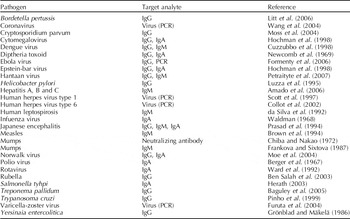
In human diagnostic medicine, a plethora of publications describing the development and application of oral fluid assays for HIV detection may be found in the literature, but the application of oral fluid diagnostics for pathogens and/or antibody has been described for several other infectious diseases (Table 1). Oral fluid offers some distinct advantages over serum-based testing. In particular, specimens are easily collected by personnel with minimal technical training, or even by patients in their homes, and samples are easily processed and stored (Chiappin et al., Reference Chiappin, Antonelli, Gatti and De Palo2007). As a result, oral fluid-based testing has facilitated the collection of large amounts of epidemiological data on significant infectious agents, e.g., HIV in Africa (Fylkesnes and Kasumba, Reference Fylkesnes and Kasumba1998; Connolly et al., Reference Connolly, Shisana, Colvin and Stoker2004) and Thailand (Frerichs et al., Reference Frerichs, Silarug, Eskes, Pagcharoenpol, Rodklai, Thangsupachai and Wongba1994) and measles in Europe (Ramsay et al., Reference Ramsay, Brugha and Brown1997), Ethiopia (Nigatu et al., Reference Nigatu, Samuel, Cohen, Cumberland, Lemma, Brown and Nokes2008), Brazil (de Azevedo Neto et al., Reference de Azevedo Neto, Richards, Nokes, Silveira, Cohen, Passos, de Souza, Brown, Pannuti and Massad1995; Oliveira et al., Reference Oliveira, Siqueira, Brown, Camacho, Faillace and Cohen1998) and Africa (Ohuma et al., Reference Ohuma, Okiro, Bett, Abwao, Were, Samuel, Vyse, Gay, Brown and Nokes2009).
Selected examples of diagnostic applications for the detection of infectious diseases of humans based on oral fluid specimens

Oral fluid diagnostics in veterinary medicine
The literature describing the detection of antibodies and pathogens in oral fluid of domesticated mammals describes core findings similar to those reported in human research. For simplicity, the veterinary research section of the review is organized by species.
Feline samples
Detection of antibodies in oral fluid
Harley et al. (Reference Harley, Gruffydd-Jones and Day1998) developed ELISA methods to quantify feline immunoglobulins in oral fluid and characterized the levels of IgG, IgM and IgA in stimulated and non-stimulated oral fluid collected from healthy cats in 12 h intervals for 4 days. They reported that oral fluid was most rich in IgA, stimulated oral fluid contained a lower concentration of immunoglobulin, and the concentration of immunoglobulin in oral fluid was generally consistent across multiple sampling points.
Detection of pathogens in oral fluid
The detection of feline leukemia virus (FeLV) antigen in feline oral fluids was first reported in the late 1980s (Lewis et al., Reference Lewis, Wright, Lafrado, Shanker, Palumbo, Lemoine and Olsen1987; Lutz and Jarrett, Reference Lutz and Jarrett1987) and oral fluid antigen tests are commercially available and commonly used to screen for FeLV infection. Recent publications have reported the detection of FeLV RNA in oral fluid by PCR compared to the antigen ELISA routinely used in veterinary clinics (Gomes-Keller et al., Reference Gomes-Keller, Tandon, Riondato, Gönczi, Meli, Hofmann-Lehmann and Lutz2006a, Reference Gomes-Keller, Tandon, Gönczi, Meli, Hofmann-Lehmann and Lutzb). They reported excellent diagnostic performance and close agreement between PCR and antigen capture ELISA results.
Feline immunodeficiency virus (FIV) was first demonstrated in oral fluid collected from inoculated cats in 1988 (Yamamoto et al., Reference Yamamoto, Sparger, Ho, Andersen, O'Connor, Mandell, Lowenstine, Munn and Pedersen1988). Subsequently, Poli et al. (Reference Poli, Giannelli, Pistello, Zaccaro, Pieracci, Bendinelli and Malvaldi1992) demonstrated detectable anti-FIV IgG in oral fluid samples collected from 15 of 16 seropositive cats and 0 of 16 seronegative cats. Anti-FIV IgA was also detected in oral fluid from 13 of 16 seropositive cats and 0 of 16 seronegative cats.
In addition to feline retroviruses, PCR detection of Candidatus Mycoplasma turicensis’ (CMt) in oral fluid from cats was reported by Willi et al. (Reference Willi, Boretti, Baumgartner, Tasker, Wenger, Cattori, Meli, Reusch, Lutz and Hofmann-Lehmann2006). Dean et al. (Reference Dean, Helps, Gruffydd Jones and Tasker2008) corroborated the original report and also detected Mycoplasma haemofelis (Mhf) and Candidatus Mycoplasma haemominutum (CMhm) in oral fluid.
Canine samples
Detection of antibodies in oral fluid
Immunoglobulins in canine oral fluid have been described by Heddle and Rowley (Reference Heddle and Rowley1975) and in greater detail by German et al. (Reference German, Hall and Day1998). As in other species, canines produce local IgA and IgM, but oral fluid also contains serum-derived IgG and albumin. German et al. (Reference German, Hall and Day1998) found little variation in total immunoglobulin concentration between multiple oral fluid samples collected from the same animal over time. In dogs experimentally challenged with Taenia pisiformis, specific IgG and IgA were detectable for 12 weeks post-inoculation, at the end of the study (Kinder et al., Reference Kinder, Carter, Allan, Marshall-Clarke and Craing1992). The authors suggested oral fluid specimens could serve as an alternate method of detecting intestinal helminth infection and replace the traditional, but relatively diagnostically insensitive, process of detecting proglottides or eggs in fecal samples.
Detection of pathogens in oral fluid
Of public health interest, some zoonotic pathogens are detectable in canine oral fluid. Rabies virus is commonly transmitted via animal bites and has been demonstrated in canine oral fluid (Côrtes et al., Reference Côrtes, de Oliveira and Peixoto1979; Fekadu et al., Reference Fekadu, Shaddock and Baer1982; Kasempimolporn et al., Reference Kasempimolporn, Saengseesom, Lumlertdacha and Sitprija2000). Several strains of Bartonella spp. have been detected in canine saliva by PCR (Duncan et al., Reference Duncan, Maggi and Breitschwerdt2007).
Equine and ovine samples
In equines, local production of immunoglobulins in parotid and submandibular salivary glands has been demonstrated (Genco et al., Reference Genco, Yecies and Karush1969; Hurlimann and Darling, Reference Hurlimann and Darling1971). In sheep, IgA is the major immunoglobulin in oral fluid (Smith et al., Reference Smith, Dawson, Wells and Burrells1975). Experiments using radio-labeled iodine demonstrated that the majority of IgA in sheep oral fluid is of local origin, with IgM selectively transported into respiratory secretions and saliva (Scicchitano et al., Reference Scicchitano, Sheldrake and Husband1986). Diagnostically, equine oral fluid has been used to test for the presence of performance altering chemicals in race horses (Morgan and Gellhorn, Reference Morgan and Gellhorn1947; Horner, Reference Horner1976), but no reports of the use of oral fluid for the detection of infectious diseases were found for equines or ovines.
Bovine samples
While oral fluid samples have been used to isolate and culture bacterial pathogens (Escherichia coli and Salmonella) from feedlot cattle (Smith et al., Reference Smith, Gray, Moxley, Younts-dahl, Blackford, Hinkley, Hungerford, Milton and Klopfenstein2004, Reference Smith, Moxley, Clowser, Folmer, Hinkley, Erickson and Klopfenstein2005; Renter et al., Reference Renter, Visser, McFall, Smith, Berg, Silasi, Berezowski and Sorensen2004; Stanford et al., Reference Stanford, Bach, Marx, Jones, Hansen, Wallins, Zahiroddini and McAllister2005), the detection of foot-and-mouth disease virus (FMDV) has been the major focus of research in bovines. The earliest work on oral fluid reported the presence of anti-FMDV neutralizing antibody, presumably locally produced, in calves following nasal inoculation with live type 01 FMDV (Figueroa et al., Reference Figueroa, Ohlbaum and Contreras1973). Archetti et al. (Reference Archetti, Amadori, Donn, Salt and Lodetti1995) measured total anti-FMDV immunoglobulin and IgA in oral fluid and probang (oropharyngeal fluid) samples of vaccinated or FMDV-infected animals. Vaccinated cattle did not generate an antibody response detectable in either sample type, but cattle exposed to, or inoculated with, live vaccine produced detectable IgA in oral fluid. Later work led to the concept that secretory IgA in oral fluid may be used to differentiate vaccinated from FMDV-infected animals (Parida et al., Reference Parida, Anderson, Cox, Barnett and Paton2006). Parida et al. (Reference Parida, Anderson, Cox, Barnett and Paton2006) developed an ELISA to detect IgA antibody against structural proteins (SP) in oral fluid under the premise that vaccines used in response to an FMDV outbreak would not include non-structural proteins (NSPs). Considering their work preliminary, they reported a diagnostic specificity of 99.4% and suggested a potential application for detecting persistently infected, sub-clinically infected, or vaccinated animals.
Porcine samples
Detection of antibodies in oral fluid
The first report of antibodies in oral fluid from pigs was published in 1976 when Corthier (Reference Corthier1976) reported that intranasal vaccination of pigs with the Thiverval strain of classical swine fever virus (CSFV) resulted in detectable antibody in pharyngeal secretions. They subsequently inoculated pigs via intranasal and intramuscular routes and measured the antibody response in serum and oral fluid (Corthier and Aynaud, Reference Corthier and Aynaud1977). Strong responses were measured in both sample types, indicating both systemic and local immune responses. The strongest local response (in oral fluid) was observed after intranasal inoculation at the highest dose used.
DeBuysscher and Dubois (Reference DeBuysscher and Dubois1978) inoculated pigs with E. coli strain 1261 via oral or Thiry-Vella loop routes and examined the submandibular and sublingual salivary glands post mortem by staining for anti-E. coli plasma cells of the IgA, IgM and IgG isotypes. They reported IgA-secreting plasma cells to be most numerous followed by approximately equal numbers of IgM- and IgG-secreting cells, but found no significant differences in the number and isotype of plasma cells in submandibular and sublingual salivary glands between pigs inoculated orally or via Thiry-Vella loops.
A few years later DeBuysscher and Berman (Reference DeBuysscher and Berman1980) performed essentially the same experiment, but with transmissible gastroenteritis virus (TGEV). They observed substantial increases in the number of IgA-secreting cells in salivary glands, followed by IgM-secreting cells, and a small increase in the number of IgG-secreting cells in pigs inoculated orally or via Thiry-Vella loops. These findings supported the conclusion that corollary increases in antigen-specific plasma cells in intestinal mucosa and distant secretory tissues, i.e., salivary glands, result from the secretory component receptor–conveyor mechanism.
Loftager et al. (Reference Loftager, Eriksen and Nielsen1993) collected samples from pigs intranasally inoculated with Actinobacillus pleuropneumoniae (APP) and pigs naturally infected with APP. IgA concentrations in oral fluid and serum collected over time were measured using a whole-cell ELISA. IgA was detectable in oral fluid before it appeared in serum and declined more rapidly than serum IgA or IgG. These investigators concluded that oral fluid IgA detection could serve as a practical method to screen for early infection with APP.
Detection of pathogens in oral fluid
Several swine pathogens are known to be present in detectable levels in oral fluid samples. Vesicular stomatitis virus (VSV) has been isolated from oral fluid, but not serum, from pigs infected via mechanical vectors or by direct contact with infected pigs (Stallknecht et al., Reference Stallknecht, Howerth, Reeves and Seal1999). In pigs inoculated with FMDV (O TAW 3/97), virus was recovered by virus isolation for up to 10 days post-infection (DPI) and by PCR for up to 11 DPI (Eblé et al., Reference Eblé, Bouma, de Bruin, van Hemert-Kluitenberg, Van Oirschot and Dekker2004). Recently, the detection of porcine respiratory and reproductive syndrome virus (PPRSV) and porcine circovirus type 2 (PCV2) by PCR was reported in swine oral fluid samples collected under both experimental and field conditions (Prickett et al., Reference Prickett, Simer, Christopher-Hennings, Yoon, Evans and Zimmerman2008a, Reference Prickett, Simer, Yoon, Kim and Zimmermanb).
Stability of PRRSV and anti-PRRSV antibody in oral fluid
The stability of anti-PRRSV antibody and PCR-detectable PRRSV is highly temperature dependent, and antimicrobial treatment provides no improvements in stability at lower temperatures (Prickett et al., Reference Prickett, Cutler, Kinyon, Naberhaus, Stensland, Yoo and Zimmerman2010). In particular, both virus and antibody were found to be stable at ⩽10°C over 12 days of storage. Conventional serum storage protocols (freezing or refrigeration at 4°C) will preserve PRRSV and anti-PRRSV antibody in oral fluid diagnostic samples.
Future applications of oral fluid diagnostics in veterinary medicine
Cumulatively, the literature strongly supports implementation of oral fluid-based diagnostics. Pathogen-specific IgA, IgM and IgG antibodies have all been demonstrated in oral fluid collected from diverse domestic animal species in response to infection. A variety of infectious agents, both local and systemic, are shed in oral fluid, including some of the most economically significant pathogens of production animals, e.g., FMDV, CSFV and PRRSV. From the perspective of animal welfare, oral fluid sampling also offers a more ‘animal friendly’, non-invasive method to collect diagnostic specimens. Taken together, the diagnostic properties of oral fluid present an opportunity to formulate diagnostically sensitive and specific oral fluid-based assays.
The application of oral fluid-based testing facilitates monitoring, surveillance and detection of disease in animal populations. For example, despite our best efforts, PRRSV, Mycoplasma hyopneumoniae, influenza, PCV2 and other endemic pathogens continue to cause significant economic losses in large U.S. swine production systems (Holtkamp et al., Reference Holtkamp, Rotto and Garcia2007). In part, the disease status quo is maintained by the lack of timely information on the circulation of pathogens. Oral fluid testing offers an opportunity to easily collect herd-level disease data on a periodic basis. Integration of longitudinal disease data with herd records (interventions, morbidity, mortality and production parameters) would provide for (1) appropriately timed and targeted interventions, (2) ‘real time’ evaluations of interventions and (3) improved, herd-specific estimates of the impact of specific pathogens on pig health and productivity. This proactive approach to disease monitoring translates diagnostic costs into improved growth performance rather than a historical vignette of past disease events.
The presence of local and systemic antibody in oral fluid suggests the possibility of isotype-specific antibody assays. That is, isotype-specific assays could provide insight into the chronology of infection, such as with the early production of IgM or the production of IgG after class switching has taken place.
Once established for the routine management of endemic diseases, an oral fluid surveillance infrastructure would facilitate rapid data collection in situations such as regional or national disease control and/or eradication programs or foreign animal disease (FAD) contingency efforts. Collection of diagnostic specimens by on-site personnel would profoundly impact the time to detection and effectiveness of control of FAD outbreaks.
The purpose of this review was to summarize the history of the development and implementation of oral fluid diagnostics for infectious diseases of human and domestic animals. A tangential objective was to promote research in diagnostic applications in veterinary medicine, with the hope that animal health can eventually realize the benefits offered by oral fluid diagnostics. At this point, research is necessary to develop and standardize collection methods, optimize diagnostic assays for the oral fluid specimens, and establish sampling protocols for pathogens of interest. Ultimately, point-of-care rapid assays, i.e., cow-side, sow-side, or pen-side tests, and access to real-time infectious disease data will revolutionize our delivery of health management services.