Introduction
It has been 100 years since Erwin Payr first developed an operation to improve the effects of a paralysed vocal fold,Reference Payr 1 and operations based on this technique are still in use today. This technique, medialisation thyroplasty, aims to improve the symptoms caused by vocal fold palsy by realigning the lateralised vocal fold into the midline.Reference Montgomery, Montgomery and Warren 2 This allows the non-paralysed vocal fold to adduct fully against the paralysed vocal fold, permitting the vocal folds to return to their normal function of lower airway protection, respiration and phonation.Reference Harries 3
Although relatively uncommon in the general population,Reference Abraham, Gonen and Kraus 4 , Reference Abraham, Bains, Downey, Korst and Kraus 5 vocal fold palsy can have a devastating effect on the basic functions of the larynx.Reference Flint, Purcell and Cummings 6 , Reference Sipp, Kerschner, Braune and Hartnick 7 It is not a disease per seReference Ahmad, Muzamil and Lateef 8 and is rarely due to diseases of the larynx; rather, it is most commonly caused by lung and oesophageal cancers,Reference Lam, Ho, Ng and Wei 9 , Reference Yumoto, Minoda, Hyodo and Yamagata 10 trauma,Reference Ahmad, Muzamil and Lateef 8 and post-operative complications.Reference Lang, Chu, Tsang, Wong and Wong 11 Some patients, especially those who are otherwise well, can compensate for a paralysed vocal fold by aligning the normal vocal fold across the midline.Reference Abraham, Bains, Downey, Korst and Kraus 5 However, this can cause significant morbidity in the generally co-morbid patients most commonly affected by vocal fold palsy. In these patients, vocal fold palsy can lead to complications including voice hoarseness, dysphagia, cough, sore throat, breathlessness, aspiration and stridor.Reference Abraham, Bains, Downey, Korst and Kraus 5 , Reference Damrose 12
Historical vocal fold palsy treatments
The importance of the larynx and vocal folds in voice production was well understood by the ancient Greeks. The author of the Hippocratic work ‘On the Flesh’ first noted that patients with a severed larynx, for example due to attempted suicide, were left without a voice.Reference Assimakopoulos, Patrikakos and Lascaratos 13 Galen, in the second century AD, also noted the importance of the recurrent laryngeal nerves, and experimentally proved their importance in voice production.Reference Kühn 14 However, vocal fold palsy could not be formally diagnosed without the ability to view the vocal folds, which was impossible until the nineteenth century.
A Frenchman, Leveret, is credited with the first attempt to examine the living larynx, in 1743, with a bent mirror and a snare for removing laryngeal polyps,Reference Alberti 15 , Reference Jahn and Blitzer 16 but his technique was not widely adopted. Both Bozzini in Germany, in 1807,Reference Jahn and Blitzer 16 and Babington in England, in 1829,Reference Assimakopoulos, Patrikakos and Lascaratos 13 , Reference Jahn and Blitzer 16 made further attempts to visualise the vocal folds, but Bozzini's device was ignored. Babbington's device was the first to be recognisable as a laryngoscope; however, he never published details of his invention. It was not until 1854 that Manuel Garcia, a Spanish voice teacher, developed the first widely known laryngoscope.Reference Jahn and Blitzer 16 Ludwig Türck and Johann Czernak became the first physicians to be recognised for viewing the larynx of living patients in 1858,Reference Jahn and Blitzer 16 at which point the field of laryngology as a whole began to greatly expand.
By 1892, vocal fold paralysis was recognised as a neurological problem of the larynx, as discussed in a textbook written by Bosworth. 17 However, treatment for vocal fold paralysis was slow to develop, with Jackson and Jackson's 1942 text on laryngology stating that treatment for vocal fold paralysis ‘is chiefly the treatment of the basic disease causing the paralysis. Nothing is of any avail locally’. 18 During this time, surgery for laryngeal cancers was beginning to develop, with Bilroth performing the first total laryngectomy in 1873.Reference Čoček 19
Brunnings, in 1911, was one of the first to document attempts to repair vocal fold palsy. He described a technique to medialise paralysed vocal folds by injecting the paralysed fold with paraffin.Reference D'Antonio, Wigley and Zimmerman 20 This method proved to be highly successful;Reference Isshiki 21 however, paraffin was found to cause severe local foreign body reactions, such as paraffinomas,Reference Isshiki, Morita, Okamura and Hiramoto 22 so experiments with injection therapy continued with other materials.Reference D'Antonio, Wigley and Zimmerman 20 Teflon was the most widely used material for injection therapy after its introduction by Arnold in 1962.Reference D'Antonio, Wigley and Zimmerman 20 , Reference Koufman 23 Although effective at producing medialisation of the paralysed vocal fold, issues such as a relatively high rate of airway compromise,Reference Netterville, Stone, Lukas, Civantos and Ossoff 24 the formation of Teflon granulomasReference Netterville, Coleman, Chang, Rainey, Reinisch and Ossoff 25 and formations of intra-thyroid masses mimicking thyroid carcinomaReference Crumley, Izdebski and McMicken 26 led to a search for alternative methods for vocal fold medialisation.
Historical medialisation thyroplasty methods
The first attempt at true medialisation thyroplasty was conducted in 1915, by a German surgeon called Erwin Payr.Reference Payr 1 Although probably best known for his work in neurosurgery, Payr had an interest in multiple areas of surgery, with Payr's disease, Payr's sign, Payr's membrane and the Payr pylorus clamp all named after him.Reference Kühnel and Seifert 27 He developed many novel surgical techniques and instruments during his career, and had particular interests in joint disorders, vascular surgery and nerve surgery.Reference Schwokowski 28
Payr's interest in thyroid surgery in particular led him to work on an operation for vocal fold palsy.Reference Payr 1 , Reference Bray, Young and Harries 29 He had noted that compression of the elastic thyroid cartilage significantly improved the voice of a young soldier with dysphonia following a thyroidectomy. In order to replicate this effect, Payr's operation involved a U-shaped incision through the thyroid cartilage, utilising an anteriorly pedicled flap of thyroid cartilage depressed inwards with a small wedge of cartilage to force the vocal fold into the midline.Reference Payr 1 However, there were several disadvantages to this technique. Lack of safe anaesthesia, blood replacement and risk of infectionReference Tucker 30 complicated all surgical techniques during this time period, but this particular operation was prone to post-operative infections and was associated with a high incidence of airway compromise.Reference Carrau, Pou, Eibling, Murry and Ferguson 31 Complications specific to Payr's surgery included the unpredictable durability of the pedicle,Reference Merati and Bielamowicz 32 especially when the thyroid cartilage was calcified,Reference Isshiki 21 as well as difficulty in determining the degree of vocal fold displacement and in fixing the cartilage in the desired position.Reference Seiffert 33
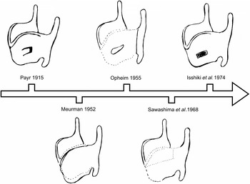
Local treatments for vocal fold paralysis were thought to be ineffective as late as 1941,Reference Woodson 34 but modifications to Payr's approach were made over the next few decades. A diagram illustrating some of the different external approaches to medialisation thyroplasty is shown in Figure 1. One of the very earliest attempts was by Seiffert in 1942, who utilised a piece of cadaveric rib cartilage implanted through a hole in the thyroid cartilage to medialise the vocal fold.Reference Seiffert 33 Meurman published on a series of 15 cases in 1952, where cartilage taken from the costal rib was implanted via an external approach between the thyroid cartilage and the inner perichondrium in order to place a paralysed vocal fold into a median position.Reference Meurman 35 A similar technique was devised by Opheim in 1955, who instead used an incised piece of the thyroid cartilage itself, inserted via an external incision at the level of the vocal folds in the inner perichondrium.Reference Opheim 36 However, these techniques and others that involved direct intervention on the soft tissue immediately lateral to the vocal fold led to post-operative oedema, which often required a tracheostomy.Reference Isshiki, Morita, Okamura and Hiramoto 22
Despite these early setbacks, the value of medialisation thyroplasty was evident and the search for better ways to surgically medialise paralysed vocal folds continued. Sawashima et al., in 1968, developed a method based on that of Meurman and Opheim, which used an incised piece of thyroid cartilage placed between the ala of the thyroid cartilage and the inner perichondrium via an incision in the external thyroid cartilage.Reference Sawashima, Totsuka, Kobayashi and Hirose 37 Kamer and Som, in 1972, used a similar technique to place a piece of thyroid cartilage incised from its lower rim in patients after traumatic vocal fold paralysis.Reference Kamer and Som 38 Tucker in 1979 and 1983 further developed a similar technique, inserting a piece of thyroid cartilage from the ipsilateral upper margin of the ala into a pocket created between the inner perichondrium and the cartilage at the level of the vocal folds.Reference Tucker 39 , Reference Tucker 40 Kressner, in 1953, devised a technique utilising two cartilage implants; one aimed to shift the paralysed vocal fold medially, whilst the other smaller piece was inserted into the cricoarytenoid joint space to lift the arytenoid and to further medialise the posterior part of the vocal fold.Reference Kressner 41 This technique was refined and used in a series of 12 patients by Westhues in 1973.Reference Westhues 42 Deneke, in 1964, used a wedge from the posterior edge of the thyroid, but kept this in place by dividing the cricoarytenoid tendon, medially displacing the arytenoid and suturing it against the cartilage wedge.Reference Deneke 43 These techniques all relied on cartilage implants, and concerns about the long-term durability of the implants limited their widespread application.
Not all of the early techniques utilised pieces of thyroid cartilage. Libersa, in 1952, developed a technique similar to that of Meurman, with the only significant difference being the use of a piece of acryl as an implant instead of cartilage.Reference Libersa 44 Morrison, in 1948, described a technique to displace the arytenoid cartilage towards the midline along the (partially removed) posterior superior border of the cricoid cartilage in order to medialise the vocal folds, which was the first attempt at correcting arytenoid displacement,Reference Morrison 45 whereas Montgomery, in 1966, fixed the arytenoid cartilage to the cricoid cartilage in a new position with a pin.Reference Montgomery 46 However, neither of these methods gained much popularity because of their technical difficulty.Reference Isshiki, Morita, Okamura and Hiramoto 22 Bernstein and Holt, in 1967, attempted to reposition the vocal fold in experimental animals by transposing the sternohyoid muscle between the thyroid cartilage and inner perichondrium,Reference Bernstein and Holt 47 whilst Mündnich, in 1970, tensed and medically shifted the vocal fold by pulling and fixing the arytenoid towards the lower horn of the thyroid cartilage.Reference Mündnich 48 However, thyroplasty did not gain widespread acceptanceReference Remacle and Eckel 49 until the development of type I thyroplasty in 1974.
A diagram illustrating some of the different external approaches to medialisation thyroplasty, from PayrReference Payr 1 in 1915 to Isshiki et al.Reference Isshiki, Morita, Okamura and Hiramoto 22 in 1974. PayrReference Payr 1 (1915) described a U-shaped incision made in the thyroid cartilage at the level of the vocal folds, which is displaced inwards to medialise the underlying vocal fold. MeurmanReference Meurman 35 (1952) reported splitting the anterior angle of the thyroid cartilage, avoiding damage to the perichondrium, and making a pocket for the cartilage implant between the perichondrium and the inner side of the thyroid ala. OpheimReference Opheim 36 (1955) described splitting the thyroid cartilage along the anterior midline using a small electric saw, avoiding damage to the inner perichondrium. A horizontal incision was made through the inner perichondrium at the level of the vocal folds, through which a small pouch was formed for the cartilage implant. Sawashima et al.Reference Sawashima, Totsuka, Kobayashi and Hirose 37 (1968) reported excising a cartilage implant from the upper part of the thyroid ala, contralateral to the paralysed vocal fold. An incision was then made through the anterior angle of the thyroid cartilage, avoiding damage to the underlying perichondrium, and a pocket was made between the thyroid cartilage and the perichondrium at the level of the vocal folds for insertion of the cartilage implant. Isshiki et al.Reference Isshiki, Morita, Okamura and Hiramoto 22 (1974) described a rectangular incision made through the thyroid cartilage at the level of the vocal folds, and the insertion of a Silastic implant shaped to the size of the incision, abutting the laryngeal mucosa.

Modern medialisation thyroplasty
Isshiki and his team at Kyoto University first described the technique now known as type I thyroplasty in 1974, in a paper describing a series of operations on dogs. Instead of operating directly on the vocal folds, which caused mucosal scarring, reducing vocal fold compliance and hindering vibration,Reference Isshiki 50 they instead aimed to alter the position and physical property of the vocal fold by actively changing the cartilaginous framework on which the vocal folds were suspended.Reference Isshiki, Morita, Okamura and Hiramoto 22 As discussed above, this was not an entirely novel idea, and shared the same basic principle as Payr's initial 1915 operation.
Type I thyroplasty involves creating a window into the anterior thyroid cartilage ala under local anaesthesia, with or without sedation, and inserting a prosthesis, which was initially made from Silastic (a type of hardened siliconeReference McCulloch and Hoffman 51 ).Reference Isshiki, Morita, Okamura and Hiramoto 22 This pushes the paralysed vocal fold medially, allowing it to make contact with a non-paralysed vocal fold on the opposite side and restoring its function.Reference Harries 3 Being able to carry out this procedure under local anaesthesia is a major benefit, as this not only enables patients otherwise unfit for general anaesthesia to undergo the procedure, but also allows for auditory feedback from the patient's own voice to ensure the best possible vocal result.Reference Harries 3 It is also a much simpler surgical procedure than those developed before it, taking around 30 minutes to complete in an uncomplicated case.Reference Harries 3
However, type I thyroplasty was not immediately widely used. Koufman noted in 1986 that Teflon injection was still the most common therapy for vocal fold palsy, despite its drawbacks.Reference Koufman 23 Koufman's 1986 paper described a slightly modified version of Isshiki's technique, wherein a Silastic implant is inserted through a window in the thyroid cartilage, and the best possible improvement in voice and glottic closure is ensured by asking the patient to phonate during the procedure and by viewing the vocal fold intra-operatively under fibre-optic examination.Reference Koufman 23
Koufman's post-operative outcomes were very good, supporting the more widespread use of medialisation thyroplasty.Reference Koufman 23 Thyroplasty started to become much more widely utilised after this review, as discussed by Netterville et al. in 1993.Reference Netterville, Stone, Lukas, Civantos and Ossoff 24 Netterville et al. reviewed a series of 116 medialisation thyroplasty operations performed on 100 patients from 1987 to 1992, and observed both the results and complication rates of the surgery. These authors found that deglutition and aspiration were universally improved, and the voices of a vast majority of patients were significantly better. The ability to adjust or reverse the procedure (by moving or removing the implant) represented a significant improvement over Teflon injections. The authors also found that it was much easier to train junior doctors to perform medialisation thyroplasty.Reference Netterville, Stone, Lukas, Civantos and Ossoff 24 In addition, the complication rate was very low, with no patients having extrusion of their implant, although one implant needed to be removed because of post-irradiation laryngocutaneous fistula formation.Reference Netterville, Stone, Lukas, Civantos and Ossoff 24
Thyroplasty started to become more widespread over the next few years, quickly becoming the treatment modality of choice in some centres,Reference Netterville, Jackson and Civantos 52 particularly when it was shown that medialisation thyroplasty led to an improved vocal result when compared to Teflon injections.Reference D'Antonio, Wigley and Zimmerman 20 A review of 84 patients by Flint et al. showed that medialisation thyroplasty improved dysphagia and reduced aspiration rates, improving outcomes when compared to conservative management alone.Reference Flint, Purcell and Cummings 6 Isshiki's technique has continued to be used and modified over the two decades following its initial development,Reference Carrau, Pou, Eibling, Murry and Ferguson 31 , Reference Isshiki 50 , Reference Maragos 53 – Reference Choi, Chung, Lim and Kim 55 and was considered the ‘gold standard’ treatment by 1999.Reference Carrau, Pou, Eibling, Murry and Ferguson 31
Currently, medialisation thyroplasty is still the gold standard and most widely used surgical technique for vocal fold medialisation. The technique continues to be modified.Reference Charous 56 The most commonly used modification involves the use of a folded piece of Gore-Tex rather than a Silastic implant,Reference Isshiki 57 which allows the degree of medialisation to be fine-tuned without having to remove and re-carve the entire implant,Reference Selber, Sataloff, Spiegel and Heman-Ackah 58 and is now becoming the most commonly used implant.Reference McCulloch and Hoffman 51 Medialisation thyroplasty is also increasingly being combined with a similar surgery, arytenoid adduction, in which the arytenoid muscle is pulled across anteriorly by a suture until it is almost parallel to the lateral cricothyroid muscle.Reference Franco 59 This is particularly helpful in closing posterior gaps between adducted vocal folds, which further improvesReference Chester and Stewart 60 the changes in voice seen after medialisation thyroplasty.Reference Abraham, Gonen and Kraus 4 , Reference Charous 56 , Reference Miller, Grady and Netterville 61
As with all surgical procedures, medialisation thyroplasty is by no means perfect. The surgery is often carried out under local anaesthesia, is of short duration and involves little risk of significant blood loss,Reference Tucker, Wanamaker, Trott and Hicks 62 allowing this operation to be carried out in frail patients with significant co-morbidities such as multiple cranial neuropathies and terminal cancer. Post-operative haemorrhage into the vocal folds can cause airway obstruction requiring tracheostomy,Reference Tucker, Wanamaker, Trott and Hicks 62 although this complication has become less common as the original technique has been refined over time.Reference Abraham, Gonen and Kraus 4 , Reference Cotter, Avidano, Crary, Cassisi and Gorham 63 A much more common complication of medialisation thyroplasty is implant extrusion, with an estimated incidence of 0.8–9.8 per cent,Reference Halum, Postma and Koufman 64 , Reference Kartha, Young and Mohan 65 which generally presents as increasing dysphonia.Reference Halum, Postma and Koufman 64 Fortunately, one of the advantages of medialisation thyroplasty is its potential reversibility. An extruded implant is generally very straightforward to remove by either an open or endoscopic approach,Reference Abraham, Gonen and Kraus 4 , Reference Tucker, Wanamaker, Trott and Hicks 62 – Reference Halum, Postma and Koufman 64 and it is possible for a patient to undergo revision thyroplasty to replace a malpositioned or extruded implant.Reference Koufman and Postma 66 , Reference Lundeberg, Flint, Purcell, McMurray and Cummings 67
Conclusion
External approach medialisation thyroplasty was not the only surgical technique developed over the last 100 years to repair vocal fold palsy.Reference Harries 68 Voice therapy together with surgical techniques can improve phonation,Reference Kasterovic, Veselinovic and Mitrovic 69 but the two main surgical alternatives to medialisation thyroplasty are vocal fold injection and vocal fold re-innervation.Reference Sipp, Kerschner, Braune and Hartnick 7
Vocal fold injection with Teflon was the most commonly used technique for several decades, but problems with vocal fold granuloma formation, lack of reversibility and over-reliance on an individual surgeon's skill decreased the popularity of this technique.Reference D'Antonio, Wigley and Zimmerman 20 , Reference Koufman 23 , Reference Netterville, Coleman, Chang, Rainey, Reinisch and Ossoff 25 Injection therapy has been employed using other materials, including collagen (autologous or from animal sourcesReference Damrose 12 , Reference Tsunoda, Baer and Niimi 70 ), autologous fatReference McCulloch, Andrews, Hoffman, Graham, Karnell and Minnick 71 , Reference Chen, Pai, Lin, Wang and Hsiung 72 or Gelfoam.Reference Anderson and Mirza 73 However, most of the new materials used for injection therapy are reabsorbed, meaning that patients will often need either repeat injections or further intervention with medialisation thyroplasty unless the vocal fold palsy resolves. This is an advantage in patients where fast, temporary intervention is needed; hence, injection therapies are still in widespread use.Reference Damrose 12 , Reference Friedman, Burns, Heaton and Zeitels 74 Research into injection therapy for vocal fold palsy is ongoing. Some research shows long-term improvement after autologous fascia transplantation,Reference Sant'Anna and Mauri 75 possibly because of the transfer of tissue stem cells and the regeneration of damaged tissues in the vocal folds.Reference Tsunoda, Baer and Niimi 70 , Reference Tsunoda, Kondou, Kaga, Niimi, Baer and Nishiyama 76 Injection medialisation is currently undergoing a resurgence in popularity because the procedure can be performed in the office under local anaesthesia. There is ongoing evidence that early injection for unilateral vocal fold palsy gives a good voice outcome.Reference Friedman, Burns, Heaton and Zeitels 74 , Reference Jang, Lee, Ahn and Son 77 This procedure is being adopted in many units where the nature of surgery exposes the recurrent laryngeal nerve, such as head and neck dissection, and cardiothoracic, cervical spinal or skull base surgery.
Another alternative approach is that of paralysed vocal fold re-innervation. Medialisation thyroplasty minimises the effect of vocal fold palsy without treating the underlying paralysis of the vocal fold; re-innervating the fold should, in theory, recreate its normal mass and tension.Reference Crumley, Izdebski and McMicken 26 Initial operations aimed to anastomose the ansa hypoglossi to the recurrent laryngeal nerve,Reference Crumley, Izdebski and McMicken 26 , Reference Tucker 78 , Reference Tucker 79 but the most common technique used in re-innervation is to anastomose the ansa cervicalis and the recurrent laryngeal nerve.Reference Su, Hsu, Chen and Sheng 80 – Reference Zur and Carroll 83 This technique is often carried out in combination with either injection therapy or laryngeal framework surgery because there is a significant delay before full improvements to the voice are seen, as it takes time for the anastomosed nerve to regenerate.Reference Crumley, Izdebski and McMicken 26 , Reference Tucker 79 , Reference Chhetri, Gerratt, Kreiman and Berke 84 , Reference Blumin and Merati 85
One hundred years after the first operation, medialisation thyroplasty remains the method of choice for treating vocal fold palsy,Reference Dursun, Boynukalin, Bagis Ozgursoy and Coruh 86 with significant benefits to both vocal performance and quality of life in the patients on whom it is performed.Reference Hajioff, Rattenbury, Carrie, Carding and Wilson 87 , Reference Billante, Clary, Childs and Netterville 88 However, with experimental advances, the rise of re-innervation and resurgence of injection medialisation, it is impossible to say whether it will remain the gold standard in another 100 years. Whatever the future holds, it will be a very interesting journey.