Introduction
There have recently been many reported cases of sensorineural hearing loss of mitochondrial origin. In such patients, the effectiveness of cochlear implantation has been recognised in those with the m.1555A>G and m.3243A>G mutations.Reference Sinnathuray, Raut, Awa, Magee and Toner1 However, the efficacy of such treatment for patients with other mitochondrial DNA mutations has not yet been defined.
Here, we present a patient with syndromic hearing loss, probably caused by a novel mitochondrial DNA mutation (m.625G>A), who gained excellent benefit from cochlear implantation.
Case report
The patient, an 11-year-old girl, was the first child of healthy and nonconsanguineous Japanese parents. There was no family history of hearing loss or epilepsy, and the patient had had no perinatal problems. Her motor and cognitive development was normal, but she displayed an abnormally short stature for her age.
The patient's hearing difficulty had first been noticed by her mother at the age of six years. Two years later, the patient had been examined by an otolaryngologist for the first time, and bilateral hearing aids had been prescribed. However, her hearing ability continued to deteriorate. There had been no previous exposure to aminoglycoside antibiotics. In addition to hearing loss, at the age of eight years the patient had begun to suffer generalised tonic seizures, uncontrolled by valproic acid. At the age of 10 years, she had been referred to our institution, as her family had moved to the locality near our hospital.
On physical examination, the patient had a height of 119.0 cm (−3.0 standard deviations (SD)), a weight of 21.9 kg (−1.7 SD) and a head circumference of 53.6 cm (+0.9 SD). Cranial nerve and cerebellar functions were normal. Hypertrichosis was observed. Although her muscle force did not decrease, she was unable to exercise for extended periods of time. Deep tendon reflexes were normal, without spasticity. She was unable to communicate verbally, although her intelligence appeared normal as she could communicate in writing and could solve age-appropriate arithmetic problems. Otitis media was not found.
Laboratory data revealed mildly elevated blood lactate levels (24.0 mg/dl (normal range, <17 mg/dl), with a pyruvate level of 1.0 mg/dl (normal range, <0.9 mg/dl)), and noticeably elevated cerebrospinal fluid lactate levels (55.8 mg/dl, with a pyruvate level of 2.0 mg/dl).
Electroencephalography revealed no distinct epileptic discharge during waking and sleeping states.
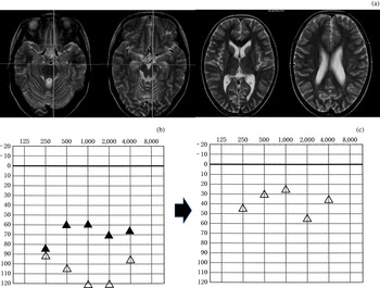
Computed tomography showed no internal ear malformations. Magnetic resonance imaging (MRI) revealed mild brain atrophy without focal lesions (Figure 1a).
(a) Axial magnetic resonance imaging brain scans, showing mild brain atrophy without focal lesions. (b) Left ear audiogram taken at 11 years, before cochlear implantation, following progression of hearing loss (hearing aids were no longer useful). (c) Left ear audiogram taken one month after implantation, showing significant improvement, with hearing thresholds of almost 25 − 45 dB. Δ = sound source 1 m away, without hearing aids; ▴ = with hearing aids in both ears
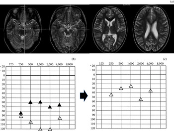
Formal pure tone audiography revealed hearing thresholds of between 90 and 120 dB at 250 through to 4 kHz. The patient's hearing aids only minimally improved her hearing thresholds (Figure 1b).
Auditory evoked potential testing showed a barely detectable auditory reaction at maximum intensity stimulation of 105 dB.
Therefore, the patient was considered to be a candidate for cochlear implantation.
Informed consent for participation in academic research was obtained from the patient and her parents.
During cochlear implantation, temporalis muscle and skin specimens were obtained.
Genomic DNA was extracted from blood, skin and muscle specimens. Sequencing of the whole mitochondrial genome was performed using the mitoSEQ resequencing system (Applied Biosystems, Foster City, California, USA). Polymerase chain reaction amplification was conducted, using forward mismatch primer (nucleotides 601–624, 5′-GCAATACACTGAAAATGTTTAGC-3′; where G = guanine, C = cytosine, A = adenine and T = thymine) and reverse primer (nucleotides 768–786, 5′-CGTTTTGAGCTGCATTGCT-3′). This enabled the m.625G>A sequence to be specifically recognised, and cut using the restriction enzyme BstOI (Promega, Madison, WI, USA). The proportion of heteroplasmy was approximately measured by using a mixture-template standard curve of wild type and mutant clones.
The activities of the mitochondrial respiratory chain complexes I, II, III and IV were assayed, using methods previously described.Reference Rahman, Blok, Dahl, Danks, Kirby and Chow2 We used the diagnostic criteria for respiratory chain disorders previously published by Bernier et al. Reference Bernier, Boneh, Dennett, Chow, Cleary and Thorburn3
Cochlear implantation and clinical course
The patient underwent left-sided cochlear implantation (using a CI24RCS device; cochlear LTD, Lane Cove, Australia) at the age of 11 years.
One month after implantation, she was able to use the telephone, clearly indicating improvement in her hearing function. Audiological data indicated a good response (Figure 1c). Her speech perception score increased to almost 100 per cent, from 0 per cent before surgery.
Twenty months after surgery, the patient and her parents were satisfied with her improved communication, and she continued to attend regular school classes. Her epileptic seizures were well controlled by carbamazepine and clonazepam. Her neurological signs and symptoms remained nonprogressive, possibly due to vitamin B1 supplementation.
Histological analysis
Unfortunately, many artifactual opaque fibers were observed in the temporalis muscle biopsy. Nevertheless, a few cytochrome c oxidase (COX) negative fibres were identified, although there were no ragged red fibres or strongly succinate dehydrogenase (SDH) reactive blood vessels (data not shown).
Genetic analysis
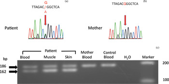
Whole mitochondrial DNA genome analysis, using peripheral blood DNA, detected two heteroplasmic base transitions: m.625G>A (Figure 2a) and m.5231G>A (data not shown).
Detection of the heteroplasmic m.625G>A transition. Diagrams represent screening of the patient's peripheral blood (a) and her mother's peripheral blood (b) for whole mitochondrial DNA genomes, and show the heteroplasmic m.625G>A transition in the patient's blood but not the mother's blood. (c) Electrophoretic strip showing that, in the presence of the m.625G>A mutation, the 186 base pair (bp) fragment was cleaved into 162 and 24 bp fragments (the latter not shown) by (BstOI is manufactured by Promega, Madison, WI, USA). This mutation was present in a heteroplasmic state in the patient's blood, muscle and skin, but was not detected in the mother's blood. Wild-type clones contained only the m.625G sequence.
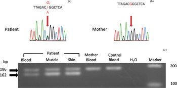
The m.625G>A transition was present in a heteroplasmic state in the patient's blood, muscle and skin, but was not detected in her mother's blood (Figures 2b). The proportion of m.625G>A in muscle and skin was higher than that in blood (the approximate mutation load was 80 per cent in muscle and skin, and 70 per cent in blood) (Figure 2c). This transition was not present in 50 healthy controls.
The heteroplasmic m.5231G>A transition was present in both the patient's and her mother's blood.
Biochemical analysis
Respiratory chain enzyme assay showed that complex III activity was markedly decreased (30 per cent relative to citrate synthase, 17 per cent relative to complex II) while complex IV activity was slightly decreased (55 per cent relative to citrate synthase, 31 per cent relative to complex II).
Discussion
Mitochondrial sensorineural hearing loss is divided into the nonsyndromic type associated with m.1555A>G and the syndromic type associated with m.3243A>G. The complex of mitochondrial encephalopathy, lactic acidosis and stroke-like episodes (known as MELAS) is representative of the latter.Reference Sinnathuray, Raut, Awa, Magee and Toner1
We considered our case to be the syndromic type, because the patient had short stature and suffered from hypertrichosis and epilepsy. Moreover, she showed high lactate levels in her blood and cerebrospinal fluid, and mild brain atrophy on MRI.
In the syndromic type of mitochondrial hearing loss, the retrocochlear auditory pathways require investigation, specifically to establish whether the auditory peripheral nerve and central nervous system (CNS) are intact or not. However, successful cochlear implantation has been reported in patients with the mitochondrial encephalopathy, lactic acidosis and stroke-like episode complex.Reference Rosenthal, Kileny, Boerst and Telian4–Reference Sue, Lipsett, Crimmins, Tsang, Boyages and Presgrave6 Sue et al. have reported successful cochlear implantation in such a patient, who had profound, bilateral hearing loss.Reference Sue, Lipsett, Crimmins, Tsang, Boyages and Presgrave6
Our case, too, underwent successful cochlear implantation, despite possible CNS disorders. In patients with many types of mitochondrial, profound, sensorineural hearing loss, we speculate that cochlear implantation may represent a promising treatment, because hearing loss associated with mitochondrial disorders is more likely to be caused by cochlear dysfunction than retrocochlear abnormalities.Reference Sinnathuray, Raut, Awa, Magee and Toner1, Reference Sue, Lipsett, Crimmins, Tsang, Boyages and Presgrave6, Reference Yamasoba, Tsukuda, Oka, Kobayashi and Kaga7 Results from a guinea pig cochlear model also suggest that chronic mitochondrial dysfunction may most predominantly affect the stria vascularis and supporting cells.Reference Yamasoba, Goto, Komaki, Mimaki, Sudo and Suzuki8 Therefore, we believe that cochlear implantation should be considered in patients with progressive sensorineural hearing loss associated with a mitochondrial disease, regardless of whether their hearing loss is syndromic or nonsyndromic.
Of course, this treatment option should be reviewed for the potential complications; it may develop contraindications on the MRI scan (unless the magnet in the receiver-stimulator has been moved), or adverse events such as post-implant meningitis due to bacterial cellulitis.Reference Papsin and Gordon9
In our patient, whole mitochondrial DNA genome analysis detected two different heteroplasmic, one-base substitutions: m.625G>A and m.5231G>A.
Although heteroplasmic single nucleotide polymorphisms are rare, the m.5231G>A transition is unlikely to be pathogenic, because it has been listed as a single nucleotide polymorphism in the Mitomap database,10 and because it was carried by our patient's healthy mother.
On the other hand, the m.625G>A transition (which involves the transfer RNA gene for phenylalanine) has not previously been reported in association with disease. This transition lies in close vicinity to the site of the m.622G>A mutation, which has been reported to be present in mild mitochondrial disease with hearing impairment.Reference Deschauer, Swalwell, Strauss, Zierz and Taylor11 Moreover, other mutations in the same transfer RNA gene for phenylalanine (e.g. m.582T>C, m.583G>A, m.606A>G, m.608A>G, m.611G>A, m.618T>C, m.636A>G and m.642T>C) have been recognised and listed in Mitomap, with deafness frequently mentioned as a clinical symptom.Reference Mancuso, Filosto, Mootha, Rocchi, Pistolesi and Murri12–Reference Valente, Piga, Lamantea, Carrara, Uziel and Cudia14 In our patient, respiratory enzyme studies revealed a significant defect in complex III and a possible slight defect in complex IV, relative to citrate synthase and complex II. These results resembled those for other mutations of the same mitochondrial transfer RNA gene for phenylalanine, such as m.622G>A and m.618T>C.Reference Deschauer, Swalwell, Strauss, Zierz and Taylor11, Reference Kleinle, Schneider, Moosmann, Brandner, Krähenbühl and Liechti-Gallati15 Moreover, m.625G>A was not identified in our patient's mother's peripheral blood DNA, implying a de novo origin of the mutation, although this is not conclusive because only blood DNA was available from the mother. Such sporadic mutations have been reported in other patients with the same mitochondrial transfer RNA phenylalanine gene mutation.Reference Mancuso, Filosto, Mootha, Rocchi, Pistolesi and Murri12, Reference Darin, Kollberg, Moslemi, Tulinius, Holme and Grönlund16
• This report describes the case of a girl with mitochondrial sensorineural hearing loss
• Cochlear implantation was effective, and improved the patient's quality of life
• The mitochondrial DNA 625G>A mutation may be pathogenic for syndromic hearing loss
Accordingly, we conclude that the m.625G>A transition may cause mitochondrial respiratory dysfunction and syndromic hearing loss. Another standard muscle biopsy and cybrid study would clarify the pathogenicity of the m.625G>A transition.
Conclusion
We report a sporadic case of progressive sensorineural hearing loss and epilepsy due to a mitochondrial disorder, successfully treated with cochlear implantation. The novel, heteroplasmic m.625G>A transition in the mitochondrial transfer RNA gene for phenylalanine may have been the pathogenic mutation in this case.
Cochlear implantation should be considered for patients with progressive, profound, bilateral, sensorineural hearing loss due to mitochondrial disease other than that due to the m.3243A>G mutation of the transfer RNA (tRNA(leu)) gene, or the m.1555A>G mutation of the 12s rivosomal RNA.
Acknowledgements
We are grateful to Dr Kei Murayama and Dr Akira Ohtake for their enzyme study and helpful comments. We also thank Dr Ichizo Nishino for muscle pathology analysis. This work was partially supported by a grant from the Ministry of Education, Science and Culture of Japan.